L21: Ovarian cycle
 Call Kai
 Learn
 Practice Test
 Spaced Repetition
 Match
 Flashcards
 Knowt Play
1/10

There's no tags or description
Looks like no tags are added yet.
11 Terms
Formation and activation of primordial follicles
After initial differentiation of the gonad:
A large number of PGCs (now named oocytes) will die
Squamous granulosa cells start to differentiate
Oocytes organized in clusters (cysts) surrounded by the squamous granulosa
Oocytes start meiosis and arrest in prophase I
Formation and activation of primordial follicles in different animals
Timing differ between species
→Rodents: Clusters (cysts) assembly occurs at birth
→In humans and cows, this process occurs mid gestation
Process is gonadotropin (LH; FSH) independent in mammals
How follicles form and develop in the ovary of a newborn mouse
Stage A – Unassembled follicles: Oocytes are grouped together in nests (cysts) in the newborn mouse's ovary.
Stage B – Primordial follicles: The cysts break down, and individual oocytes become surrounded by a layer of flat (squamous) pregranulosa cells, forming primordial follicles.
Stage C – Primary follicles: By postnatal day 4, some follicles have developed into primary follicles, where the oocyte is surrounded by cuboidal granulosa cells instead of flat cells.
Once a follicle is stimulated to become a primary follicle, the process is irreversible (it cannot go back).
During this process, many oocytes die, making it a critical stage of follicle development.
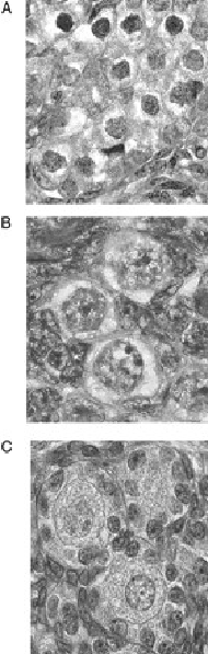
Follicle development diagram

What happens to the oocyte during the primary follical stage
Primary follicle formation: A primary follicle is identified by a single layer of cuboidal granulosa cells surrounding the oocyte.
Meiosis is paused: The oocyte is arrested (stopped) at the diplotene stage of prophase I of meiosis.
Oocyte growth: While meiosis is paused, the chromosomes decondense (uncoil) and are actively transcribed. This allows the oocyte to grow significantly.
Zona pellucida formation: A membrane begins forming around the oocyte. As zona pellucida (ZP) protein genes are expressed, this develops into the zona pellucida.
Oocyte stimulates granulosa cells: The oocyte releases factors that stimulate the surrounding granulosa cells.
Granulosa cells stimulate oocyte: In return, granulosa cells release factors that encourage the oocyte to grow.
What happens during oocyte growth
Replication of cytoplasmic organelles especially mitochondria and their DNA
Increase in ribosome, mRNA and proteins
Lots of nutrients stored in granules and vesicles
Golgi apparatus enlarge and moves to the periphery (export ZP glycoproteins; cortical granules)
Centriole disappears (the male one will be used)
Interactions between oocyte and granulosa
Granulosa cells start expressing FSH receptor and are thus now responsive to FSH (critical point for future growth and recruitment)
Communication via gap-junctions between granulosa cells and oocyte critical to prevent meiosis to proceed

How does the secondary follicle develop
Granulosa cells multiply: The number of granulosa cell layers increases significantly around the oocyte. These cells are surrounded by a basal lamina.
Theca cells develop: Theca cells begin multiplying on the outside of the basal lamina, forming two layers:
Internal theca: The inner layer.
External theca: The outer layer, which contains a muscular layer.
Blood vessels develop: Simultaneously, blood vessels begin forming within the theca layers.
Nutrient and waste exchange: There are no blood vessels inside the follicle, so nutrients must diffuse from the theca layer into the follicle, while waste products diffuse outward.

How do antral follicles develop
Cavitation occurs: A fluid-filled cavity appears inside the follicle
Antral follicles develop through four stages:
Small
Medium
Large
Preovulatory (before ovulation)
Cells continue multiplying: Both granulosa cells and theca cells increase in number as the follicle develops
Composition of theca cells
Theca externa: This is the outer layer, which contains muscle and is innervated (has nerve connections).
Theca interna: This is the inner layer, which produces androgens under the control of LH (luteinizing hormone).
