N245: Disorders of Ventilation and Gas Exchange
 Call Kai
 Learn
 Practice Test
 Spaced Repetition
 Match
 Flashcards
 Knowt Play
1/87

There's no tags or description
Looks like no tags are added yet.
88 Terms
Function of respiratory system
To transport oxygen to the tissues and carbon dioxide away from the body

Upper respiratory tract
Consists of the nose, mouth, pharynx, epiglottis, larynx, and trachea (contains resident flora)

Lower respiratory tract
Consists of the larynx, trachea, bronchi

Hypercapnia
Excessive carbon dioxide in the blood --> metabolic acidosis --> hyperventilation

Hypoxemia
Low oxygen in the blood

Chemoreceptors
Chemical sensors in the brain that detect changes in blood oxygen and blood carbon levels

Ventilation
Movement of air in and out of the lungs (breathing)

What stimulates ventilation?
Increases in blood CO2 level
Hypoxic drive definition
A backup system of respiration --> instead of CO2 levels, chronically low levels of oxygen in the blood stimulate the respiratory drive

Which patients will experience the switch to hypoxic drive?
Seen in patients with chronic lung diseases
Hypoxic drive process
Chronic elevation of CO2 --> chemoreceptors become desensitized to hypercapnia --> no increase in respiration until there is SEVERE decrease in O2

What causes hypoxemia?
1. Lack of oxygen in the air
2. Dysfunction of the respiratory system, neurological systems, or circulatory system
What is the numeric criteria for hypoxia?
Below 95%
Gas diffusion
The movement of gas across the respiratory membrane between alveoli and capillaries, and across capillary walls between blood and other tissues

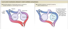
Mismatching of ventilation and perfusion
When areas of the lung are ventilated (have air) but not perfused (blood) or when areas are perfused (have blood) but not ventilated (no air)
Mild hypoxemia symptoms
Metabolic acidosis, increased BP and HR, sweating, confusion

Why does hypoxia cause acidosis?
When skeletal muscles don't have oxygen, they can metabolize using aerobic respiration --> metabolic acidosis
Chronic hypoxemia symptoms
Increased RR, vasoconstriction, increased production of RBCs, cyanosis

Why does hypoxemia lead to increased RBC levels?
Kidneys sense low O2 --> secrete erythropoietin to compensate
Pneumothorax
Air in the pleural cavity, aka a collapsed lung

Closed pneumothorax
Air enters the pleural space through an opening in the pleura that covers the lung (no opening in chest wall)

Simple/spontaneous pneumothorax
A type of closed pneumothorax where air enters the pleural space through a tear on the surface of the lung

Secondary pneumothorax
Associated with underlying respiratory disease
A type of closed pneumothorax in which air enters the pleural space due to:
1. Rupture of an emphysematous bleb on lung surface
2. Erosion of the lung by a tumor/TB
Bleb
Alveolar rupture --> development of small air blisters (blebs) on the lung surface

Open (traumatic) pneumothorax
Air enters the pleural cavity due to an injury/opening to the chest wall

"Sucking" wound
A type of open pneumothorax in which something punctures the chest wall --> air is sucked into the wound (air moves from high to low pressure) --> pneumothorax

Tension pneumothorax
A type of open pneumothorax --> air enters the pleural cavity during inspiration --> hole closes pleural cavity during expiration --> trapping air inside

S+S of pneumothorax
Atelectasis, dyspnea,, unequal breath sounds and chest expansion, hypoxia

Treatment of pneumothorax
Chest tube with suction --> pulls the air out of the pleural cavity and allows the collapsed lung to re-expand

Pleural Effusion
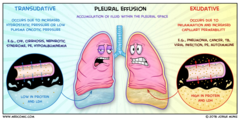
Excess fluid in the pleural cavity --> causes increased pressure in the pleural cavity

Exudative effusions
Response to inflammation (purulent drainage, blood, chyle)
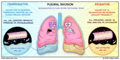
Transudate effusions
Occurs due to increased hydrostatic pressure/low colloid osmotic pressure --> watery exudate
Treatment of pleural effusion
1. Remove underlying cause
2. Thoracentesis to remove fluid

Thoracentesis
The surgical puncture of the chest wall with a needle to drain fluid from the pleural cavity
S+S of pleural effusion
Dyspnea (sudden onset), chest pain, dull sounds during percussion and absence of breath sounds

Atelectasis
Incomplete expansion of the alveoli --> collapsed lung

How can obstruction of bronchi cause atelectasis?
Obstruction prevents air from being inhaled --> alveoli cannot expand --> lung collapses
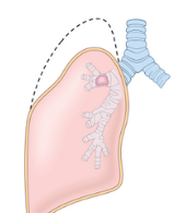
How can loss of surfactant cause atelectasis?
Surfactant decreases surface tension within alveoli --> lack of surfactant will cause alveoli to collapse in on themselves

How can fibrotic tissue cause atelectasis?
Fibrotic scar tissue cannot expand --> restricts expansion of lungs

Postoperative atelectasis
Can occur after surgery --> shallow ventilation due to pain, slow respirations due to anesthetics

S+S of atelectasis
Dyspnea, increased HR and RR, chest pain

Treatment of atelectasis
Goal is to reduce obstruction and increasing lung expansion (O2, mucous thinners, bronchodilators)

Chronic Obstructive Pulmonary Disease
A group of chronic respiratory disorders --> causes progressive and irreversible damage to lungs
1. Asthma
2. Emphysema
3. Chronic bronchitis

Cor pulmonale
Right-sided ventricular hypertrophy and heart failure due to COPD

Cor pulmonale pathophysiology
Poorly ventilated alveoli --> pulmonary arterioles vasoconstrict to divert blood --> too many alveoli poorly ventilated --> pulmonary hypertension --> hard for right ventricle to pump blood into lungs --> right ventricular hypertrophy

Asthma
Bronchial obstruction due to a type 1 hypersensitivity in hypersensitive bronchioles

What does the parasympathetic nervous system do to bronchial tone?
Parasympathetic nervous system --> vagus stimulation --> bronchiole constriction
What does the sympathetic nervous system do to bronchial tone?
Sympathetic nervous system --> adrenaline binds to β2 adrenergic receptors --> bronchiole dilation
What causes asthma
Hypersensitive airways due to interplay between genetics and the environment

Extrinsic asthma
Stimulated by allergies (type 1 hypersensitivy reaction), onset in children

Intrinsic asthma
Not an allergic reaction --> onset in adulthood --> triggered by infection, exercise, stress
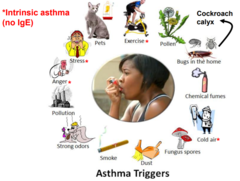
Pathophysiology of asthma
Inflammation --> edema --> bronchospasm and constriction of bronchiole --> obstruction

Asthma attack
Exposure to allergen --> sensitized mast cells release histamine --> inflammation and bronchoconstriction --> more WBCs are called --> more inflammation

S+S of asthma
Wheezing, increased residual volume, dyspnea, fatigue, anxiety

Long term affects of asthma
Smooth muscle walls thicken, fibrosis develops (making expansion more difficult), hyperplasia of mucous glands

Status asthmaticus
A prolonged, extremely severe, life-threatening asthma attack
Asthma pH Changes
Hyperventilation --> respiratory alkalosis
Air trapping --> respiratory acidosis
How is asthma diagnosed
Pulmonary function test, flow rate measurement, signs and symptoms, family history

How is asthma treated (short term)
Inhalers (B2 agonists or corticosteroids)

How is asthma treated (long term)
Inhalers (B2 agonists, cromlyn sodium to prevent inflammation, leukotriene modifiers)

Xolair
Drug that binds to and deactivates IgE antibodies (treatment for severe asthma)

Emphysema
Hyperinflation of air sacs with destruction of alveolar walls

Risk factors of emphysema
1. Genetic factors (deficiency of alpha-1 antitrypsin)
2. Smoking

What is the function of alpha 1-antitrypsin
Alpha 1-antitrypsin is a protein that protects elastase from breaking down lung tissue
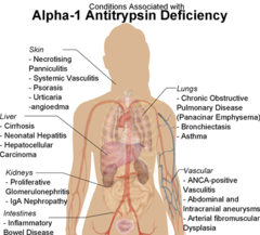
Pathogenesis of emphysema
Smoking --> inflammation --> WBCs release elastase --> decreased alpha 1-antitripsin --> destruction of elastic fibers in lung --> emphysema

What is elastase?
A protein produced by white blood cells; breaks down elastic fibers in the lungs
Breakdown of alveolar walls results in:
1. Loss of surface area for gas exchange (decreased oxygen intake)
2. Loss of pulmonary capillaries
3. Loss of elastic fibers (reduced ROM)

Fibrosis of lungs
Smoking can cause scar tissue to develop in the lungs --> narrowed airways + less lung expandability

S+S of emphysema
Barrel chest due to hyperinflation of the lungs, flattened diaphragm, "pink puffer"

Why are emphysema patients referred to as "pink puffers"?
Since air becomes trapped inside the lungs and the airways are narrowed, pts with emphysema have to use accessory muscles to breathe --> pursing the lips assists with expiration

Advanced emphysema may cause:
Pneumothorax, cor pulmonale
Treatment of emphysema
1. Stop smoking
2. Alpha 1-antitrypsin injections
3. Oxygen
4. Bronchodilators
5. Lung transplant

Chronic bronchitis
Inflammation of the bronchi persisting over a long time

Causes of chronic bronchitis
Cigarette smoking (90%)
Repeated airway infections

How do bronchiole cells change during chronic bronchitis
There is an increase in goblet cells, and ciliary epithelial cells change into squamous cells
Why are chronic bronchitis patients referred to as "blue bloaters"?
Chronic bronchitis can cause severe difficulty breathing --> decreased oxygen in the body --> cyanosis

Treatment of chronic bronchitis
1. Stop smoking/treat infection
2. Bronchodilators
3. Oxygen

Bronchiectasis
What: Abnormal dilation of the bronchi with accumulation of mucus (occurs after infection)
S+S: Chronic cough with sputum, rhonchi, chest pain, shortness of breath

Cystic Fibrosis
An autosomal recessive genetic disorder that is present at birth and affects both the respiratory and digestive systems

CFTR
Cystic fibrosis transmembrane conductance regulator

What does the CFTR gene do?
The CFTR protein moves chloride ions OUT of mucous cells --> chloride attracts water --> helps thin the mucous on the lung surface
The CFTR protein moves chloride ions INTO of cells of sweat glands --> prevents salt loss when sweating

What happens to the CFTR gene in individuals who have cystic fibrosis?
Mutation of CFTR protein --> chloride (salt) becomes trapped in mucous cells --> causes exocrine glands to produce a large amount of thick mucous
Mutation of CFTR protein --> chloride (salt) becomes trapped outside sweat cells --> causes sweat glands to produce lots of salty sweat

How does CF affect the digestive system?
Mucus causes obstructions of the bile duct and pancreatic duct --> prevents the release of digestive enzymes that break down food and absorb nutrients --> malnutrition

How does CF affect the reproductive system?
Mucous prevents sperm from traveling through the vas deferens and blocks the cervix --> causing infertility
How does CF affect the sweat glands?
Sweat becomes very salty --> dehydration

S+S of cystic fibrosis
Salty skin, malnutrition, chronic cough, infections, stunted development

How is CF diagnosed?
Sweat test will reveal elevated NaCl

How is CF treated?
1. Pancreatic enzyme replacement
2. High caloric diet/multivitamins
3. Mucolytics
4. Bronchodilators