Diagnostic Imaging 2- Exam 3- Tumor 1 Malignant Notes- part 2
 Call Kai
 Learn
Learn Practice Test
 Spaced Repetition
 Match
Match Flashcards
 Knowt Play
1/105

There's no tags or description
Looks like no tags are added yet.
Name | Mastery | Learn | Test | Matching | Spaced | Call with Kai | Chat |
|---|
No analytics yet
Send a link to your students to track their progress
106 Terms
What is the most common places for metastases to occur
Lungs, liver and bone
What is the third most common site for metastatic disease
Bone
What are the types of cancer that most often spread to bone first
Breast and prostate
Metastatic bone disease likes to go where
Places with red marrow (pelvis, spine, humerus, skull, ribs)
Metastic bone disease arises from
A primary extra skeletal site
What represents 70% of all malignancies
Metastatic bone disease
Most common primary sites of metastatic bone disease are from where?
Breast
Lung
Prostate
Kidney
Thyroid
Bowel
Metastic bone disease may be what types
Blastic
Lytic
Mixed
Expansile
What are responsible for 80% of bone metastasis?
Breast, lung and prostate cancer
What percentage of people with breast, lung and prostate cancer will develop bone metastasis
Over half of them
What is the most common bone metastasis for females? Where does it come from and what type is it?
Breast, most of it being lytic
What is the most common bone metastasis for males? Where does it come from and what type is it?
Prostate, most of it being blastic
What type of cancer in males most commonly causes metastasis lytic changes
Lung cancer
What is the most common pathway for metastasis to bone
Hematogenous dissemination (blood) via Batsons Venous plexus (vertebral venous plexus)
What shows up in testing when it is blastic metasis to bones
Alkaline phosphatase
What shoes up in testing when it is prostate cancer
Acid phosphatase and/or PSA antigen
Most patients with metastatic disease are what age
Past 4th decade of life
Clinical features of metastatic disease
Recent weight loss
Cachetic (look dead)
Fever
Initial sign of metastatic disease may be
Pain and pathological fracture
When a patient has pain with metastatic disease, what does it look like
Persistent and nocturnal
Common sites for metastasis are where
Areas rich in red marrow
-spine
-ribs and sternum
-pelvis and sacrum
-proximal extremities
-skull
Where is it rare to get metastasis
Distal to knees and elbow
Lytic Mets distal to elbow and knees most common to hands/feet is called_______ metastasis and is most likely due to
Acral metastasis
From lung primary
Osteolytic signs are most likely from what types of cancer in males and females
Females= breast
Males= lungs
Osteoblastic signs are most likely form what types of cancer in males
Prostate
Blow out radiographic signs of metasis in bone are typically from
Renal and thyroid
Blastic Mets example

Lytic Mets example

Blown out Mets example

Favorite spot of metasis is? Why?
Spine cause of the vertebral venous plexus
Radiographic features of vertebral metastasis
Ivory vertebra
Pedicab destruction
Pathological collapse
Focal osteoporosis of body
Malignant Schmorl`s node formation
Most common reason for a missing pedicle
Lytic metastasis
Missing pedicle etiologies
Congenital
Destructive
-Tumor (primary- ABC, osteoblastoma. Soft tissue- neurofibroma. Secondary- lytic Mets
-infection
Surgical
Missing pedicle with contralateral hypertrophy means
Developmentally absent
Example of missing pedicle with contra lateral hypertrophy

Missing pedicle without contralateral hypertrophy so it means its most likely form lytic Mets

Reasons for pathological collapse of vertebra
Lytic metastatic carcinoma
Multiple myeloma
Traumatic fracture (osteoporosis)
Reasons for pathological collapse of vertebra listed from most to least likely
Osteoporosis (traumatic fracture)
Lytic Mets
Multiple myeloma
-remember from last exam!!
Pathological collapse at L5, also list the reasons from most likely to least likely
Osteoporosis
Lyitc mets
Multiple myeloma

There is a 40 year old female with a pathological fracture in the spine, what is not likely the cause and why? List the causes for this case in order of most to least likely
Most likely not osteoporosis cause she's only 40
-lytic Mets
-multiple myeloma
-osteoporosis
Order of things need to be done if the pathological fracture looks like its from osteoporosis
Nothing more
Order of things need to be done if the pathological fracture if from lytic Mets
Bone scan
MRI
Biopsy
Order of things need to be done if the pathological fracture is from multiple myeloma
PEP (protein electrophoresis)
Skeletal survey
MRI
Biopsy (need >10% of plasma cells)
Most likely cause of a pathological fracture in a kid
Eosinophilic granuloma
Another name for pathological fracture
Vertebra plana
Pathological fracture in kid, name what it is most likely from
Most likely from eosinophilic granuloma

If a fracture is dark on T1 then it means
It's a new fracture
Old fractures on T1 look
Bright (same as the normal bones)
Rope 3 causes that could cause a solitary ivory vertebra
Paget's disease
Hodgkin's lymphoma
Osteoblastic metastasis
If an ivory vertebra is enlarged than it is most likely from what
Paget's disease
Example of ivory vertebra from Paget's disease

What is the most common reason in a 20-40 age range for ivory vertebra
Lymphoma
Example of ivory vertebra

Different joint diseases
Degenerative joint disease/OA
Inflammatory- seropostives (RA) seronegatives (AS, psoriatic, reactive)
Metabolic- Gout, CCPD
Infection
What type of joint diseases show shiny corner sign
Seronegative (AS< psoriatic, reactive)
-all have HLAB27
Endplate sclerosis exaggerated by DJD is called
Hemispherical spondylosclerosis

Shiny corner (reactive sclerosis)

Sclerosis of the left ischium and femoral neck

Sclerosis of the anterior aspect of the left 6th rib
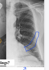
There is sclerosis in the pelvis, the pelvis is a male so this means most likely?
It's blastic Mets from prostate cancer
Extensive blastic Mets on a bone scan

Extensive blastic Mets on an X ray in a pelvis

Osteoblastic metastases in a woman with breast cancer

What is well differentiated mature bone tissue within the marrow
Bone island

What is another name for a bone island
Enostosis
Bone island

Name for multiple bone islands
Osteopoikilosis

Osteopoikilosis loves what location
Ischial tuberosities of the pelvis
If blastic Mets is a clinical possibility when looking at an X ray and you cant tell if its that or a bone island than a blood test will look for?
Alkaline and acid phosphatase levels as as well as PSA and a digital rectal exam
Bone island in the proximal humerus

Mixed lytic and blastic Mets from breast primary cancer

Expansile lytic destructive changes in the glenoid region of the scapula..... what is the most likely cause too
Thyroid or renal cancer

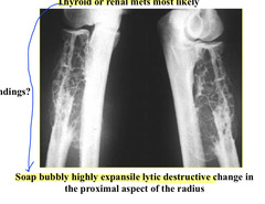
Soap bubbly highly expansive lytic destructive changes in the proximal aspect of the radius..... this is most likely from
Thyroid or renal Mets
What MRI modality is the most sensitive to finding metastasis
T1 (it will be a low signal)
What MRID modality is decent at finding metastasis and how does it look
T2 is variable, ( it will be a high signal)
What do you look for on MRI with metastasis
Lesion that is oblique, angled or round
Pedicular involvement (lytic Mets likes to destroy pedicels)
Extradural mass, multiplicity
No pathological fracture and degenerative signal is linear, _______ on T1 and ______ on T2
Low on T1
High on T2
What is the T1 rule of thumb
White is right concerning bone marrow
On T1 MRI, CSF should be what color
Dark
Lytic Mets shown on T1 MRI

Another example of lytic Mets shown on T1 MRI

Primary cause for lytic Mets in females is
Breast
Most bone malignancies are
Secondary
Most common metastasis to bone is what type
Lytic
What accounts for 30% of all malignancies
Primary malignant neoplasia
What is the order of primary malignant neoplasia when it comes to most to least common
Multiple myeloma
Osteosarcoma
Chondrosarcoma
Ewings sarcoma
What are the primary malignant neoplasia for adults
Multiple myeloma a
Chondrosarcoma
What is the most common primary malignant neoplasia for adults
Multiple myeloma
What is the second most common primary malignant neoplasia for adults
Chondrosarcoma
What is the most common primary malignant neoplasia for young people
Osteosarcoma
What is the second most common primary malignant neoplasia in young people
Ewings sarcoma
What is a malignant proliferation of plasma cells, which infiltrate the bone marrow
Multiple myeloma
-blood tumor that infiltrates the bone marrow
What is the most common primary malignant tumor of bone
Multiple myeloma
What is the third most prevalent blood cancer that happens to occur in the bone
Multiple myeloma
Multiple myeloma usually occurs at what age
5-8th decade
Most between ages 50-70 (mid to older life)
Malignant proliferation of plasma cells
Multiple myeloma
Multiple myeloma is what type of tumor
Round cell tumor (rebound nuclei and rounded lesions)
Multiple myeloma is 2x more common in ________ and lowest in _________
More common in African Americans
Asian Americans
Clinical features of multiple myeloma
Pain is initial symptom
Unexplained weight loss
Anemia
Renal disease
Respiratory disease
Why do you get renal problems with multiple myeloma
Lots of calcium deported into blood (from bone destruction)