Exam 3 (lec 16-22)
 Call Kai
 Learn
 Practice Test
 Spaced Repetition
 Match
 Flashcards
 Knowt Play
1/129

There's no tags or description
Looks like no tags are added yet.
130 Terms
Bisubstrate rxns
2+ substrates
Single displacement - random or ordered
Double displacement
>60% of biochem rxns
Single displacement Random
both substrates bund ro enzyme b4 product is formed
AEB to PEQ is rate limiting
creatine Kinase- random SD
direction determined by [] of ATP, ADP, Cr and CrP and eq constant
Most kinases have this mechanism
Single Displacement Ordered
AEB to PEQis rate limiting
ordered release of products
no B will bind to E in the absence of A
no turn over until both substrates bind
Single Displacement Bisubstrate
saturation when curve no longer shifts
resembles noncompetitive inhibitors
higher V max
increasing concentration = lower slope
Double Displacement
2 substrates bind and react separately in a ping pong manner
burst kinetics w P being seen before Q
E’ covalently modified enzyme intermediate
product of enzyme’s rxn w A is released prior to rxn of enzyme w B
sub bind independently to 1 another
Double Displacement Bisubstrate
resembles uncompetitive inhibitors
slope is constant
increasing saturation = lower slope
Glutamate:aspartate Aminotransferase
enzyme conforming to a double displacement bisusbtrate mechanism
Kinetic analysis
Lineweaver-Burk plot to distinguish if mechanism is
single or double displacement
parallel = double
non-parallel = single
Substrate binding assay
to determine if substrate addition is random or ordered
macrocalorimetry
no heat = ordered, specificity
heat = random
Exchange reactions
attempt reverse reaction in absence of one substrate
double displacement will allow the reaction
single displacement will not
Exchange reactions example
Sucrose phosphorylase and Maltose phosphorylase both cleave disaccharides and add Pi to one product
Mix 32P tracers with G-1-P and add either enzyme…
Sucrose phosphorylase does catalyze the exchange
DOUBLE DISPLACEMENT
Maltose phosphorylase does not catalyze the exchange
SINGLE DISPLACEMENT
transition state (X‡)
all rxns go thru
short-lived, unstable
high free energy and at least 1 partial bond
NOT AN INTERMEDIATE
rxn rate : [reactant that X‡
Enzymes lower ΔG‡
enzymes bind to transition state more tightly than substrate
lower free energy
Don’t want Ks to be too tight as the enzyme needs to bind to X better than S (Kt)
X‡ needs to be stabilized more than S
loss of entropy = ES needs to be destabilized
How ES is destabilized
structural strain
desolvation
electrostatic effects
structural strain
occurs bc of induced fir binding
lock and key hypothesis
1st explanation for specificity
induced fit hypothesis
more accurate description of specificity
favors formation of transition state
Destabilization of ES
charged groups are desolvated and more active
unfavorable interactions between like charges
How do enzymes work
by speeding attainment of Keq, from 107 to 1014 times faster
Keq is not changed
delta Grx is not changed
But activation energy is lowered
Binding of Transition-State Analogs
Kt may be 10-20 to 10-26 M (very tight)
TSAs are stable mlcs that are chemically and structurally similar to transition sates
act as competitive inhibitors and make good drugs
outcompete substrates
Would TSAs bind better or worse than competitive inhibitors?
better
enzymes have a higher affinity to transition state than substrate
TSA in medicine
Enalapril and Aliskiren lower blood pressure
statins lower serum cholesterol
protease inhibitors are AIDS drugs
Juvenile hormone esterase in insects is a pesticide target
tamiflu is a viral neuraminidase inhibitor
Statins
like lipitor
powerful cholesterol lowering drugs
they are TSAs of HMG-CoA reductase, key enzyme in the biosynthetic pathway for cholesterol
prolonged use = neurological deficits
Tamiflu
neuraminidase inhibitor and antiviral agent
based on the transition state of the neuraminidase reaction
Neuraminidase
major glycoprotein on influenza virus
essential for viral replication and infectivity
curved arrow
shows movement of an electron pair from a filled orbital to an empty one
full arrowhead
represents an electron pair
half arrowhead
represents a single electron
bond-breaking event
the arrow begins in the middle of the bond
Nucleophiles
nucleus loving
electron rich groups that can donate a pair of electrons
Electrophiles
electron poor groups that can receive electrons
leaving group
anything attached to the electrophile b4 rxn
Proton Transfer
can change a nucleophile into an electrophile
protonation state of substrate and active site residues
dependent on pKa and pH
Histidine
pKa values
can change in active site microenvironment
Mechanisms of Catalysis
catalysis by proximity (or formation of near-attack
complexes)
Covalent catalysis
General acid-base catalysis
Metal ion catalysis
Low barrier hydrogen bonds
Catalysis by Proximity
enzymes bring substrates in active site close enough and in the proper orientation so collisions occur at higher frequency
active site is preorganized into these
near attack complexes (NACs)
reacting atoms are in van der waals contact at an angle resembling the bond in the transition state
Near-attack Complexes
characterized as having reacting atoms w/in 3.2 A and an approach angle of +- 15o of the bonding angle in transition state
Proteins are constantly moving
bonds vibrate,
side chains bend and rotate,
backbone loops wiggle and sway,
whole domains move as a unit
Active site conformation changes can:
Assist substrate binding
Bring catalytic groups into position
Induce formation of NACs
Assist in bond making and bond breaking
Facilitate conversion of substrate to product
Covalent Catalysis
active site residues form a temporary covalent bond w substrate
bond is broken to regenerate enzyme
occurs due to nucleophilic attack by AA side chains on electrophilic groups on substrates
can involve prosthetic groups
facilitates electron transfer
most have unknown mechanisms
E’
covalently modified enzyme intermediate
double displacement
Trypsin , Chymotrypsin
react w serine
Glyceraldehyde-3-P dehydrogenase
react w cysteine
Phosphoglucomutase
react w serine
phosphoglycerate mutase, Succinyl-CoA synthetase
react w Histidine
Aldolase, Pyridoxal phosphate enzymes
react w Lysine and other amino groups
General Acid-Base Catalysis
transfer of a proton in transition state
facilitates proton transfer (back and forth)
specific = H+ or OH- that has diffused into the active site
Transferring an H+ in acid-base catalysis may:
activate nucleophiles
stabilize charged groups
improve electrostatic interactions that stabilize TS
Candidate amino acids for acid-base catalysis (Catalytic triad)
Glu
Asp
His
Histidine Often Plays a Role
might normally be protonated
can be deprotonated by another group and act as a base, accepting protons from substrate
Water Often Plays a Role
H2O can act as acid/base at active site thru proton transfer w an assisting active site residue
Shifts in pKa for Residues in Active Sites Create Catalytic Agents
microenvironments play role in pKa of active sites
primary: His, Cys, Asp, Glu, Arg, Lys, Tyr, Ser, Thr, Asn, Gln
Secondary Roles
~1/2 of AA engage directly in catalytic effects in enzyme active sites
Other residues may function in secondary roles in the active site:
Raising or lowering catalytic residue pKa values
Orientation of catalytic residues
Charge stabilization
Proton transfers via hydrogen tunneling
Metal Ion Catalysis
eg: zinc, magnesium, iron
metal atoms lose electrons easily and exist as cations
basics
Positive charge allows metal atoms to:
stabilize transient and intermediate structures
assist in forming strong nucleophilic species (image)
hold substrate inside the active site
stabilize charge
Low-Barrier Hydrogen Bonds (LBHBs)
As distance between heteroatoms becomes smaller <.25nm, H bonds become stronger (almost covalent in nature)
stabilization energy can approach 60kJ/mol in solution
pKa values of the 2 electronegative atoms must be similar (Asp and His)
energy released in forming an LBHB
can assist catalysis
typical H-bond strength
10-30 kJ/mol
typical O-O separation
.28 nm
LBHBs
.25 nm to <.25 nm
Trypsin, chymotrypsin, elastase
igestive enzymes secreted as proenzymes/zymogens
Thrombin
blood clotting cascade
Subtilisin
bacterial enzyme
Plasmin
cleaves fibrin polymers of blood clots
Tissue plasminogen activator
cleaves plasmin proenzyme plasminogen and is administered to prevent heart attack
Acetylcholinesterase
NOT a protease but mechanistically similar in breaking down acetylcholine
Similar sequences, structures and mechanisms
evolutionary relatedness
Trypsin, Chymotrypsin and Elastase function
proenzymes/ zymogens
all cleave polypeptide chains
identical sequences/aa are conserved for functionality
Structure of chymotrypsin
His is flanked by Asp and Ser
Ser is close to the peptide that would be cleaved in the rxn
The Catalytic Triad of the Serine Proteases
catalytic triad at the active site of chymotrypsin
His, Ser, Asp
Trypsin, Chymotrypsin and Elastase specificities
similar structures and mechanism, but DIFF specificities
Trypsin- C side of basic (EXCEPT WHEN FOLLOWED BY PROLINE bc of fixed phi angle)
Chymotrypsin- C side of aromatic
Elastase- C side of small, neutral residues
DIFF specificities
given by active site
Enzymatic Fragmentation Trypsin
4 cut sites = 5 peptides
product is a mix of peptide fragments w C terminal Arg/Lys + single peptide from the C terminal end of the polypeptide
Trypsin Substrate-binding Pocket
cuts after basics
negatively charged
Chymotrypsin Substrate-binding Pocket
cuts after aromatics
hydrophobic (ser)
rings themselves are hydrophobic
Elastase Substrate-binding Pocket
cuts after small mlcs
bulky residues like Thr and Val at top and shallow
Chymotrypsin Kinetics
speed
assayed using artificial substrate
useful as nitrophenolate product absorbs at 400nm
observed burst kinetics, suggesting first fast step second slow step
Serine Proteases Display Burst Kinetics
quick cut
slow serine binding
Serine Protease Mechanism
mix of covalent + general acid-base catalysis
Asp orients His and forms LBHB
His acts as acid/base
Ser forms covalent bond w peptide to be cleaved
trigonal C into tetrahedral C
Tetrahedral oxyanion intermediate stabilized by backbone NH groups of Gly + Ser
Transition-State Stabilization
chymotrypsin involves 2 tetrahedral oxyanion transition states
these are stabilized by a pair of amide groups termed oxyanion hole
the oxyanion hole
O- in tetrahedral oxyanion is stabilized by interaction w the backbone NH groups of Ser + Gly
Catalytic Triads In General
found in several hydrolases + transferase enzymes
occur via divergent + convergent evolution
Residues far away in primary structure
Catalytic Triads Include:
cid that orients and stabilizes the base (Asp, Glu, His)
Base that polarizes the nucleophile ( His, rarely Lys)
Nucleophile that attacks substrate (Ser, Cys, sometimes Thr)
Aspartic Proteases
DIFFERENT structure and mechanism than serine proteases
active site = 2 Asp
cleave peptide bond btw 2 hydrophobic aa
No covalent catalysis
Structures of HIV-1 protease and pepsin
HIV-1 protease - homodimer
pepsin- monomer
Each lobe contributes one catalytic aspartate to the active site.
pH-dependence of asp proteases
mechanism requires that one Asp be protonated and the other Asp be deprotonated when the substrate binds
active at acidic pH
A Mechanism for the Aspartic Proteases
LBHB allowing “hydrogen tunneling)
LBHBs play a role in states E, ES, ET’, EQ’, and EP’Q.
Protease Inhibitors for AIDS Patients
HIV-1 protease selectively inhibited = new HIV particles cannot form
structure based drug design has developed several inhibitors
HIV-1 Protease
novel asp protease
cleaves polyprotein products of HIV genome
remarkable imitation of mammalian asp proteases
homodimer- more genetically economical
active site = 2 fold symmetric flaps
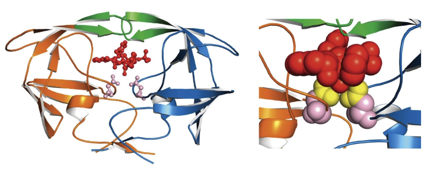
Protease Inhibitors Block the Active Site of HIV-1 Protease
HIV-1 protease complexed with the inhibitor Crixivan (red)
made by Merck. The “flaps” that cover the active site are
green; the catalytic active-site Asp residues are violet.
enzyme regulation overview
rxns to match cell requirements
natural physiological phenomenon
focus on activity
Achieved by regulating enzyme:
abundance
activity
abundance
controlled by gene expression (not treated)