Week 1 - Nervous System

0.0(0)Studied by 0 people
Build your Mastery score
multiple choiceMultiple Choice & FRQ
call kaiCall Kai
Card Sorting

1/19

flashcard set

Earn XP

Description and Tags

This set covers the following topics: the function and divisions of the nervous system, neurons, properties of neurons and neuroglia, and the action potential.

Last updated 9:22 PM on 10/6/26
Name
Mastery
Learn
Test
Matching
Spaced
Call with Kai
Chat

No analytics yet

Send a link to your students to track their progress

20 Terms

1
New cards

Functions of the Nervous System

  • Through the nervous and endocrine systems, two key internal bodily communication systems, multicellular organisms are able to maintain homeostasis.

  • Nervous tissue contains highly specialized cells, in which neurons play a principal role. They are characterized by their ability to generate an action potential and conduct it for a long distance.

  • Through the use of neurons and neurotransmitters, a type of chemical messenger, the nervous system is a key communicative and control system in the body.

  • Sensory function: receive stimuli

    • Sensory receptors gather information and send it to the CNS.

  • Integration: process the stimuli

    • The CNS creates sensations, thoughts, decisions, and memory.

  • Motor function: initiates a response to stimuli 

    • Decisions are acted upon, and impulses are sent to effector organs.


2
New cards

Divisions

  • CNS = Central Nervous System

    • Brain and spinal cord

  • PNS = Peripheral Nervous System

    • Cranial and spinal nerves + ganglia


<ul><li><p><span style="background-color: transparent;">CNS = Central Nervous System</span></p><ul><li><p><span style="background-color: transparent;">Brain and spinal cord</span></p></li></ul></li><li><p><span style="background-color: transparent;">PNS = Peripheral Nervous System</span></p><ul><li><p><span style="background-color: transparent;">Cranial and spinal nerves + ganglia</span></p></li></ul></li></ul><p></p>
3
New cards

Neurons and nerves

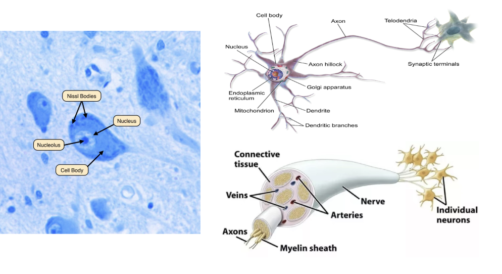
  • Nervous tissue is primarily made of neurons and neuroglia/”glia”

  • Nissl bodies: clumps of RER & ribosomes

  • Neurons are organized into discrete nerves in the PNS and grey & white matter in the CNS.

  • Nerves are cable-like bundles of nerve fibers (axons)

<ul><li><p><span style="background-color: transparent;">Nervous tissue is primarily made of neurons and neuroglia/”glia”</span></p></li><li><p><span style="background-color: transparent;">Nissl bodies: clumps of RER &amp; ribosomes</span></p></li><li><p><span style="background-color: transparent;">Neurons are organized into discrete nerves in the PNS and grey &amp; white matter in the CNS.</span></p></li><li><p><span style="background-color: transparent;">Nerves are cable-like bundles of nerve fibers (axons)</span></p></li></ul><p></p>
4
New cards

Grey and white matter

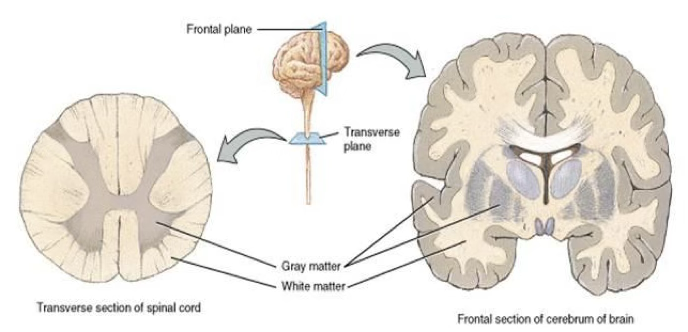
  • Tissue in the CNS defined by its color 

    • Grey matter: contains unmyelinated neurons, neuronal cell bodies, dendrites, and glia

    • White matter: contains a large number of myelinated axons. Found in deeper parts of the brain and superficially on the spinal cord

<ul><li><p><span style="background-color: transparent;">Tissue in the <u>CNS</u> defined by its color&nbsp;</span></p><ul><li><p><span style="background-color: transparent;">Grey matter: contains <em>unmyelinated</em> neurons, neuronal cell bodies, dendrites, and glia</span></p></li><li><p><span style="background-color: transparent;">White matter: contains a large number of <em>myelinated</em> axons. Found in deeper parts of the brain and superficially on the spinal cord</span></p></li></ul></li></ul><p></p>
5
New cards

Structural neuron types

  • Unipolar: single structure extending from the cell body which contains one axon with dendrites.

  • Pseudounipolar: one process that extends from the cell body which then divides into two structures, axon and dendrite.

  • Bipolar: one axon and one dendrite extending from the cell body.

  • Multipolar: one axon and multiple dendrites extending from the cell body; most common type of neuron


<ul><li><p><span style="background-color: transparent;">Unipolar: single structure extending from the cell body which contains one axon with dendrites.</span></p></li><li><p><span style="background-color: transparent;">Pseudounipolar: one process that extends from the cell body which then divides into two structures, axon and dendrite.</span></p></li><li><p><span style="background-color: transparent;">Bipolar: one axon and one dendrite extending from the cell body.</span></p></li><li><p><span style="background-color: transparent;">Multipolar: one axon and multiple dendrites extending from the cell body; most common type of neuron</span></p></li></ul><p></p>
6
New cards

Functional neuron types

  • Sensory/Afferent Neurons: neurons that collect sensory information from sensory receptors and conduct this info to the CNS

  • Interneurons: neurons that connect sensory and motor neurons in the CNS

  • Motor/Efferent: neurons that conduct nerve impulses away from the CNS to effector organ(s)


<ul><li><p><span style="background-color: transparent;">Sensory/Afferent Neurons: neurons that collect sensory information from sensory receptors and conduct this info to the CNS</span></p></li><li><p><span style="background-color: transparent;">Interneurons: neurons that connect sensory and motor neurons in the CNS</span></p></li><li><p><span style="background-color: transparent;">Motor/Efferent: neurons that conduct nerve impulses away from the CNS to effector organ(s)</span></p></li></ul><p></p>
7
New cards

Macroscopic nervous structures

  • A collection of cell bodies (soma)

    • CNS – nucleus/nuclei

    • PNS – ganglion/ganglia

  • A collection of axons

    • CNS – tracts

    • PNS – nerves


8
New cards

Ganglia

  • Ganglia/ganglion: clusters of nerve cell bodies found throughout the body

  • Part of the PNS. Helps carry nerve signals to and from the CNS.


<ul><li><p><span style="background-color: transparent;">Ganglia/ganglion: clusters of nerve cell bodies found throughout the body</span></p></li><li><p><span style="background-color: transparent;">Part of the PNS. Helps carry nerve signals to and from the CNS.</span></p></li></ul><p></p>
9
New cards

The nerve plexus

  • A branching network of nerves that originate from the same anatomical area and serve specific parts of the body 

  • Nerves in a plexus are made of afferent and efferent fibers that come from the merging spinal nerves

  • Nerve fibers from different spinal nerves are then sorted and recombined into one nerve that goes to a specific body part – similar to an electrical junction box


<ul><li><p><span style="background-color: transparent;">A branching network of nerves that originate from the same anatomical area and serve specific parts of the body&nbsp;</span></p></li><li><p><span style="background-color: transparent;">Nerves in a plexus are made of afferent and efferent fibers that come from the merging spinal nerves</span></p></li><li><p><span style="background-color: transparent;">Nerve fibers from different spinal nerves are then sorted and recombined into one nerve that goes to a specific body part – similar to an electrical junction box</span></p></li></ul><p></p>
10
New cards

Neuroglia

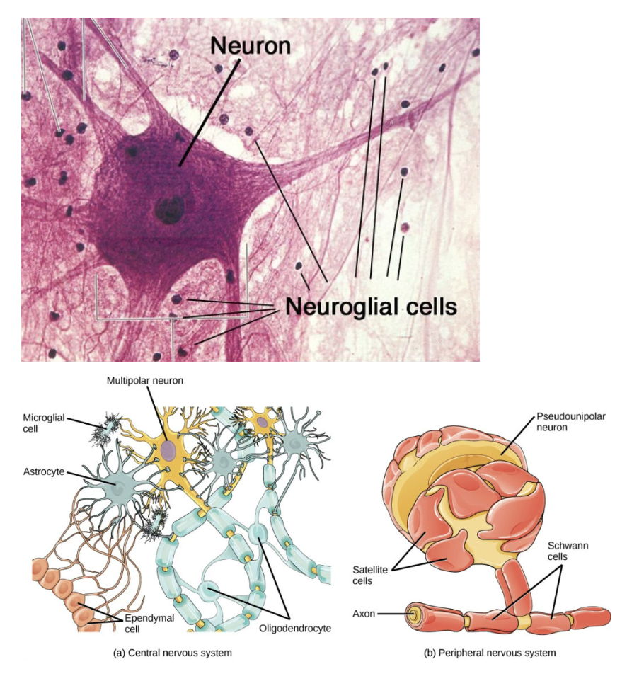
  • Neuroglial cells function to maintain a stable environment for neurons in the nervous system. They protect nervous tissue and assist in nerve function.

    • Ex. in the PNS and CNS, glial cells provide scaffolding on which the nervous system is built. They help neurons line up closely for neuronal communication, provide insulation to neurons, transport nutrients and waste, and mediate immune responses

  • Neuroglia in the CNS: astrocytes, microglial cells, ependymal cells, oligodendrocytes

  • Neuroglia in the PNS: satellite and Schwann cells

<ul><li><p><span style="background-color: transparent;">Neuroglial cells function to maintain a stable environment for neurons in the nervous system. They protect nervous tissue and assist in nerve function.</span></p><ul><li><p><span style="background-color: transparent;">Ex. in the PNS and CNS, glial cells provide scaffolding on which the nervous system is built. They help neurons line up closely for neuronal communication, provide insulation to neurons, transport nutrients and waste, and mediate immune responses</span></p></li></ul></li><li><p><span style="background-color: transparent;">Neuroglia in the CNS: astrocytes, microglial cells, ependymal cells, oligodendrocytes</span></p></li><li><p><span style="background-color: transparent;">Neuroglia in the PNS: satellite and Schwann cells</span></p></li></ul><p></p>
11
New cards

Schwann cells and myelin

  • Schwann cells: a glial cell that surrounds some neurons, keeping them alive and sometimes covering them with a myelin sheath

    • Major glial cell type in PNS. Vital in the development, maintenance, function, and regeneration of peripheral nerves.

  • Myelin: fatty substance produced by Schwann cells in PNS and oligodendrocytes in CNS

    • Insulating, non-conductive material. Myelinating a nerve cell results in better conduction of nerve impulses. Specifically prevents Na+ from leaving the neuron so that the Na+ concentration will be strong enough to maintain depolarization until the next node of Ranvier.


<ul><li><p><span style="background-color: transparent;">Schwann cells: a glial cell that surrounds some neurons, keeping them alive and sometimes covering them with a myelin sheath</span></p><ul><li><p><span style="background-color: transparent;">Major glial cell type in PNS. Vital in the development, maintenance, function, and regeneration of peripheral nerves.</span></p></li></ul></li><li><p><span style="background-color: transparent;">Myelin: fatty substance produced by Schwann cells in PNS and oligodendrocytes in CNS</span></p><ul><li><p><span style="background-color: transparent;">Insulating, non-conductive material. Myelinating a nerve cell results in better conduction of nerve impulses. Specifically prevents Na+ from leaving the neuron so that the Na+ concentration will be strong enough to maintain depolarization until the next node of Ranvier.</span></p></li></ul></li></ul><p></p>
12
New cards

Synapse

  • Synapse, synaptic cleft, presynaptic neuron, postsynaptic neuron, neurotransmitter


<ul><li><p><span style="background-color: transparent;">Synapse, synaptic cleft, presynaptic neuron, postsynaptic neuron, neurotransmitter</span></p></li></ul><p></p>
13
New cards

Neurotransmitters

  • Acetylcholine (ACh): induces skeletal muscle contraction, slows cardiac muscle contraction rate, affects mood and memory

  • Dopamine: reward-motivated behavior, controls movement and motion, love, and pleasure

  • Epinephrine and norepinephrine: speed the heart rate, dilate pupils and the airways to the lungs, slow gut contractions, and increase anxiety 

  • Serotonin: elevates mood, appetite, and sleep, and plays a role in memory and learning

  • Endorphins: dull pain, elevate mood, and promote feelings of well-being


<ul><li><p><span style="background-color: transparent;">Acetylcholine (ACh): induces skeletal muscle contraction, slows cardiac muscle contraction rate, affects mood and memory</span></p></li><li><p><span style="background-color: transparent;">Dopamine: reward-motivated behavior, controls movement and motion, love, and pleasure</span></p></li><li><p><span style="background-color: transparent;">Epinephrine and norepinephrine: speed the heart rate, dilate pupils and the airways to the lungs, slow gut contractions, and increase anxiety&nbsp;</span></p></li><li><p><span style="background-color: transparent;">Serotonin: elevates mood, appetite, and sleep, and plays a role in memory and learning</span></p></li><li><p><span style="background-color: transparent;">Endorphins: dull pain, elevate mood, and promote feelings of well-being</span></p></li></ul><p></p>
14
New cards
<p>Neuron properties</p>

Neuron properties

  • Neurons contain properties of:

    • Excitability: the ability to respond to different stimuli (chemical, electrical, mechanical)

    • Conductivity: ability to conduct electrical charges


15
New cards

Action potential

  • The following terms are to be defined through this process: resting membrane potential, threshold, action potential, depolarization, repolarization, sodium-potassium pump, propagation, refractory period

  • Action potential: a sudden rise and then fall in neuronal membrane voltage in response to stimuli. These are only generated when the RMP reaches a threshold value of -55mV. 

    • The generation of an action potential is a series of orderly events that can be explained in 3 stages: depolarization, repolarization, and hyperpolarization.


<ul><li><p><span style="background-color: transparent;">The following terms are to be defined through this process: resting membrane potential, threshold, action potential, depolarization, repolarization, sodium-potassium pump, propagation, refractory period</span></p></li><li><p><span style="background-color: transparent;">Action potential: a sudden rise and then fall in neuronal membrane voltage in response to stimuli. These are only generated when the RMP reaches a threshold value of -55mV.&nbsp;</span></p><ul><li><p><span style="background-color: transparent;">The generation of an action potential is a series of orderly events that can be explained in 3 stages: depolarization, repolarization, and hyperpolarization.</span></p></li></ul></li></ul><p></p>
16
New cards

Resting membrane potential

  • Resting membrane potential (RMP): electrical (potential) difference across a neuronal cell’s plasma membrane when the cell is at rest and not actively conducting electricity

    • Caused by the uneven distribution of ions across the cell membrane

    • RMP is negatively charged in a neuron. The inside of a neuron is about 70 millivolts more negative than the outside. 

    • At rest, neurons are negatively charged since there are more positively charged Na+ ions outside of the cell than inside. Specifically, at rest, there are more Na+ ions outside the neuron and more K+ inside.


<ul><li><p><span style="background-color: transparent;">Resting membrane potential (RMP): electrical (potential) difference across a neuronal cell’s plasma membrane when the cell is at rest and not actively conducting electricity</span></p><ul><li><p><span style="background-color: transparent;">Caused by the uneven distribution of ions across the cell membrane</span></p></li><li><p><span style="background-color: transparent;">RMP is negatively charged in a neuron. The inside of a neuron is about 70 millivolts more negative than the outside.&nbsp;</span></p></li><li><p><span style="background-color: transparent;">At rest, neurons are negatively charged since there are more positively charged Na+ ions outside of the cell than inside. Specifically, at rest, there are more Na+ ions outside the neuron and more K+ inside.</span></p></li></ul></li></ul><p></p>
17
New cards

Depolarization

  • Depolarization: sudden change in the neuron’s RMP from a negative to a positive internal charge due to the influx of Na+ ions into the cell

    • During depolarization, sodium channels open in response to a signal received by dendrites. When depolarization reaches about -55mV, the neuron fires an action potential. 

    • With the opening of the sodium channels, a rapid passive influx of sodium ions into the cell makes the MP more positive. This moves the MP past zero.


<ul><li><p><span style="background-color: transparent;">Depolarization: sudden change in the neuron’s RMP from a negative to a positive internal charge due to the influx of Na+ ions into the cell</span></p><ul><li><p><span style="background-color: transparent;">During depolarization, sodium channels open in response to a signal received by dendrites. When depolarization reaches about -55mV, the neuron fires an action potential.&nbsp;</span></p></li><li><p><span style="background-color: transparent;">With the opening of the sodium channels, a rapid passive influx of sodium ions into the cell makes the MP more positive. This moves the MP past zero.</span></p></li></ul></li></ul><p></p>
18
New cards

Repolarization

  • Repolarization: restoring the negative internal charge and MP following depolarization

    • Starts with the inactivation of the sodium channels and the opening of voltage-gated potassium channels

    • Since potassium ions are more concentrated inside the neuron than outside, the opening of potassium channels results in a rapid passive efflux of potassium to the outside of the cell. This efflux causes the MP to return toward the -70mV value of the resting MP


<ul><li><p><span style="background-color: transparent;">Repolarization: restoring the negative internal charge and MP following depolarization</span></p><ul><li><p><span style="background-color: transparent;">Starts with the inactivation of the sodium channels and the opening of voltage-gated potassium channels</span></p></li><li><p><span style="background-color: transparent;">Since potassium ions are more concentrated inside the neuron than outside, the opening of potassium channels results in a rapid passive efflux of potassium to the outside of the cell. This efflux causes the MP to return toward the -70mV value of the resting MP</span></p></li></ul></li></ul><p></p>
19
New cards

Refractory period

  • Refractory period: the brief period after a neuron has generated a nerve impulse and another cannot be generated

    • For the neuron to fire again, the RMP must be re-established. The sodium-potassium pump must re-establish the electrolytes in their correct positions on either side of the membrane for another action potential to propagate.

    • Because an area of a neuron has a refractory period, propagation of the action potential can only occur in one direction


<ul><li><p><span style="background-color: transparent;">Refractory period: the brief period after a neuron has generated a nerve impulse and another cannot be generated</span></p><ul><li><p><span style="background-color: transparent;">For the neuron to fire again, the RMP must be re-established. The sodium-potassium pump must re-establish the electrolytes in their correct positions on either side of the membrane for another action potential to propagate.</span></p></li><li><p><span style="background-color: transparent;">Because an area of a neuron has a refractory period, propagation of the action potential can only occur in one direction</span></p></li></ul></li></ul><p></p>
20
New cards

Saltatory conduction

  • Saltatory conduction: describes the way action potentials travel down myelinated neurons

    • Myelin insulates neurons, preventing Na+ from going out in that zone, so that the Na+ concentration will be strong enough to stimulate depolarization until the next Ranvier node

    • The action potential jumps between each Node of Ranvier

    • This mechanism is ten times faster at getting action potentials to travel down an axon with less energy expenditure


<ul><li><p><span style="background-color: transparent;">Saltatory conduction: describes the way action potentials travel down myelinated neurons</span></p><ul><li><p><span style="background-color: transparent;">Myelin insulates neurons, preventing Na+ from going out in that zone, so that the Na+ concentration will be strong enough to stimulate depolarization until the next Ranvier node</span></p></li><li><p><span style="background-color: transparent;">The action potential jumps between each Node of Ranvier</span></p></li><li><p><span style="background-color: transparent;">This mechanism is ten times faster at getting action potentials to travel down an axon with less energy expenditure</span></p></li></ul></li></ul><p></p>