[MOD5LEC] Muscular System
 Call Kai
 Learn
Learn Practice Test
 Spaced Repetition
 Match
Match Flashcards
 Knowt Play
1/54

There's no tags or description
Looks like no tags are added yet.
Name | Mastery | Learn | Test | Matching | Spaced | Call with Kai | Chat |
|---|
No analytics yet
Send a link to your students to track their progress
55 Terms
What are the general functions of the skeletal muscular system?
Controls voluntary movement of the head, face, trunk, limbs, etc.
Makes the maintenance of the position of the body possible
Provides 85% of the body’s heat through glucose catabolism
Accounts for approximately 40% of the body’s mass
Participates in involuntary spinal cord reflexes
What are some important characteristics of skeletal muscle tissue?
It can also be referred to as voluntary muscle tissue or striated muscle tissue
Muscles attach to various bones of the skeleton by tendons
What are universal characteristics of muscles?
Responsiveness - all living cells are excitable
muscle cells respond to stimuli in a unique way by producing an electric current that travels along the plasma membrane and triggers contraction of the cell
Conductivity - stimulation of a muscle cell produces more than just a local effect
local electrical change triggers a wave of excitation that travels rapidly along the cell and initiates processes leading to contraction
Contractility - unique in their ability to shorten substantially when stimulated
enables muscle cells to exert tension on other tissue to which they are mechanically coupled to create movement
Extensibility - able to stretch back after contraction
most cells rupture if they are stretched even a little, but skeletal muscle fibers can safely stretch to as much as 3 times their contracted length
Elasticity - when muscle cells are stretched and released, they recoil to a shorter length
different from the ability to stretch, which is a trait of extensibility
Why are muscle and nerve cells regarded as electrically excitable cells?
Because their plasma membranes exhibit voltage changes in response to stimulation
Electrical activity of cells is related to differences in the concentration of ions in the intracellular fluid (ICF) and extracellular fluid (ECF) adjacent to the plasma membrane
Explain why a membrane is polarized as a result of the concentration of ions in the intracellular and extracellular fluid
The ICF contains a greater concentration of negative anions than the ECF does, such as negatively charged proteins, nucleic acids, and phosphates
The ICF also contains a great excess of potassium ions (K+), whereas the ECF contains a great excess of sodium ions (Na+)
The difference in electrical charge from one point to another is called an electrical potential, or voltage
What is resting membrane potential (RMP)?
About 90 millivolts (mV), a number that is critically important to life
Resting membrane potential is maintained by the sodium-potassium pump
![<p>Sarcolemma [Microscopic Structure Of Skeletal Muscle Cells]</p>](https://assets.knowt.com/user-attachments/837c5137-5606-45e1-831f-a5988f6a56e9.png)
Sarcolemma [Microscopic Structure Of Skeletal Muscle Cells]
Specialized plasma membrane specific to skeletal muscle cells

![<p>Sarcoplasm [Microscopic Structure Of Skeletal Muscle Cells]</p>](https://assets.knowt.com/user-attachments/fef499a6-b4f5-4022-a835-3ed0c857f02f.png)
Sarcoplasm [Microscopic Structure Of Skeletal Muscle Cells]
Specialized type of cytoplasm specific to skeletal muscle cells, that contains an abnormally higher amount of mitochondria than other cells for the high energy demands of muscle

![<p>Sarcoplasmic reticulum (SR) [Microscopic Structure Of Skeletal Muscle Cells]</p>](https://assets.knowt.com/user-attachments/a1358786-8ecb-450f-ae19-8820421da5c4.png)
Sarcoplasmic reticulum (SR) [Microscopic Structure Of Skeletal Muscle Cells]
Specialized smooth endoplasmic reticulum (ER) parallel to myofibrils
The sarcoplasmic reticulum of muscle cells stores calcium, which is vital in the process of muscle contraction and relaxation


Terminal cisternae
A perpendicular terminal, which is an enlarged portion of sarcoplasmic reticulum (SR)
A reservoir for calcium ions
Has gated channels in its membrane that can release a flood of calcium into the sarcoplasm

![<p>Transverse tubules or T-tubules [Microscopic Structure Of Skeletal Muscle Cells]</p>](https://assets.knowt.com/user-attachments/ca2fde62-ee9e-4e52-a20e-db2366090576.png)
Transverse tubules or T-tubules [Microscopic Structure Of Skeletal Muscle Cells]
A hollow elongated tube
Carries an electrical current from the extracellular fluid and sarcolemma to myofibrils
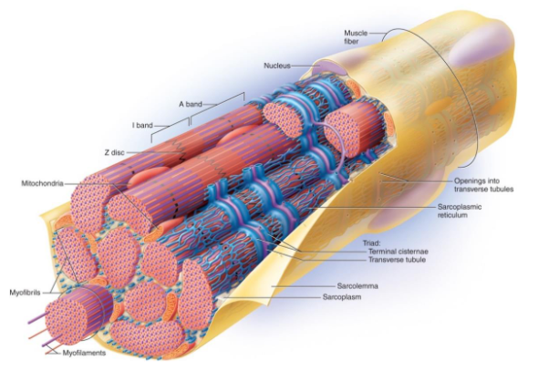
![<p>Triad [Microscopic Structure Of Skeletal Muscle Cells]</p>](https://assets.knowt.com/user-attachments/d20d59f6-8301-4c2d-b513-5e754f9880b8.png)
Triad [Microscopic Structure Of Skeletal Muscle Cells]
Refers to an arrangement of a T-tubule and terminal cisternae on each side

![<p>Myofibrils [Microscopic Structure Of Skeletal Muscle Cells]</p>](https://assets.knowt.com/user-attachments/00254710-2455-4854-a790-6c301d6f6fa8.png)
Myofibrils [Microscopic Structure Of Skeletal Muscle Cells]
The many threadlike rods that extend and make up the full length of a muscle fiber
Fill up most of the muscle cell
Composed of protein myofilaments
Myofilaments refer to two types of filaments; thick filaments and thin filaments
PICTURE: Thick filaments are purple, thin filaments are red

![<p>Thick filaments [Types Of Myofilaments]</p>](https://assets.knowt.com/user-attachments/37967bc1-65f9-4c65-9773-261242865f37.png)
Thick filaments [Types Of Myofilaments]
A type of myofilament made of the protein myosin
Myosin molecules are shaped like golf clubs, with a shaft-like tail and a globular head
A thick filament consists of 200 to 500 myosin molecules, with their heads directed outward in a spiral array around the bundle
Heads on one half of the thick filament angle to the left, while the heads on the other half angle to the right
There is an area in the middle of thick filaments where there is no myosin heads present, and is referred to as a bare zone or an H-zone

![<p>Thin filaments [Types Of Myofilaments]</p>](https://assets.knowt.com/user-attachments/46436447-5821-4c86-b9a0-71340ae6a63a.png)
Thin filaments [Types Of Myofilaments]
A type of myofilament made of two intertwined strands. These strands are made out of the protein known as fibrous actin
Fibrous actin is referred to as F-actin
Each F-actin molecule is a string, which is made of bead-like subunits called globular actin or G-actin
Each G-actin has an active site that binds to the heads of myosin molecules on thick filaments
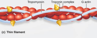

Tropomyosin
A long strand-like protein that covers and blocks the active sites of thin filaments
Tropomyosin occupies the spiral groove along the length of an F-actin molecule


Troponin
A globular molecule that binds with tropomyosin and G-actin at the active site
Troponin contains a calcium (Ca++) receptor


How do tropomyosin and troponin work to regulate muscles?
They act as a “switch” to determine when a muscle can and cannot contract
Tropomyosin molecules have troponin bound to them at regular intervals

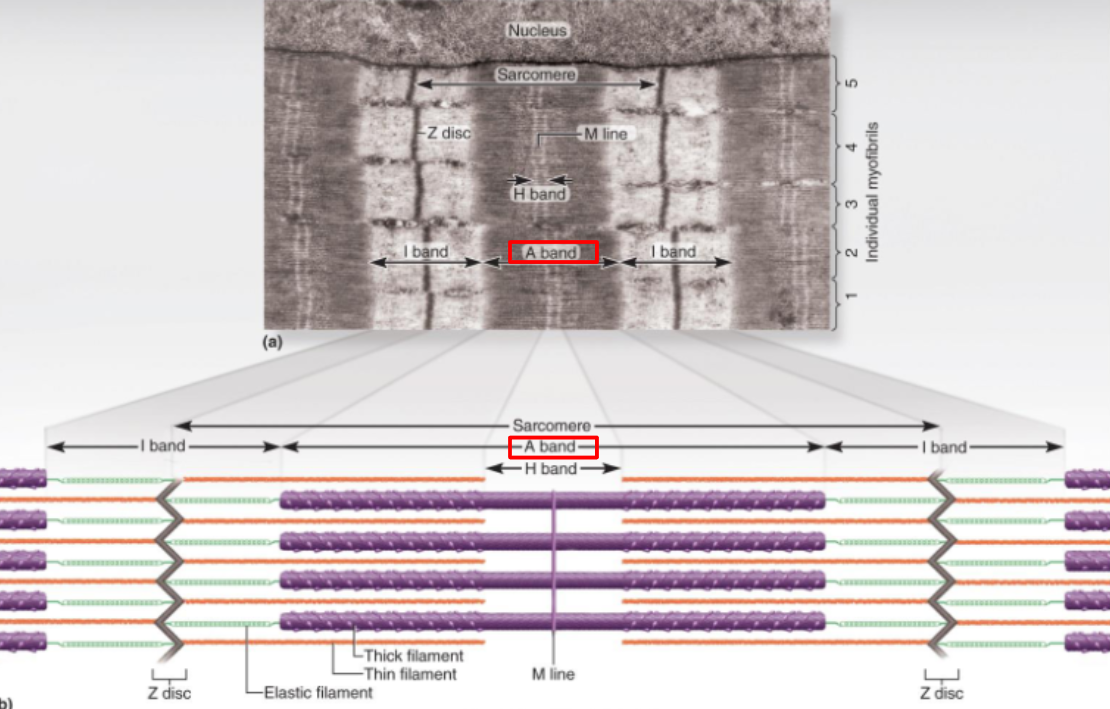
A-bands
Refers to the darkest area of a muscle fiber, where myosin and actin myofilaments overlap
These are the primary reason that cross-striations are visible


H-band
The area of an A-band that is only composed of myosin (thick filament)
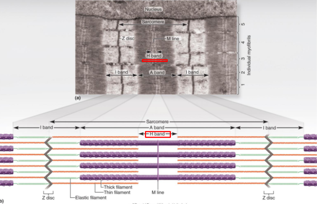

I-bands
Lightest area between A-bands composed of actin only
Z-discs bisect the light colored I-bands

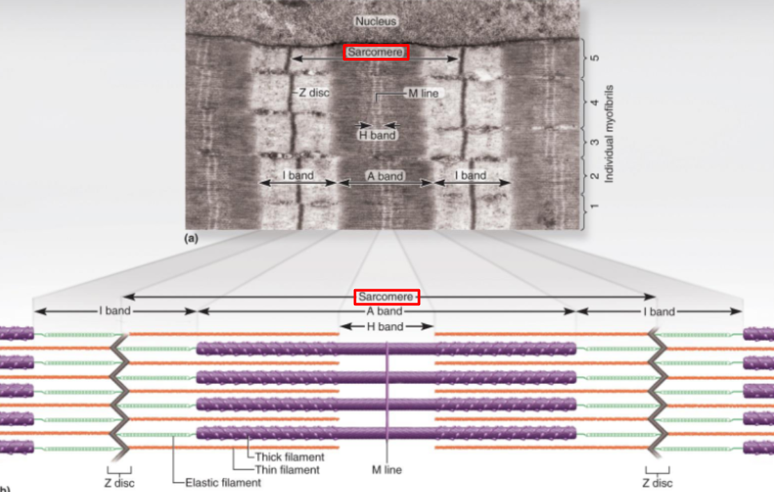
Sarcomere
The functional unit of the myofibril
Refers to the portion of a myofibril from one Z-disc to the next
Constitutes one contractive unit
Muscles shorten because the individual sarcomeres shorten, thus pulling the Z-discs closer together
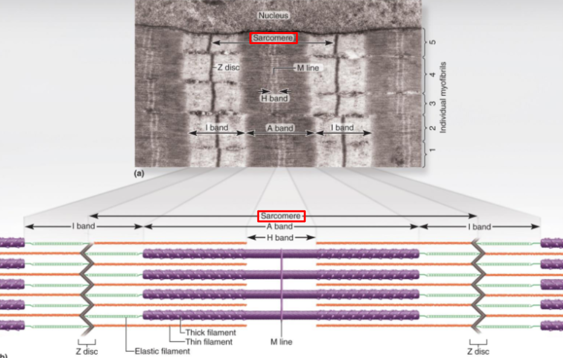
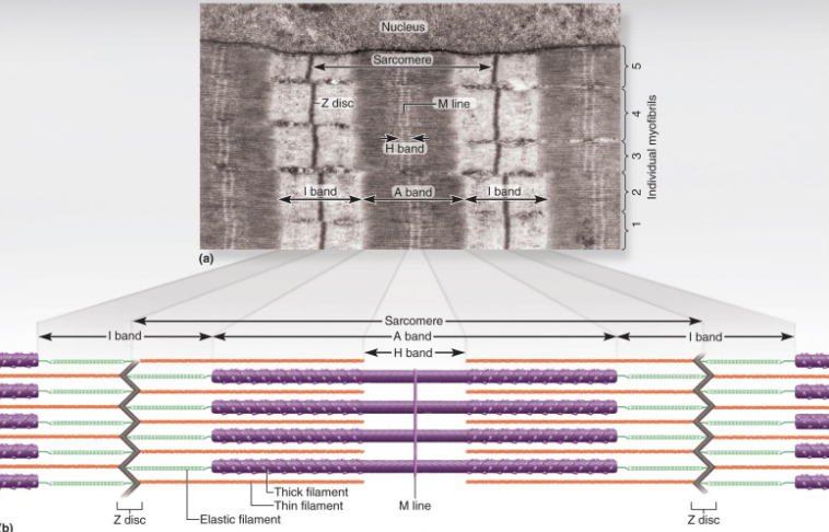
Z-disks
Dark peripheral boundaries of sarcomere

Striations
The appearance given off by actin and myosin fibers, which are organized into geometrically precise arrays
Striated muscle consists of dark A-bands alternating with lighter I-bands

What is the structure of skeletal muscle from its smaller units to its larger units?
A bundle of muscle fibers is a fascicle
Each muscle fascicle is surrounded by perimysium on all sides
A group of muscle fascicles is collectively surrounded by epimysium
Epimysium is then covered by fascia

What are the types of fascia?
Subcutaneous fascia - lies deep to the skin
Subserous fascia - covers organs and body cavities
Deep fascia - surrounds and penetrates whole muscles

Describe what a neuron is and what structures they’re made of
A neuron is a nerve cell that the nervous system uses to communicate
Soma - control center of the neuron; contains the nucleus
Dendrites - branching structures only found on the soma that receive signals from other neurons
Axon - emerges from the axon hillock of the soma, specialized for rapid conduction of nerve signals to points far from the soma
Nerve impulses only travel in one direction through a neuron:
Dendrites → through the soma → to the axon hillock → out through the axon → other neurons or effectors

What is a motor unit? [Neuromuscular Junctions]
One nerve fiber and all the muscle fibers innervated by it
There is an average of 200 muscle fibers that connect to a motor neuron
The muscles behave as a single unit
What is the location of a neuromuscular junction? What is the function of a motor neuron? [Neuromuscular Junctions]
Neuromuscular junctions are located wherever motor neurons meet muscle fibers
The function of motor neurons is to relay nerve impulses to a muscle fiber, where it then becomes a motor impulse
What structures make up a motor neuron? [Neuromuscular Junctions]
Synaptic knob - contains synaptic vesicles
Synaptic vesicles - contain the neurotransmitter acetylcholine (ACh)
![<p>Describe the <strong>synaptic cleft</strong> [Neuromuscular Junctions]</p>](https://assets.knowt.com/user-attachments/a9dfadf4-5c6f-4de1-b2f2-35415769268a.png)
Describe the synaptic cleft [Neuromuscular Junctions]
The gap between a motor neuron and a muscle fiber at a neuromuscular junction
The muscle fiber contains about 50 million acetylcholine (ACh) receptors
Junctional folds - infoldings of a muscle cell’s sarcolemma that increases the surface area and allows more ACh receptor sites to be present
Acetylcholine is released from the synaptic knob of motor neurons and travels to the surface of the muscle (sarcolemma) to be retrieved by acetylcholine receptors

What are the four major phases of muscle contraction and relaxation?
Excitation - action potentials in the nerve fiber lead to action potentials in the muscle fiber
Excitation-Contraction Coupling - the events that link the action potentials on the sarcolemma to activation of the myofilaments, preparing them to contract
Contraction - muscle fibers develop tension and may shorten
Relaxation - when its work is the done, a muscle fiber relaxes and returns to resting length
Excitation [Phases Of Muscle Contraction And Relaxation]
Action potentials in the nerve fiber lead to action potentials in the muscle fiber
The dendrites of a motor neuron are stimulated
An action potential (nerve impulse) travels along the neurilemma of an axon to the synaptic end-foot
Synaptic vesicles in the synaptic knob release acetylcholine (ACh)
ACh diffuses across the synaptic cleft towards a muscle fiber
ACh binds to an ACh-receptor site and stimulates a motor impulse
Acetylcholine receptors are ligand-gated ion channels
SUMMARY: A nerve impulse travels through a motor neuron to the synaptic knob, prompting the synaptic vesicles to release ACh that move across the synaptic cleft and bind to ACh receptors to stimulate a motor impulse
During the excitation phase, what happens when ACh bind to ACh receptors? [Excitation]
Sodium (Na+) quickly flows into the muscle cell and potassium (K+) flows out
Results in the reversing of polarity of sarcolemma
Voltage jumps from -90 mV to a peak of +75 mV as sodium (Na+) enters
Voltage drops back to -90 mV as potassium (K+) exits
ACh is a ligand, and the ACh receptors on a muscle cell are ligand-gated ion channels; open or close when a ligand binds to it
During the excitation phase, what do voltage-gated ion channels do? [Excitation]
Some are specific to sodium (Na+) and allow it to leave the cell
Some are specific to potassium (K+) and allow it to leave the cell
The movement of these ions creates an action potential, which excites the muscle fiber
Voltage-gated ion channels open or close in response to a change of voltage
Excitation-Contraction Coupling [Phases Of Muscle Contraction And Relaxation]
The events that link the action potentials on the sarcolemma to the activation of the myofilaments, preparing them to contract
Wave of action potentials spread from the endplate of a synaptic knob in all directions
Motor impulse travels into a muscle fiber via T-tubules
Motor impulses stimulate the sarcoplasmic reticulum to release calcium (Ca++) ions
Ca++ ions diffuse out of terminal cisternae into the sarcoplasm
Ca++ ions bind to troponin molecules causing them to change shape. This change in shape causes tropomyosin molecules to be pushed out of the way and exposes the active sites on G-actin molecules
SUMMARY: Action potentials travel into a muscle cell through the T-tubules, and stimulate the sarcoplasmic reticulum to release Ca++ ions. These ions diffuse through terminal cisternae into the sarcoplasm, and bind to troponin. Troponin changes shape and pushes tropomyosin molecules, and exposes the active sites on G-actin molecules, thus allowing muscle contraction to take place
When the active sites on G-actin molecules are blocked, muscles cannot contract
Contraction [Phases Of Muscle Contraction And Relaxation]
Muscle fibers develop tension and may shorten
Cross-bridges form between myosin heads and active sites
Power stroke; ATP breaks the cross-bridge and allows myosin heads to pull actin molecules towards the center of the sarcomere
Sarcomere and thus the muscle fiber shortens and contraction has occurred
Relaxation [Phases Of Muscle Contraction And Relaxation]
When its work is done, the muscle fiber relaxes and returns to resting length
Nerve signals stop arriving at the neuromuscular junction
Synaptic knob stops releasing acetylcholine (Ach)
ACh disassociates or leaves its receptor
Acetylcholinesterase (AChE) stimulates the break down of ACh
Ca++ ions are actively transported away from the troponin molecules they were bound to, and move back into the sarcoplasmic reticulum
Cross-bridges between myosin and actin myofilaments are broken
The active sites on tropomyosin molecules are once again blocked
Muscle fibers lengthen; relaxation has occurred
Muscle twitch
Refers to a single muscular contraction and relaxation
Threshold stimulus
Minimal stimulus required for a single muscle twitch
Latent period
Delay of about 2 milliseconds between the onset of a stimulus and the onset of the twitch
All-or-none response (phenomenon)
Muscle fibers may respond to their fullest extent when exposed to only threshold stimulus
Graded muscle response
An entire muscle will contract to varying degrees depending on the nature of the stimulus
Recruitment
A given amount of muscle fibers or motor units that can be activated by a stimulus
Treppe
Increased strength of stimuli causes an increase in the strength of contractions
Maximal stimulus
Stimulus causing maximum contraction of a muscle
Supramaximal stimulus
Any stimulus greater than maxial stimulus
Summation
When an increased frequency of stimuli does not allow a muscle to completely relax before the next contraction occurs; wave summation
This results in fluttering contractions; incomplete tetany
Complete tetany
When muscle contraction lacks even a partial relaxation, and contractions fuse into a smooth, prolonged contraction
Muscle tone
The small amount of sustained muscular contraction occurring in most muscles at rest
Necessary for maintaining posture, breathing, etc.
ATP (adenosine triphosphate)
All muscle contraction depends on ATP; no other source can serve in its place
The supply of ATP depends on the availability of oxygen, glucose, and fatty acids
The two main pathways through which ATP is created are:
Aerobic respiration - cells produce ATP in the presence of oxygen
and
Anaerobic fermentation - cells produce ATP without oxygen, but produce much less; generates lactic acid
Describe how ATP is produced and utilized when the body needs immediate energy (PART 1)
In examples such as an 100 meter dash:
Myoglobin in a muscle fiber supplies oxygen so that aerobic respiration can occur in a limited amount
Oxygen supply is quickly depleted
Until the respiratory and cardiovascular systems catch up with the heightened oxygen demand, the muscle meets most of its ATP requirements by borrowing phosphate groups (Pi) from other molecules and transferring them to ADP
Describe how ATP is produced and utilized when the body needs immediate energy (PART 2)
Two enzymes control the phosphate group (Pi) transfer to ADP
Myokinase - breaks off a Pi from one ADP to another ADP, converting the latter to ATP
Creatine kinase - obtains Pi from a phosphate-storage molecule, creatine phosphate
donates it to ADP to make ATP
ATP and creatine phosphate are collectively called the phosphagen system, and provide nearly all the energy used for short bursts of intense activity
Describe how ATP is produced and utilized when the body needs short-term energy (CONNECTED TO NEXT SLIDE)
When the phosphagen system is exhausted, the muscles transition to anaerobic fermentation until the heart and lungs can begin to produce more oxygen required for aerobic respiration
During this period, muscles obtain glucose from the blood and their own stored glycogen
The pathway from glycogen to lactic acid, called the glycogen-lactic acid system, produces enough ATP for about 30 to 40 seconds of maximum activity
Describe how ATP is produced and utilized when the body needs long-term energy (CONNECTED TO PREVIOUS SLIDE)
After 40 seconds or so, the respiratory and cardiovascular systems have caught up and deliver oxygen to the muscles fast enough for aerobic respiration to begin taking up ATP production
Typically another 30 ATP per glucose
It is a very efficient means of meeting the ATP demands of prolonged exercise