adapted from quizlet jessiebeedmd Pediatric SSCE Exam Flashcards
 Call Kai
 Learn
 Practice Test
 Spaced Repetition
 Match
 Flashcards
 Knowt Play
1/98

There's no tags or description
Looks like no tags are added yet.
99 Terms
What two factors are considered moderate risk for 0-5 yo pt?
Child is a recent immigrant
Child has special health care needs
⭐️What five factors are considered moderate risk for pts 6 yrs or older?
child is a recent immigrant
pt uses hypo-salivary medication(s)
pt has special healthcare needs
pt wears intraoral appliance
pt has defective restorations
⭐️Guidelines for prescribing radiograph initial exam primary dentition:
2 posterior BW (#0)
2 occlusal (#2)
If proximal surfaces cannot be visualized or probed
Selected PA (#0)
⭐Guidelines for prescribing radiograph initial exam mixed dentition:
2 posterior BW (#1,2)
1 panoramic
Selected PA (#1,2)
⭐Guidelines for prescribing radiograph initial exam permanent dentition:
4 posterior BW (#2)
1 panoramic
Selected PA (#2)
⭐Guidelines for prescribing radiograph recall for clinical caries, increased risk:
Repeat BW every 6-12 months or until no caries evident for primary, mixed, and permanent dentition
⭐Guidelines for prescribing radiograph recall for no clinical caries or increased risk:
Repeat BW every 12-24 months for primary and mixed dentition
⭐Guidelines for prescribing radiograph recall for clinical caries, increased risk:
Repeat BW every 18-36 months for permanent dentition
⭐Guidelines for prescribing radiograph recall for perio disease or hx of perio tx
Individualized radio graphic examination PA and BW
what are disease indicators for caries in a child who is 0-5yrs old? (3)
noncavitated caries lesions (incipient/white spot)
visible caries lesions
recent restorations or missing teeth due to caries
these pts are high caries risk
what are the disease factors against caries for children >6yrs old (4)?
presence of interproximal caries lesion(s)
new noncavitated lesions (white spot lesions)
patient has new cavitated lesions or lesions into dentin radiographically
patient has restorations that were placed in the last 3 years (new patient) or in the last 12 months (pt of record)
what are the oral hygien instructions for an infant or child with no teeth?
wipe with clean cloth & water
the following are the oral hygiene instructions for what age group?
brush 2x/day
smear layer of toothpaste
supervise child
brush tongue
less than 3 yrs old
the following are the oral hygiene instructions for what age group?
brush 3x/day
pea size of fluoridated toothpaste
supervise child
brush tongue
3-6 yrs old
A child should be supervised when brushing until what age?
10 - 11 years old
when should a child begin flossing (or when should the caregiver begin flossing a child's teeth)?
as soon as you see contact between the teeth
⭐what are the post op instructions for fluoride varnish? (4)
can eat & drink immediately
no crunchy or sticky foods
no hot beverages
no brushing for at least 6 hours
what is a proactive, developmentally based counseling that focuses on the needs of patient at a particular stage of life.
anticipatory guidance
what does anticipatory guidance include? (6)
-diet
-oral hygiene
-fluoride exposure
-non-nutritive habits
-injury prevention
-dental & oral development
the following are the dietary recommendations for an infant of which age?
-avoid allowing the infant to sleep or nap with a bottle
-avoid frequent, on-demand breastfeeding at night --> develop a feeding schedule so the infant takes several ounces every few hours rather than feeding continuously
birth to 6 months
the following are the dietary recommendations for an infant of which age?
avoid putting to sleep with the bottle
wean off the bottle by 12 months, & introduce a regular cup
no carbonated beverages
learn about appropriate snacks to avoid cariogenic foods between
6 -12 months
the following are the dietary recommendations for an infant of which age?
discontinue bottle-feeding, revisit the use of a sippy cup
avoid constant sipping from a cup unless it contains only water
limit cariogenic snacks between meals
12-24 months
the following are the dietary recommendations for an infant of which age?
consume cariogenic foods with meals, not as snacks
revisit sippy-cup
evaluate juice exposure & carbohydrates
24-36 months
the following are the oral hygiene recommendations for an infant of which age?
after each feeding & before bed, clean gums & newly erupted teeth with wet, soft, clean washcloth/gauze pad/water-soaked toothbrush
no fluoride supplementation
birth - 6months
the following are the oral hygiene recommendations for an infant of which age?
-clean teeth with a soft child's toothbrush & smear amount of toothpaste
-teething-place a damp, cold washcloth
-fluoride supplementation after a fluoride assessment done by pediatric dentist or pediatrician
6 -12 months
the following are the oral hygiene recommendations for an infant of which age?
-child participation in oral hygiene
-a smear amount of fluoride toothpaste
12-24 months
the following are the oral hygiene recommendations for an infant of which age?
-use pea-sized amount of Fluoride toothpaste
-use dental floss
-continuation of parental supervision
24-36 months
⭐which teeth grow in 30 days after birth?
neo-natal teeth
⭐which teeth are present at birth?
natal teeth

⭐what does the following describe?
whitish-yellow nodules located on the gums or roof of mouth (Found along the midline of the hard palate)
epstein pearls

⭐Found along the gum line (the alveolar ridges, on the cheek-side or tongue-side) rather than the center of the palate
bohn’s nodules
⭐what does the following describe?
dentinal union of 2 embryologically developing teeth
more common primary dentition 0.5%
2 separate pulpal chambers & separate or fused canals
clinical diagnosis = missing tooth
fusion
⭐what does the following describe?
division of single tooth germ during the proliferation stage
common in primary dentition 0.5 - 2.5%
the tooth appears as a bifid crown
single root & pulp chamber
clinical diagnosis = extra crown
germination

⭐which primary tooth molar relationship?
straight

⭐which primary tooth relationship?
distal

⭐which primary tooth relationship? (right side)
mesial
In Class I molar occlusion, the MB cusp of maxillary 1st molar contacts the _____________ of the 1st mandibular molar
anterior buccal groove
class II MB cusp is more M
class III MB is more D
the following are the indications of which restorative treatment?
deep retentive pits & fissures
stained pits & fissures w/ decalcification or opacification
no radiographic/clinical caries interproximally
good isolation can be achievable
pit & fissure sealants
what is to be considered for treatment planning pit & fissure sealants?
caries risk → high caries risk is a good indication
age at placement → not based on how long ago tooth was erupted, but whether or not a sealant is necessary to prevent caries
teeth to be sealed = tooth at risk of developing caries
patient's cooperation
the following are the indications for what treatment?
all surfaces of anterior teeth, including those with developmental or acquired defects
up to 2 surfaces of posterior primary or permanent teeth
restorations w/ adhesive materials
the following are the contraindications of what treatment?
where the tooth cannot be isolated to obtain moisture control
in patients needing large multiple surface restorations in the posterior primary dentition
in high caries risk patients
restorations w/ adhesive materials
⭐indications for stainless steel crown (ssc)
extensive carious lesion that involves a cusp or part of a cusp is missing
extensive carious lesion where more than 2 surfaces are connected
interproximal caries extending beyond line angles
following a pulpectomy or pulpotomy
patients w/ increased risk who' s cooperation is affected by age, behavior, medical hx, or require general anesthesia for treatment
intermediate restoration of fractures tooth
restoration of hypoplastic or hypocalcified primary or permanent tooth
restoration of teeth w/ hereditary anomalies
failure of available restorative materials
True or False: Stainless steel crowns cannot be used to restore hypoplastic, hypocalicified, or teeth w/ hereditary anomalies
False ==> SSC can restore all of these
what are the objectives of a pulpotomy of a primary tooth?
preserve the vitality of radicular pulp w/o adverse signs & symptoms
no radiographic evidence of pathologic external root resorption or canal calcification / internal root resorption should be self-limited and stable
no harm to succedaneous tooth
what are the indications for a pulpotomy on a primary tooth? (2)
mechanical or carious exposure in a primary tooth w/ normal pulp or reversible pulpitis
exposure due to trauma
the following are contraindications of what treatment?
hx of unprovoked/spontaneous pain
presence of a fistula or swelling
evidence of necrotic pulp
uncontrolled pulpal hemorrhage
periapical or bifurcation radiolucency
pathological resorption of the pulp
dystrophic calcification
more than 1/3 external root resorption, internal root resorption
pulpotomy
what are the steps for a pulpotomy on primary teeth?
1) local anesthesia
2) rubber dam isolation
3) caries removal
4) access to pulp chamber and evaluate bleeding (bleeding clinical indication of pulp status)
5) amputation of coronal pulp tissues
6) achieve hemostasis w/ cotton pellet
7) apply selected medicament on remaining radicular pulp tissues
8) remove cotton pellet with medicament & fill chamber with ZOE (zinc oxide eugenol)
9) restore tooth w/ SSC = most effective long-term
which restoration is most effective longterm following a pulpotomy?
stainless steel crown (SSC)
which medicament is used to fill the chamber during pulpotomy?
zinc oxide eugenol (ZOE)
What are the 3 medicaments used in vital pulp therapy? (3)
formocresol
ferric sulfate
mineral trioxide aggregate (MTA)
which medicament for vital pulp therapy?
Fixative Agent with progressive fibrosis
Bactericidal
Calcific changes evident , no dentinal bridging
Preserves the cell detail
Inhibits autolytic changes and bacterial growth
formocresol
which medicament for vital pulp therapy?
Hemostatic Pulpotomy agent
Blood reacts with Ferric and Sulfate Ions within the acidic PH of the solution
Agglutination of proteins forms plugs that occlude the capillary orifices
No antibacterial action (must assume the radicular pulp is healthy) Concentration 15-20%
Low Toxicity
ferric sulfate
which medicament for vital pulp therapy?
Root end fillings
Direct pulp caps
Furcation
Apexification
Pulpotomies
Properties: Excellent biocompatibility, Antimicrobial, Alkaline ph -Radiopacity, High Sealing Capacity, Ability to induce Dentin formation, cement, bone
MTA (mineral trioxide aggregate)
what are the two therapies for non-vital primary teeth?
pulpectomy (necrotic and abscessed) and extraction
what are the indications for pulpectomy of primary teeth?
Restorable Tooth (contraindicated when damaged beyond restoration or mobile)
Non-Vital Pulp diagnosed with irreversible pulpitis or necrosis
Tooth planned for a Pulpotomy, but the radicular pulp clinically shows signs of irreversible pulpitis or necrosis
Adequate Root Remaining, minimal to no root resorption (contraindicated when more than 2/3 resorption)
Cooperative Patient
contraindication to pulpectomy of primary teeth?
pt severely immunocompromised
objectives of pulpectomy of primary teeth
Resolution of Infective Process
No Signs or Symptoms
Radiographic Success
pounds to kg
/2.2
⭐how to calculate max dose amoxicillin suspension?
40mgs x kg x day → gives you mg per dose given every 8 hours for 7 days
then choose which approximates your dose better 125 mg/5ml or 250mg/5ml (see pic) = sig aka directions
then calculate x3/day and 7 days for disp aka dispense

what constitutes space loss?
w/in first 6 mo or days premature primary tooth loss due to mesial drift of perm 1st M and maybe distal drift of teeth mesial to an extraction site (especially the mandibular cuspids when incisors erupting)
what does LLHA stand for?
lower lingula holding arch
indications of which space maintenance appliance?
To preserve space created by loss of one tooth in a single quadrant
Unilateral/ Bilateral loss of a 1st or 2nd primary molar
After the eruption of permanent lateral incisors replace with LHA
band and loop

which space maintenance appliance?
band and loop
which space maintenance appliance?
A distal extension with horizontal and vertical components, cantilevered from the first primary molar
Indication: loss of 2nd primary molar BEFORE eruption of permanent first molar
Disadvantages:
Difficulty in the construction of the appliance needs a couple of radiographs and precise placement into the tissue
Presence of constant foreign body in a sensitive area of mouth
Possible route of infection between oral and submucosal areas where it enters the tissue due to incomplete epithelialization
Periodic re-evaluations for correct positioning needed
Breakage of appliance can result in trauma or infection
Must remove and place other appliance upon eruption of 1st permanent molar
distal shoe

which space maintenance appliance?
distal shoe
which space maintenance appliance?
Stabilizes mandibular arch
Unilateral and bilateral space loss
Prevents mesial tipping of the molars and lingual tipping of the incisors while maintaining the arch length
Permanent first molars, central and lateral incisors must be present
Rests on the cingulum of the lower anterior teeth
LLHA
which space maintenance appliance?
INDICATIONS:
Premature loss of a mandibular first primary molar (prevents loss of arch dimension and permits disto-lateral movement of primary canine)
Premature loss of a mandibular second primary molar (prevents loss of arch dimension and prevents mesial tipping of first primary molar)
ADVANTAGES
Incorporate resistance of several teeth (good anchorage)
Allows free individual movement of teeth while maintaining space in the desired areas
Causes little or no inconvenience to the patient
Less bulky than a removable appliance
SM for more than one succedaneous tooth in the arch
LLHA

indications of which space maintenance appliance?
LLHA
which space maintenance appliance?
Designed to retain maxillary molar position
Acrylic button rests on the palatal rugae (serves as a brace against anterior wall of palatal vault)
Prevents mesial movement/tipping and rotation over the palatal root
INDICATIONS
Either primary/mixed dentition, as it does not interfere with eruption of permanent maxillary incisors
For premature loss of:
Unilateral 1st/2nd or both primary molars
Bilateral primary molars
nance

indications of which space maintenance appliance?
nance
true or false: Space loss not critical after premature loss of primary incisors
True!
There may be rearrangement of space between remaining incisors, but no net loss of space
If primary canine lost and no space maintainer placed, a midline shift to affected side (and lingual shift of mandibular incisors) will occur when permanent incisors erupt
what are the indications for anterior space maintenance?
Space maintenance (loss of primary canine)
Esthetics
Speech (if child has not acquired speech skills)
Masticatory function (feeding usually not a problem)
Prevents tongue habit
Emotional development
More than six months before eruption of permanent central incisors
Acceptable behavior
what makes something an uncomplicated trauma to permanent dentition? complicated?
uncomplicated if it’s in only E and/or D
complicated if there is pulp involvement
treatment for uncomplicated trauma to permanent dentition
resin restoration (if exposed dentin w/in 0.5mm of pulp place CaOH2 and GI)
⭐treatment for complicated trauma to permanent dentition
Evaluate apical development
Treatment:
Open apex: Preserve pulp vitality (Pulp Cap/Pulpotomy) CaOh2 Mature apical development – RCT
Apexogenesis (vital)⭐
Apexification (non vital)⭐
⭐what is a root fracture?
Coronal segment mobile and displaced
Tender to percussion
Bleeding from gingival sulcus
False negative (initially)
Monitor pulp Status
Crown discoloration (red,gray)

⭐radiographic findings for root fracture
Horizontal plane fracture → Cervical Third fractures → Radiograph: PA 90
Oblique plane Fracture → Apical third fractures (common) → Radiograph: occlusal

⭐root fracture treatment
Reposition if displaced ASAP
Verify position with radiographs
Stabilization flexible splint 4 weeks (apical third and middle third)
Root fracture near cervical area (up to 4 months) longer period is beneficial
Monitor healing (up to 1 year)
RCT if necrosis
On mature teeth optimal repositioning of fragment increases frequency of hard tissue healing
On immature teeth, favorable pulp revascularization, high repair for young cells, healed even sub-optimal repositioning
⭐what are some findings for concussed tooth? treatment?
Tender to touch or tapping
No displacement
No mobility
Sensibility test: +
no radiographic findings
tx: monitor pulp for a year
minor traumatic injury where a blow to the mouth damages the supporting periodontal ligaments and bruises the inner dental pulp, without knocking, loosening, or displacing the tooth

⭐what are some findings for subluxated tooth? treatment?
Tender to touch or tapping
No displacement
Increased mobility
Bleeding from gingival crevice
Sensibility test: -
Radiographs: no findings
tx: No treatment normally Stabilization for patient comfort if excessive mobility, Monitor normal pulpal response
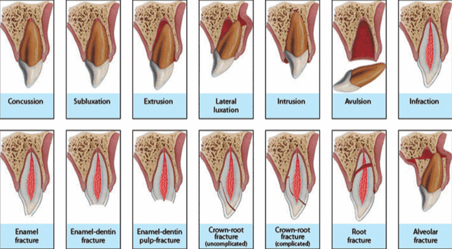
⭐what are some findings for extrusive luxation? treatment?
Tooth appears elongated Excessively mobile
Sensibility test: (-)
Radiographic Findings: Increased PDL space apically
tx: Reposition gently into socket Stabilization: 2 weeks, In mature teeth where pulp necrosis is anticipated or if signs and symptoms indicate that pulp (immature or mature teeth) became necrotic RCT is indicated

⭐what are some findings for lateral luxation? treatment?
Tooth is displaced in palatal/lingual or labial direction
Non-Mobile
Percussion (high metallic sound)
Alveolar process fracture
Sensibility test: (-)
Radiographic Finding: Widening of PDL space (occlusal)
tx: Reposition tooth digitally or with forceps to disengage from bony lock, Stabilization 4 weeks, Monitor pulpal condition If pulp becomes necrotic RCT indicated to prevent resorption

⭐what are some findings for intrusive luxation? treatment?
Tooth displaced axially into alveolar bone
Immobile High metallic sound at percussion
Sens: (-)
Radiographic Findings: PDL space absent from all or part of root, CEJ located more apically

⭐tx for intrusive luxation w/incomplete root formation
Allow eruption w/o intervention. If no movement in 4 weeks, begin ortho repositioning
⭐tx for intrusive luxation w/complete root formation
If intruded <3mm, allow to re-erupt with no intervention
If no movement within 8 weeks, reposition surgically and splint for 4 weeks
Alternatively, reposition orthodontically before ankylosis begins
If the tooth is intruded 3-7mm, reposition surgically (preferably) or orthodontically before ankylosis develops
If intruded >7mm, reposition surgically
Pulp almost always becomes necrotic->start RCT at 2weeks using a corticosteroid-antibiotic or CaOH2 in the cana
⭐what are some considerations for avulsion management?
Time elapsed to replantation
Stage of root development
Presence of crown or alveolar bone fractures
Time and type of storage medium (Best is HBSS- Hanks Balanced Salt Solution)
Contamination of PDL
Splinting
Occlusion
Root canal therapy
which storage medium is best for avulsion?
HBSS (Hanks Balanced Salt Solution)
⭐⭐why is there NO replantation for avulsed primary teeth?
high risk of ankylosis
⭐what are some findings and treatment for avulsion for immature tooth with dry time less than 60 minutes?
Change transport medium HBSS
Clean root surface, apical foramen w/saline
Administer anesthetic
Irrigate socket with saline
Replant and verify with radiographs
Splint 7-10 days up to 2 weeks
Monitor
If no REVASCULARIZATION, RCT recommended

⭐what are some findings and treatment for avulsion for mature tooth with dry time less than 60 minutes?
Change transport media HBSS
Clean root surface, apical foramen w/saline
Administer anesthetic
Irrigate socket with saline
Replant and verify with xray
Splint 7-10 days up to 2 weeks
Monitor
Start RCT within 10 days w CaOH2
what makes something an uncomplicated trauma to permanent dentition? complicated? treatments?
Uncomplicated fracture: enamel/dentin Resin restoration
Complicated fracture: w/pulp involvement
Radiographic evaluation
Pulpotomy or pulpectomy
EXT: if unrestorable or uncooperative patient
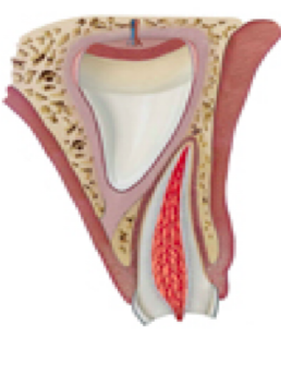
crown/root fracture trauma to primary dentition findings and tx?
Fragment Removal: only if fracture involves small part of root
Restoration: ONLY if fragment is stable and large enough for coronal restoration
EXT: In ALL other instances

findings and treatment for root fracture primary dentition?
The coronal fragment might be mobile and displaced
Fracture is usually located (mid root, apical third)
Treatment:
No displacement: NO TREATMENT
Coronal Displacement*
Mild mobility → spontaneous reposition
Excessive mobility and occlusal interference → EXT
Gentle reposition and splint 4 weeks
findings and treatment for alveolar fracture primary dentition?
Fracture involves alveolar bone and may extend to adjacent bone
Segment mobility and dislocation are common findings
Occlusal interference noted
Treatment
Radiograph evaluation (horizontal line to the apices of primary teeth and the successors will be disclosed)
Reposition displaced segments
Stabilization for 4 weeks
Monitor fracture line

findings and treatment for concussion and subluxation primary dentition?
Similar clinical findings as in permanent dentition
No treatment on primary dentition only observation and use of CHX
Color change alone does not indicate need for pulp therapy or extraction
Additional signs and symptoms of infection (mobility, radiolucency swelling, fistula) indicate further treatment

findings and treatment for extrusive luxation in primary dentition?
Partial displacement of tooth out of the socket
Tooth appears elongated
Excess in mobility
Increase PDL space
Treatment:
No interference → allow spontaneous reposition
Excessive mobility > 3mm EXT
⭐findings and treatment for lateral luxation in primary dentition?
Tooth displaced palatal/lingual Or labial
Immobile
Radiograph: occlusal X-ray, Widening PDL space apically
Treatment:
No Occlusal interference → allow to reposition spontaneously
Severe Occlusal Interference → EXT
Reposition tooth and splint 4 weeks
⭐findings and treatment for intrusive luxation in primary dentition?
Tooth displaced through buccal plate
Treatment:
Radiographic evaluation according to the location of displacement of the tooth it can appear shorter or longer
If apex displaced through buccal bone plate OR If apex is displaced into the developing tooth: allow spontaneous repositioning (takes up to 6 months)
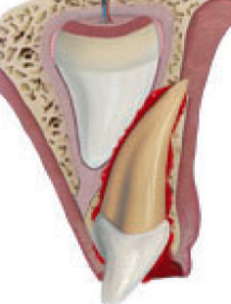
⭐what are some complications on permanent tooth after primary tooth displacement?
hypoplasia, hypocalcification, crown root dilaceration, disruption in eruption sequence