BMOL30090 L4 cellular components of innate IS
 Call Kai
 Learn
Learn Practice Test
 Spaced Repetition
 Match
Match Flashcards
 Knowt Play
1/50

There's no tags or description
Looks like no tags are added yet.
Name | Mastery | Learn | Test | Matching | Spaced | Call with Kai | Chat |
|---|
No analytics yet
Send a link to your students to track their progress
51 Terms
neutrophil function
bacteria killing, phagocytosis and cytokine production
eosinophil function
bacteria and parasite killers
macrophage function
cytokine production, antigen presentation and phagocytosis
mast cell function
vasoactive peptide production, microbe killing
natural killer cell function
bactericidal and cancer cell killing
dendritic cell function
antigen presentation
monocyte function
bactericidal and parasite killers
immune cell migration
how IICs arrive to site of infection
makes immune cells differ from other body cells
can migrate from blood into tissues, be homed to these tissues, and recirculate into blood/lymph
immune responses: combo of systemic and local events
immune cell infiltration
process occurs due to a succession of tightly coordinated phases. Many molecule types contrib to this process

vasodilation: histamines
histamine is a biogenic amine synthesised from L-histidine by L-histidine decarboxylase
mainly found in mast cells
induces vasodilation
vasodilation: prostaglandins (PGs)
PGs are made locally after injury, from arachidonic acid (cell memb. lipids) by COX
PGE2 can be produced from a myriad of cells, immune and non-immune (incl. endothelial cells)
have similar effects to histamine
ibuprofen inhibits COX and inhibits prod of PGs (pain)
chemotaxis
movement of cell in response to chemical signal/gradient
how IICs move to where they are needed
what does chemokine stand for?
chemokine - chemotactic cytokine
chemokines and rolling
released during inflam, injury and infection
engage chemokine receptors on surface of immune cells
in IICs, chemokine binding to their receptors promotes:
movement towards area of high gradient
increased binding affinity of integrins for their adhesion molecule
sources of chemokines
can be prod by immune cells themselves, epithelium and stroma
prod in response to PRR engagement or to cytokine stimulation
some receptors can bind to more than one chemokine - only from corresponding type (some CCs bind to different CCRs, some CXCs to different CXCRs) - COMPLEXITY
chemokine roles
CXCL1-CXCR2, CXCL8-CXCR1, CXCR2 - attract neutrophils
CCL2-CCR2 - attract monocytes
CCR7-CCL21/CCL19 - key for DC migration to LNs
selectins
carbohydrate-binding transmembrane proteins in the surface of immune and endothelial cells
mediate relatively low binding in the initial phase of migration
expression/conformation is increased in response to cytokines and vasoactive peptides
capture and rolling: selectins image

integrins and adhesion molecules
integrins are proteins expressed in surface of immune cells, which interact with adhesion molecules on the surface of endothelial and stromal cells
integrins are dimers formed by alpha and beta chains
transmembrane with intracellular end anchored to the cytoskeleton of the cell
interaction betw. integrins and adhesion molecules = anchor circulating immune cells to blood vessel wall, allowing rolling and crawling
integrins and adhesion molecules: ligands
ICAM-1 - neutrophils ligand
neutrophils need to be stuck to blood vessel in order to go to site of inflammation

reg. of integrins/adhesion molecules by inflam, factors: image

Step 1.
reg. of integrins/adhesion molecules by inflam, factors
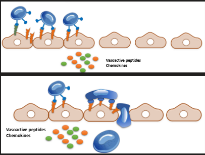
vasoactive substances - blood vessel dilation —> blood flow slows
Step 2.
reg. of integrins/adhesion molecules by inflam, factors
large leukocytes move away from the centre of blood vessels, closer to vessel walls
Step 3.
reg. of integrins/adhesion molecules by inflam, factors
selectin expression —> initial binding
selectins just recognise carbohydrates in cell surface initially
Step 4.
reg. of integrins/adhesion molecules by inflam, factors
cytokine/chemokine promote expression and conformational changes in adhesion molecules/integrins
Step 5.
reg. of integrins/adhesion molecules by inflam, factors
leukocytes crawl on endothelial cells where they find abundant expression of adhesion molecules to tether
Step 6.
reg. of integrins/adhesion molecules by inflam, factors
these abundanrt connections initiate processes inside the endothelial cells that reduce paracellular connections
rearrangement of endothelial cell cytoskeleton, possible favours their contraction
phosphorylation of VE-cadherin and destabilisation of cadherin-dependent junctions (triggered by abundant ICAM-1/VCAM-1-integrin connections
Step 7.
reg. of integrins/adhesion molecules by inflam, factors
paracellular route: immune cells move across endothelial walls using these gaps
e.g. distance between each cell is bigger to neutrophils can cross
image: reg. of integrins/adhesion molecules by inflam, factors
SUMMARY: reg. of integrins/adhesion molecules by inflam, factors
vasoactive mediators slow down local flow
leukocytes adhere and roll over the vascular wall:
first contact via selectins, rolling via integrins
circulating leukocytes in local capillaries come near vascular walls and sense chemokines
cytokines, chemokines and PGs stimulate expression or strengthen binding of adhesion molecules (vascular endothelium) and integrins (immune cells)
adhered leukocytes crawl over the vascular wall. adherent interactions become tighter
histamine, PGs and adherent binding relaxes connections between adjacent vascular endothelial cells. Oedema is facilitated
leukocytes move between vascular endothelial cells and into tissues: diapedesis
summary image

oedema
swelling/build up of fluid in a body tissue
cadherin
cell adhesion molecules important in forming adherens junctions that let cells adhere to each other
depend on Ca ions to function
leukocytes/IICs
leukocytes aka IICs
Q: proteins expressed on the leukocyte are ___
integrins
Q: proteins expressed on endothelium are ____
selectins

Q: this leukocyte stimulates gaps between endothelial cells via ____
tethering to adhesion molecules
Q: the 2 main types of vasoactive mediators are ___
histamine & PGs

Q: the mediators that attract the monocytes are ___
chemokines
recirculation of DCs
recogn. of PAMPs and exposure to cytokines at sites of infection trigger phagocytosis and Ag processing in DCs
leads to DC maturation
mature DCs migrate to nearby lymph nodes where they ‘educate/train’ the AIS
migration is coordinated by CCL21 released from lymphatic endothelium
DCs sense this via CCR7 receptor and migrate towards lymph nodes
inflammation
a natural response of our organism against injury or infection that helps clear pathogens and promotes tissue repair
chronic inflammation
a deleterious response whereby excessive or sustained activation of the IS causes tissue destruction or abnormal repair. Self-perpetuatingauto
autoimmune disease
disease where certain components of the IS actively attack host tissues
part 1. physiologic inflammation and tissue repair
epithelial barrier is breached - PAMPs and DAMPs sensed
resident ICs release early mediators: chemokines ( circulating ICs attached), histamine and PGs (vasodilation, slower blood flow)
cytokines/chemokines: induction of selectins/adhesion molecules (vascular endothelium), Ag presentation (local)
infiltration of monocytes: degranulation and bacterial killing
DCs, Ag capture, presentation and recirculation
part 2. physiologic inflammation and tissue repair
infection is cleared
granulocytes have mostly been destroyed during bacterial killing
monocytes and DCs may recirculate
remaining and resident MPs stimulate healing
stromal cells, such as fibroblasts are activated to prod extracellular matric to restore the basal membrane
epithelial cells are regenerated to repair the normal tissue architecture
phase 1 physiological image

phase 2 physiological image

phase 1. pathologic inflammation: tissue destruction or scarring
persistent activation of IICs. Can be triggered by:
genetic defects (e.g. NOD2 muts. in IBD)
loss of tolerance towards commensal microbes
defective epithelial barriers
abnormal adaptive immunity (e.g. autoantibodies)
continuous or enhanced recruitment of granulocytes: degranulation leads to epithelial damage
accum. of MPs with enhanced prod. of cytokines causing further activation of other immune and structural cells
MP prods can also damage epithelium
AICs also recruited and activated, incr. cytokine prod
phase 2. pathologic inflammation: tissue destruction or scarring
local structures such as fibroblasts are overactivated
incr. motility of fibroblasts leads to tissue accum.
in part due to constant activation fro MP-derived prods (transforming growth factor-beta1)
overprod. and deposition of ECM may lead to fibrosis and tissue strictures
or excess damage of epithelial wall can cause ulcerations or fistulas
in most patients w/ chron. inflam. D this abnorm. tissue remodelling is ultimate cause of severe disease = surgery (often)
phase 1 pathological image

phase 2 pathological image
