LAB 8
 Call Kai
 Learn
 Practice Test
 Spaced Repetition
 Match
 Flashcards
 Knowt Play
1/17

There's no tags or description
Looks like no tags are added yet.
18 Terms
Explain the compartmentalization of a whole skeletal muscle.
Epimysium: Surrounds the entire muscle.
Perimysium: Surrounds fascicles - which are groups of muscle fibers bundled.
Within a given fascicle, we have muscle cells aka fibers. These long cylindrical cells often span the entire length of muscle.
Endomysium: Surrounds each individual muscle fiber aka cell.

Muscle —> Fascicle —> Muscle Fiber —> Myofibril —> Sarcomere —> Myofilaments
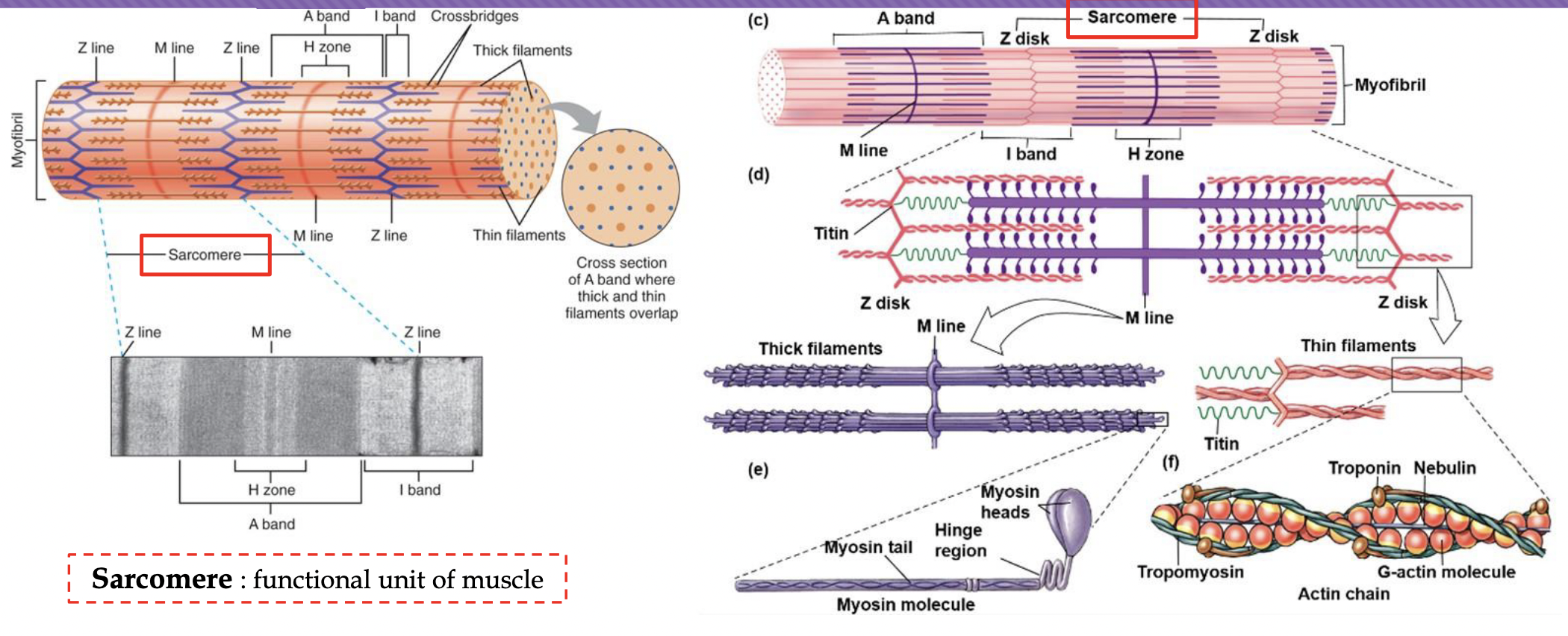
Within an individual muscle fiber we'll find specialized (and most common) contractile organelles called myofibrils. Myofibrils are made up of repeating sarcomeres (functional units of muscle).
Myofilaments are the smaller protein strands (specifically actin thin filaments and myosin thick filaments) that bundle together to build those myofibrils and make contraction possible.

Describe the basic anatomy of sarcomere
Actin (Thin Filaments): Protein filaments attached directly to the Z-lines that extend toward the center of the sarcomere. They contain the binding sites where myosin attaches during contraction.
Myosin (Thick Filaments): Heavier protein filaments located in the center of the sarcomere containing heads that interact with actin to form cross-bridges and pull the filaments together.
(and more)
Excitation Contraction Coupling
• Starting from the release of Ach to calcium being release
Calcium Entry into the Neuron: An action potential travels down the somatic motor neuron and reaches the axon terminal. This causes voltage-gated calcium channels to open, and calcium (Ca2+) rushes into the axon terminal.
ACh Release: The influx of calcium triggers synaptic vesicles to fuse with the neuron's membrane, releasing acetylcholine (ACh) into the neuromuscular junction via exocytosis.
ACh Binding: ACh diffuses across the synaptic cleft and binds to ligand-gated channels on themotor end plate.
End-Plate Potential (EPP): The opening of these channels allows sodium (Na+) to rush in, depolarizing the membrane and creating an end-plate potential.
Muscle Action Potential: Once this local depolarization reaches threshold, a full action potential fires and propagates rapidly across the sarcolemma.
T-Tubule Propagation: The action potential travels down the T-tubules deep into the muscle fiber.
Calcium Release from the SR: As the action potential passes through the T-tubules, it signals the adjacent sarcoplasmic reticulum (SR) to open its calcium channels, releasing stored Ca2+ into the sarcoplasm to initiate the cross-bridge cycle.

Muscle Contraction & Relaxation Sequence
Action Potential Propagation: Once an action potential is generated on the sarcolemma, it travels deep into the muscle fiber via T-tubules, triggering the release of stored calcium ions (Ca2+) from the sarcoplasmic reticulum (SR).
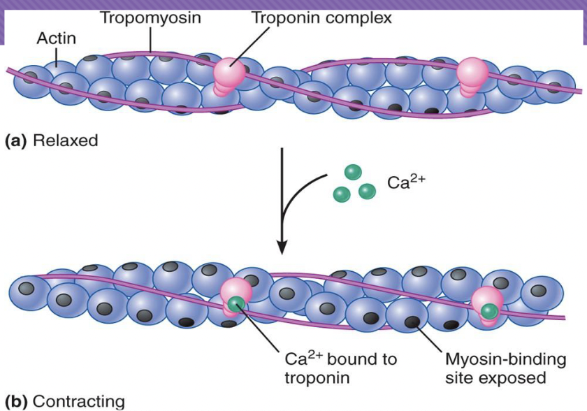
Calcium Binding: Released Ca2+ binds reversibly to troponin.
Troponin-Tropomyosin Shift: This binding causes the troponin complex to change shape, facilitating the rotation of tropomyosin.
Binding Sites Exposed: The movement of tropomyosin uncovers the myosin-binding sites on the actin filament.
Cross-Bridge Formation & Contraction: The myosin head attaches to actin (forming a cross-bridge), pivots to pull actin toward the center of the sarcomere, and shortens the muscle (sliding filament theory).
Relaxation: Ca2+ is actively pumped back into the sarcoplasmic reticulum. Without calcium bound to troponin, tropomyosin slides back to cover the myosin-binding sites, allowing the muscle to relax.

Sarcolemma
cell membrane
of a muscle cell
&
propagates APs
When acetylcholine (ACh) binds to receptors on the motor end plate, what is the direct and immediate
result?
Opening of ligand-gated ion channels, which allows sodium ions Na+ to rush into the muscle cell
What is the direct and immediatempurpose of AP as it propagates down the T-tubule?
to signal the sarcoplasmic reticulum (SR) to release stored calcium ions (Ca2+) into the sarcoplasm, which initiates muscle contraction.
Describe how calcium allows for myosin and actin to interact
The Blockade: At rest, protein strands called tropomyosin sit right over the myosin-binding sites on the actin filament, physically blocking myosin from attaching. Tropomyosin is held in this blocking position by the troponin complex.
Calcium Binding: When an action potential triggers the sarcoplasmic reticulum to release calcium (Ca2+) into the sarcoplasm, the calcium ions bind reversibly to troponin.
The Structural Shift: This binding causes troponin to change shape. Because troponin is physically linked to tropomyosin, this shape change forces tropomyosin to rotate and slide away, clearing the way.
Interaction Enabled: With the tropomyosin blockade removed, the myosin-binding sites on actin are uncovered. This allows the high-energy myosin heads to bind firmly to actin, forming cross-bridges and starting the contraction cycle.
(When calcium is actively pumped back away at the end of the contraction, troponin and tropomyosin slide right back to block the sites once again!)
ACh receptors on the Motor End Plate are destroyed
—>
Impairment in normal communication
between neurons and muscle fibers
ex: Myasthenia gravis (Mg)
Acetylcholine receptors on end plate are destroyed. There is no communication between nerves and muscle. Neuromuscular deficits – loss of motor control, muscle weakness.
Cross-bridge cycle
The mechanical "pulling" of actin is driven by a repeating chemical and physical sequence called the cross-bridge cycle.
Exposing the Binding Site (Preparatory Step):
At rest, tropomyosin blocks the myosin-binding sites on actin.
When an action potential triggers the release of Ca2+ from the sarcoplasmic reticulum, Ca2+ binds to troponin. This shifts tropomyosin out of the way, exposing the binding sites.
Cross-Bridge Formation:
With the site exposed, the high-energy myosin head binds firmly to actin, forming a cross-bridge.
The Power Stroke:
The myosin head pivots, bending and pulling the actin filament toward the center of the sarcomere (the M-line).
During this movement, stored energy is released, and ADP and Phosphate ion are detached from the myosin head.
Detachment (The Role of ATP):
A new ATP molecule binds to the myosin head.
This binding causes myosin to break its bond with actin, releasing the cross-bridge. (Without fresh ATP, muscles stay locked in a state of contraction, known as rigor mortis).
Resetting the Myosin Head (Cocked Position):
The attached ATP is quickly split into ADP and Pi by the enzyme ATPase on the myosin head.
The energy released from this hydrolysis "re-cocks" the myosin head back to its high-energy, upright position, ready to bind to a new site further down the actin chain and repeat the cycle.


The position of tropomyosin is
regulated by troponin complex,
by binding reversibly to ( ? )
Ca2+
Sliding filament theory
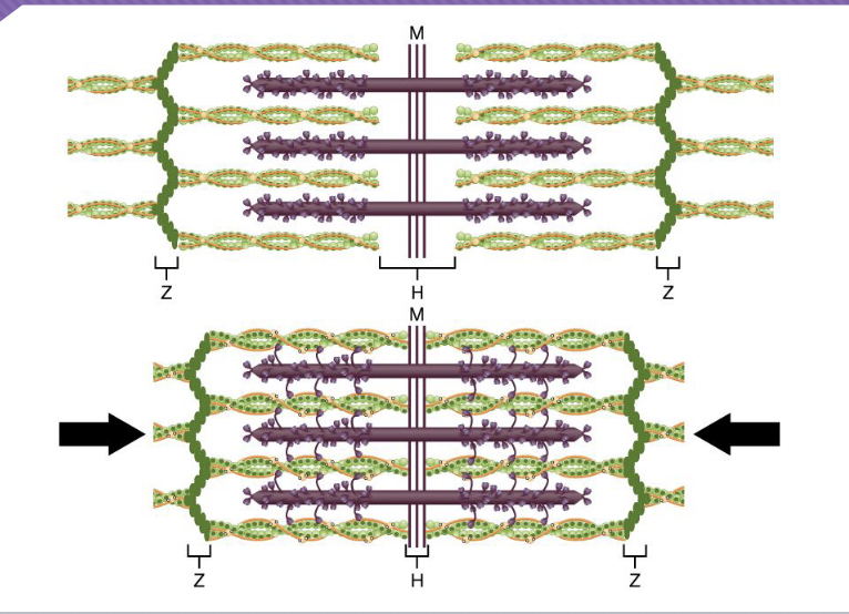
explains that muscle fibers shorten not because the individual protein filaments shorten, but because they slide past one another.
The Filaments: Thin filaments (actin) are pulled toward the center of the sarcomere (the M-line) by thick filaments (myosin).
The Result: As actin overlaps more and more with myosin, the Z-lines are pulled closer together, causing the entire sarcomere—and consequently the whole muscle cell—to shorten.
An action potential in a motor neuron triggers contraction of
all the muscle cells connected to that neuron
Muscle Twitch
a short submaximal amount of force caused by a single action potential
Explain what causes a muscle fiber twitch, summation, and tetanus.
Frequency of Stimulation
If a muscle fiber is stimulated by a single action potential, it produces a weak, brief contraction called a twitch.
If the fiber is stimulated again before it fully relaxes, the twitches add together (summation).
At very high stimulation frequencies, the muscle has no time to relax at all, fusing into a smooth, maximum sustained contraction known as tetanus, which generates peak force.

Size of motor unit depends on
# of muscle fibers
Factors Affecting Force Generation
*Number of Active Motor Units: A motor unit consists of a somatic motor neuron and all the muscle fibers it innervates. To lift a heavier load, the nervous system recruits more motor units to bring more muscle fibers into action.
*Number of Active Cross-Bridges: The more myosin heads that successfully bind to actin and pull, the greater the tension generated.
Frequency of Stimulation: At very high stimulation frequencies, the muscle has no time to relax at all, fusing into a smooth, maximum sustained contraction known as tetanus, which generates peak force.
Fiber Diameter: Larger-diameter muscle fibers contain more myofibrils packed side-by-side. Because they have more parallel contractile machinery (actin and myosin), thicker fibers can generate significantly more force than thinner ones.
Size Principle
By combining high stimulation frequencies, optimal fiber thickness, and the systematic recruitment of motor units via the size principle, the neuromuscular system can seamlessly grade force from a light twitch to maximum power.
Size Principle
motor units are recruited in order from smallest to largest (in terms of motor neuron cell body size and threshold) as the demand for muscle force increases.