APHY 160 - LO3 LS2 - Describe the Bones of the Axial Skeleton and Their Major Features
 Call Kai
 Learn
 Practice Test
 Spaced Repetition
 Match
 Flashcards
 Knowt Play
1/124

Earn XP
Description and Tags
All information taken from bright space
125 Terms
what is the cranium (skull)
it’s the skeletal structure of the head that supports the face and protects the brain
what are the two main subdivisions of the cranium (skull)
the facial bones and the brain case (or cranial vault)

what are the main functions of the facial bones
underlie facial structures, form the nasal cavity, enclose the eyeballs, and support the teeth of the upper and lower jaws

what are the main functions of the brain case
it surrounds and protects the brain and houses the middle and inner ear structures
how many cranial bones make up the adult skull
8 cranial bones
how many facial bones are in the adult skull
14 facial bones
what is the only moveable bone of the skull
the mandible (lower jaw)
how many bones are in the entire skull
28 total bones (8 cranial bones, 14 facial bones, and 6 auditory ossicles)
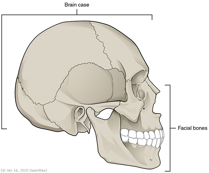
how many bones make up the cranium/brain case
8 bones

what are the eight cranial bones
1 frontal bone, 2 parietal bones, 1 occipital bone, 2 temporal bones, 1 sphenoid bone, & 1 ethmoid bone
how many bones are in the face
14 bones

the facial bones are all paired except for
the mandible and the vomer bones

the frontal bone forms what parts of the head
the forehead, part of the floor of the cranium, & most of the upper orbital (eye socket) roof

what structures are contained inside the frontal bone
two frontal sinuses

what does the parietal bones form
the lateral (side) wall of the cranium (skull)
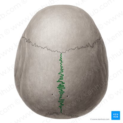
which suture separates the two parietal bones from each other
the sagittal suture

which suture separates the parietal bones from the frontal bone
the coronal suture

the occipital bone forms what part of the skull
the back and base

what is the foramen magnum
a large opening in the occipital bone that allows the spinal cord to pass through
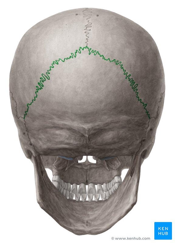
which suture separates the occipital bone from the parietal bones
the lambdoidal suture

the temporal bones form what parts of the skull
the sides and base

what is the external auditory meatus
a passageway inside the temporal bone leading to middle and inner ear structures
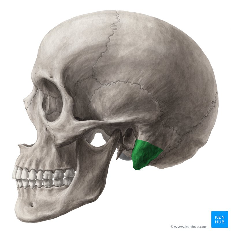
what is the mastoid process
a protuberance (bump) behind the ear of air spaces

what is the glenoid fossa
a depression on the lower portion of each temporal bone

what does the glenoid fossa do
holds the condyle of the mandible to form the temporomandibular joint (TMJ)

what is the function of the styloid processes on the temporal bones
serve as attachment points for muscles

where are the squamosal sutures located
between the parietal and temporal bones

what shape is the sphenoid bone
a bat-like shaped bone

what part of the cranium (skull) does the sphenoid bone form
the central part of the floor of the cranium
what features are found on the sphenoid bone
sinuses behind the eye, medial and lateral pterygoid plates for muscle attachment, and a depression for the pituitary gland

what part of the cranium (skull) does the ethmoid bone form
part of the floor of the cranium (skull), the orbit, and the nasal cavity

what does the ethmoid bone contain and help form
it contains sinuses, forms the upper part of the nasal septum (perpendicular plate), and the superior & middle conchae of the nasal cavity
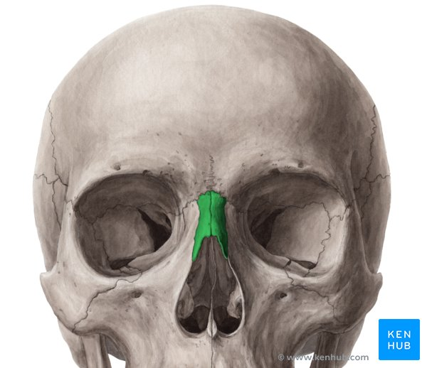
what do the nasal bones form
the bridge of the nose

what does the maxillary bones form
the upper jaw and anterior (front) part of the hard palate

what connects the two maxillary bones
the maxillary suture

where is the maxillary tuberosity located
on the outer posterior surface of the maxillary bones
what nerve is located near the maxillary tuberosity
the posterior superior alveolar nerve
why is the maxillary tuberosity useful in dental anatomy
it serves as a landmark for locating the posterior superior alveolar nerve

what structure on the maxillary bones supports the upper teeth
the alveolar process

where is the incisive foramen located
between the two maxillary bones

what parts of the cranium (skull) do the zygomatic bones form
the cheekbones and the lateral (outer) and inferior (lower) parts of the eye orbit

what is the purpose of the zygomatic arch
forms the prominence of the cheek and serves as the attachment point for the masseter muscle

what do the lacrimal bones form
the medial (inner) part of the eye orbit

what do the palatine bones form
the posterior (back) roof of the mouth and the lateral and inferior parts of the nasal cavity

the greater palatine foramen is an opening for what
the greater palatine nerve
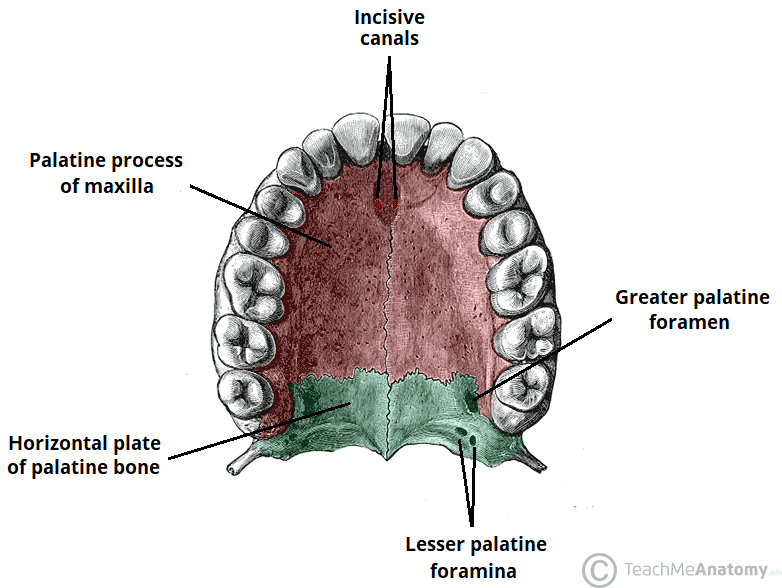
the lesser palatine foramen is an opening for what
the lesser palatine nerve

what do the inferior nasal concha bones form
the lateral walls of the nasal cavity

what does the vomer bone form
the lower part of the nasal septum

what is the mandible
a u-shaped bone forming the lower jaw that articulates with the temporal bones

what makes the mandible unique compared to other facial bones
it is the only freely moveable bone of the face

where is the hyoid bone located, and what makes it unique
it is a u-shaped bone in the neck that does not join to any other bone
what is the function of the hyoid bone
serves as an attachment site for the tongue and other muscles

where are the auditory ossicles located
within the temporal bone

which three bones make up the auditory ossicles
the malleus, incus, and stapes

what is a suture
an immobile joint located between adjacent bones of the skull
what fills the narrow gap between bones in a suture
dense, fibrous connective tissue

why are the long sutures of the brain case twisting and irregular instead of straight
to tightly interlock the adjacent bones, adding strength to the skull for brain protection

where are the coronal and sagittal sutures located
on the top of the skull

where does the coronal suture run
from side to side across the skull within the coronal plane

which bones are joined by the coronal suture
the frontal bone and the right and left parietal bones

where is the sagittal suture located
along the midline at the top of the skull in the sagittal plane, extending posteriorly from the coronal suture
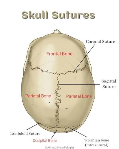
which bones are united by the sagittal suture
the right and left parietal bones
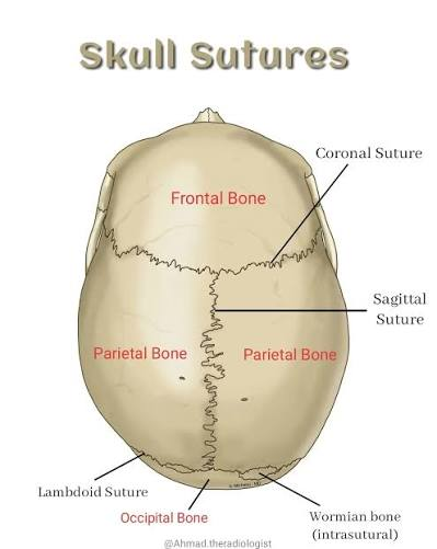
how does the sagittal suture end on the posterior skull
by joining the lambdoid suture
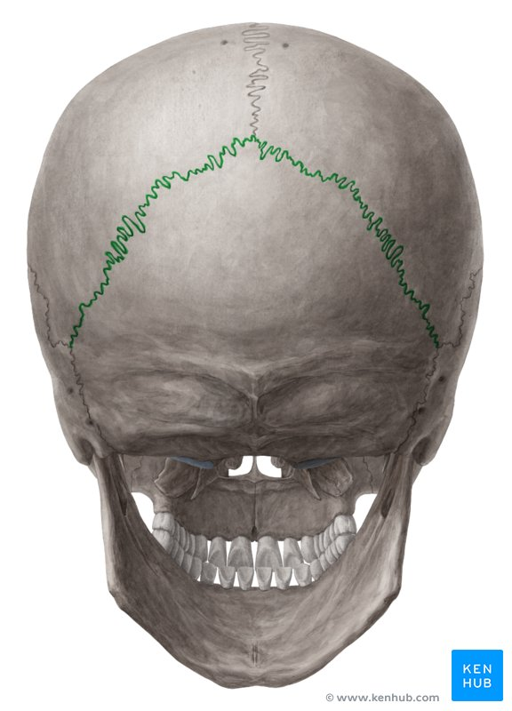
where does the lambdoid suture extend
downward and laterally to either side from its junction with the sagittal suture

which bones does the lambdoid suture join
joins the occipital bone to the left & right parietal and temporal bones
why is the lambdoid suture named that way
for its upside-down "V" shape that resembles the capital Greek letter lambda (Λ)

where is the squamous suture located
on the lateral (side) of the skull
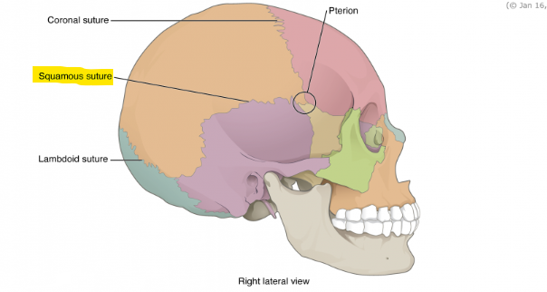
which bones are united by the squamous suture
the squamous portion of the temporal bone and the parietal bone

where is the intermaxillary suture located
on the face between the two fused portions of the maxillae

where is the median palatine suture located
down the middle of the roof of the mouth

where does the transverse palatine suture run
between the two palatine bones and the maxilla
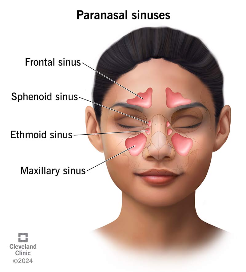
what are the paranasal sinuses
they are hollow, air-filled spaces located within certain bones of the skull
what do the words "paranasal" mean
it means "next to nasal cavity”
what lines the paranasal sinuses
they are lined with nasal mucosa
what are two main functions of the paranasal sinuses
they reduce bone mass to lighten the skull and add resonance to the voice
why does your voice sound different when you have a cold
swelling and excess mucus block the narrow passages between the sinuses and the nasal cavity
what causes pain and pressure in the sinuses during an illness
blocked passages let the sinuses fill with fluid, creating painful pressure
how are the paranasal sinuses named
they are named after the skull bone that each occupies

where is the frontal sinus located
just above the eyebrows within the frontal bone

which paranasal sinus is the most anterior (front)
the frontal sinus
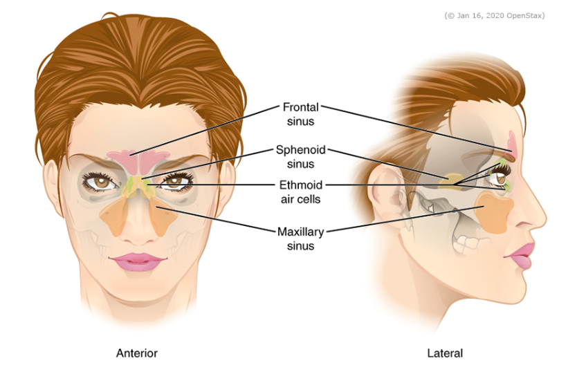
which is the largest paranasal sinus
the maxillary sinus

where are the maxillary sinuses located
in the right and left maxillary bones just below the orbits (eye sockets)
which sinuses are most involved during sinus infections
the maxillary sinuses
why are the maxillary sinuses difficult to drain
their connection to the nasal cavity is high on their medial wall

where is the sphenoid sinus located
within the body of the sphenoid bone

which is the most posterior (back) sinus
the sphenoid sinus, which is a single, midline space
what are ethmoid air cells
they are multiple small spaces separated by thin bony walls in the lateral aspects of the ethmoid bone

what is the shape and location of the sphenoid bone
a bat-like shaped bone located at the base of the skull

what structures on the sphenoid bone serve as attachment points for chewing muscles
the medial and lateral pterygoid plates, which attach to the medial and lateral pterygoid muscles of mastication

what is the hamulus
a hook-like structure on the medial pterygoid plate that helps guide the tendon of a small muscle

where is the mandibular fossa located
on the bottom of the temporal bone

what does the mandibular fossa articulate with
it articulates with the mandible (lower jaw)

what part of the skull does the occipital bone form
it forms most of the bottom back of the skull

what opening is found in the occipital bone and what is its function
the foramen magnum, which is the opening that allows the spinal cord to exit the brain case

what bone makes up most of the face and the front two-thirds of the hard palate
the maxillary bone
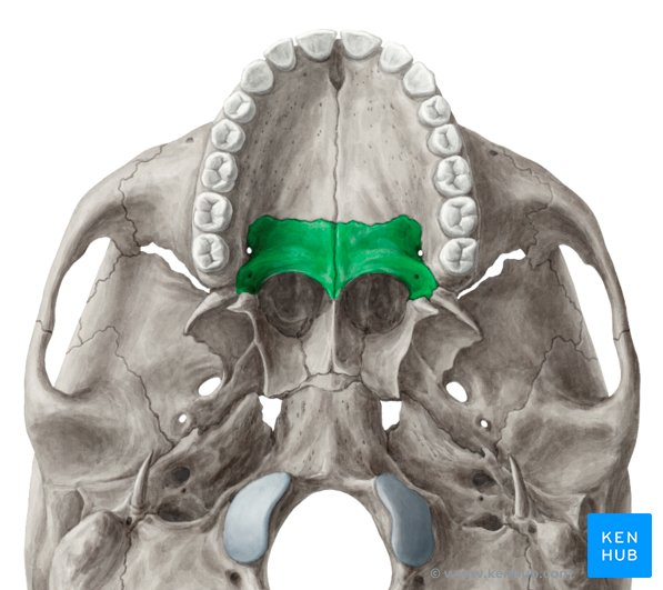
what bones form the back one-third of the hard palate
the two palatine bones

where is the incisive foramen located
at the center of the front of the hard palate
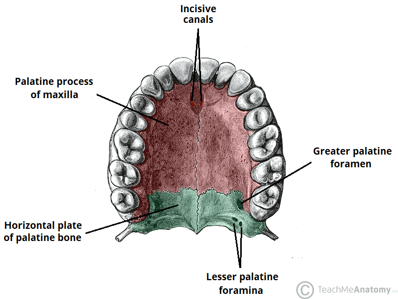
where are the greater palatine foramina located
on either side at the back of the hard palate