Equip/Image - Resolution/Distortion (Unit 2-4)
 Call Kai
 Learn
 Practice Test
 Spaced Repetition
 Match
 Flashcards
 Knowt Play
1/131

There's no tags or description
Looks like no tags are added yet.
132 Terms
What are the two geometric properties of image quality?
Spatial Resolution
Distortion
Spatial Resolution
Degree of geometric sharpness or accuracy of the structural lines actually recorded in the image.
Definition, sharpness, recorded detail
Poor resolution = ___________ sharp definition of fine detail.
Lack of
Poor resolution is caused by unacceptable levels of _________ (edge unsharpness around periphery of the image)
Penumbra
Primary film-screen unit of resolution is _______________________.
Line pairs per millimeter (lp/mm)
Most human visual acuity is limited to the range of _________ (each line is ___ mm wide and seen by the human eye).
5 lp/mm
0.1
The higher the lp/mm, the __________ the resolution.
Better
Digital imaging spatial resolution is determined primarily by 3 factors:
Matrix size
Pixel size
Grayscale bit depth
Digital imaging spatial resolution is commonly described as _______________.
Spatial Frequency
Higher frequency represents pairs of lines that can be visualized __________________ = _________________. It is good for _______ objects.
Very close together
High resolution
Smaller
Lower frequency represents pairs of lines that are __________________ = _________________. It is good for ______ objects.
Further apart
Low resolution
Larger
Pixel Pitch
Distance from the center of one pixel to the center of the next one in the display.
A _______ lp/mm value indicates better resolution.
Higher
What is the formula for calculating resolution based on pixel pitch?

Modulation Transfer Function (MTF)
Mathematically expresses spatial resolution on a scale of 0-1.
0 MTF
No signal, no image
1 MTF
Highest resolution possible.
In medical imaging, the goal is to produce an image with an MTF value as close as possible to ______.
1 or 100%
Sampling frequency is expressed as ____________.
Pixels/mm
Digital imaging requires that the spatial resolution frequency be sampled ______________.
Twice per cycle
_____________ sampling 2x/cycle produces an accurate, high-frequency digital signal.
Nyquist Frequency
A higher sampling frequency __________ image resolution (fidelity) because more information is detected and captured for accuracy.
Increases
Aliasing
Occurs when the Nyquist criterion is violated, and sampling occurs less than twice per cycle.
Aliasing produces an _________, fluctuating, ___-frequency digital signal.
Inaccurate
Low
Aliased data create a _________ artifact.
Moire pattern
Spatial resolution is often improved through factors that ________________.
Increase patient dose
Resolution problems should be approached in this order: (4)
Eliminate motion
Reduce OID
Reduce focal spot size
Increase SID
_________ of the beam is the most important factor for spatial resolution.
Geometry
Because the x-ray emanates from a small point (the focal spot), the photons ________ as they move further from their source.
Diverge
SID=___ + ___
SOD + OID
SOD=____-____
SID - OID
Resolution is improved when OID ___________ and degraded whenever it ____________.
Decreases
Increases
Affected side or part of interest is always positioned _____________ to decrease OID.
As close to IR as possible
Resolution is improved when SID ___________ and degraded when it ____________.
Increases
Decreases
Focal Spot
Area of the focal track that is impacted by the electron beam at one time.
Actual Focal Spot
The physical area of the focal track that is impacted; a for arrival.
Effective Focal Spot
The area of the focal spot that is projected out of the tube toward the object being radiographed; e for exit.
Line Focus Prinicple
Minimizes the effective focal spot while maximizing the actual focal spot to absorb the heat.
Focal spot size is a major controller of spatial resolution because it controls __________.
Penumbra
As the focal spot decreases in size, penumbra ____________, and resolution ___________.
Decreases
Increases
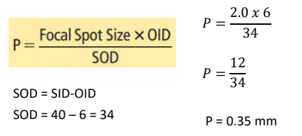
The width of penumbra can be calculated by:

Penumbra decreases when: (3)
Focal spot decreases
OID decreases
SID increases
Penumbra happens because x-rays _______.
Diverge
Penumbra almost always happens in diagnostic x-ray because overall general shape of human anatomical structures are _____________.
Round or oval
_______________ can improve spatial resolution by reducing part thickness and decreasing OID and penumbra.
Part compression
________ affects spatial resolution because it fails to permit enough time for a well-defined image to form.
Motion
As a result of motion, no ________ can be perceived.
Fine detail
Voluntary Motion
Motion that is under the direct control of the patient.
Involuntary Motion
Motion not under the conscious control of the patient.
The first choice of who should hold a patient is _____________, followed by ____________ or __________.
Non-pregnant relative
Hospital employee
Radiographer
Increase in SID causes _______ in spatial resolution.
Increase
Increase in OID causes ________ in spatial resolution.
Decrease
Increase in patient thickness causes _________ in spatial resolution.
Decrease
Increase in focal spot size causes _______ in spatial resolution.
Decrease
Increase in motion causes ________ in spatial resolution.
Decrease
As focal spot size decreases: (select all that apply)
Penumbra decreases
Sharpness increases
Spatial resolution increases
Penumbra decreases
Sharpness increases
Spatial resolution increases
Calculate the penumbra for an image taken with a 2.0 mm focal spot, at a 40” distance, and OID of 6 in.
0.35 mm
Distortion
Misrepresentation of the size or shape of the structures being examined.
Magnification is _____ distortion.
Size
Elongation and foreshortening are _______ distortion.
Shape
Magnification
Size distortion that occurs during exposure; controlled by SID, OID.
Reduced magnification size distortion ________ the spatial resolution.
Increases
______________ is the exception and requires a special machine with a tiny focal spot to magnify and elevate very subtle skeletal anomalies.
Magnification Radiography
Magnification size distortion is controlled by positioning the body part and tube to maximize ____ while minimizing ____.
SID
OID
The greater the SID, the __________ the mag.
Smaller
Magnification Factor Formula

If the SID is 40" and the OID is 2", what is the magnification factor?
1.05

Actual Size Formula

If object B is 2" in diameter on the image and is measured to be 3" from the image receptor, what is the actual size if the SID is 40"?
1.85”

% Mag Formula

Shape Distortion
Misrepresentation by unequal mag of the actual shape of the structure being examined.
Elongation
Projects object so it appears to be longer than it really is.
Foreshortening
Projects it so it appears shorter than it really is.
Elongation occurs when the _______________ is improperly aligned.
Tube or the IR
Foreshortening occurs _________________ is improperly aligned.
Only when the part
Changes in the tube angle cause __________, never ____________.
Elongation
Foreshortening
Increase in SID causes _________ in magnification.
Decrease (size)
Increase in OID causes _________ in magnification.
Increase (size)
Increase in improper alignment causes _______ in elongation or foreshortening.
Increase (shape)
If a projected image measures 5.5 in and the magnification factor is 1.25, what is the size of the acutal object?
4.4”

Which of the following occurs when the IR is misaligned with the body part?
Elongation
Image Critque Steps
Classify the image
Determine the cause of the problem
Determine appropriate corrective action
The first step in classifying the image is to:
Evaluate the entire image and determine if it is within the diagnostic acceptance limits (is it repeatable?).
Common Repeatable Issues
C: Central Ray is centered or angled wrong
R: Radiation exposure factors (technique) is wrong
A: Anatomy is clipped
P: Positioning is wrong
Slowly narrow the search by first classifying the problem into one of three major categories:
Technical factor problem
Procedural factor problem
Equipment malfunction problem
Exposure Index (EI)
Actual Exposure
Target EI (EIt)
Ideal Exposure
Deviation Index (DI)
% deviation from target. DI value of 2 or more needs to be evaluated for repeat.
Geometric problems with detail include:
Spatial resolution
Distortion problems
Spatial resolution problems are the result of:
Geometry of the beam (focal spot size, SID, or OID)
IR (matrix, pixel size)
Patient motion
Distortion problems are a result of:
Size distortion (magnification)
Shape distortion
Size distortion problems are the result of:
SID or OID
Shape distortion problems are the result of ___________________________ and result in ______________________.
Improper tube, par, and/or IR alignment
Elongation, foreshortening
A ________________ factor problem is one that involves ___________________ or _____________________ for the x-ray examination.
Procedural
Patient positioning
Patient preparation
The “classify” step of image critique …
Is step 2 of the image critique process
Involves classifying the image as acceptable or repeatable.
Involves classifying the cause of a repeatable image
Involves classifying the correctable action needed

Match the causes of imaging problems with their corresponding letter.
_____ Procedural Factor Problems
_____ Technical Factor Problems
_____ Geometric Problems
_____ Equipment Malfunction
_____ Photographic Problems

Quality Assurance
Activites that ensure that radiology service is consistently high-quality.
__________________ regular evaluation of equipment to determine its safe and accurate operation.
Quality Control (QC)
Linearity
Given mAs value, using different mA stations with appropriate exposure time adjustments to equal the same mAs, will provide consistent intensity mGya/mAs.
The max acceptable variation in linearity is _____ from one mA station to an adjacent mA station.
10%