Life-threatening injuries
 Call Kai
 Learn
Learn Practice Test
 Spaced Repetition
 Match
Match Flashcards
 Knowt Play
1/58

There's no tags or description
Looks like no tags are added yet.
Name | Mastery | Learn | Test | Matching | Spaced | Call with Kai | Chat |
|---|
No analytics yet
Send a link to your students to track their progress
59 Terms
In the time following a major trauma, at what 3 points is the patient most likely to die?
At the time of the trauma.These patients usually can’t be saved as their death is instantaneous, or their injuries are unsurvivable.
1-2 hours later. These patients are the ones who initially compensated for trauma, (eg. increasing HR, shutting off peripheral circulation etc.) but have now used up their physiological reserves.
Weeks later. This is when patients experience sequelae of trauma such as infection and multi-organ failure.
The ‘golden hour’
The first 60 minutes after a major trauma that determines a patient’s outcome. Advanced Trauma Life Support (ATLS) targets patients in this hour.
ATLS Assessment map
ABCDE - gives priority to injuries and problems that will kill a patient quickest.
ABCDE
Airways
Breathing
Circulation
Disability
Exposure
Assessing airways
Can they respond?
Gurgling, wheezes and snoring noises are more worrying
Examine the patient’s face, mouth and nose - severe injuries and physical blockages can put the airway at risk.
How to open blocked airways
Suctioning fluids
Remove any foreign bodies
Basic airway manoeuvres (lift chin)
Airway adjuncts
High flow O2
What should all trauma patients get?
High flow O2
C-Spine control
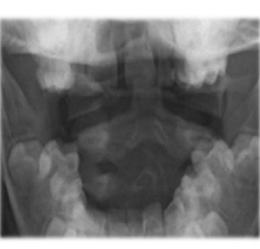
What does this radiograph show?
Fracture of the dens on the second cervical vertebra. This injury is quite common in high energy trauma, so every patient should be assumed to have a cervical fracture and get C-spine care until proven otherwise.
What are the two main issues with breathing in trauma patients?
Lung cavity fills with something else (usually air or blood)
Thoracic cage is damaged
Tension pneumothorax typical mechanism (open)
Penetrating trauma makes a hole in the chest wall, allowing air to be drawn into the thoracic cavity when the patient breathes in, and sealing shut when the patient breathes out. Every time the patient breathes in, more air enters, collapsing the lung and increasing the pressure inside.
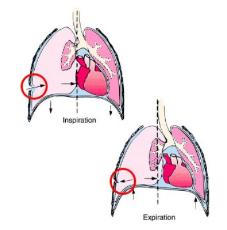
What happens as pressure rises in a patient’s thoracic cavity (tension pneumothorax)?
The mediastinum is pushed over to the other side, squashing the remaining lung and further compromising breathing.
Venous return will reduce as the pressure becomes equal to the pressure in the great veins of the heart, eventually causing heart arrest.
How to spot a tension pneumothorax
Decreased breath sounds on one side.
Increased percussion note (hyperresonant)
Engorged neck veins
Reduced lung expansion
Deviation of trachea to opposite side
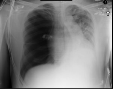
Why should you never see a radiograph that looks like this?
A tension pneumothorax this severe will have all clinical signs present, so treatment should not be delayed for a radiograph.
Treatment of a tension pneumothorax
High flow oxygen
Needle decompression - only a temporary measure
Chest drain insertion within 30 mins
Needle decompression
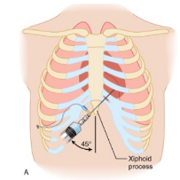
Insertion of a large bore needle through the chest wall to allow air from the thoracic cavity to escape.
Where is needle decompression done?
Adults: 4th or 5th intercostal space at the mid-axillary line (a line from the middle of the armpit). Same location as chest drain insertion.
Children: 2nd intercostal space (below 2nd rib), in line with the middle of the clavicle.
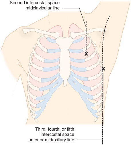
Open pneumothorax
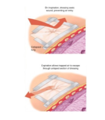
When the patient breathes in, air enters the thoracic cavity via their wound and bypasses the lungs entirely. When the patient breathes out, the air is forced out again via the wound.
Air enters the thoracic cavity via the path of…
least resistance
What are the consequences of an open pneumothorax?
The blood is not oxygenated properly because there is a lack of effective breathing.
Treatment of an open pneumothorax
High flow O2
Place a 3 sided dressing over the wound to create a makeshift external one-way valve.
Chest drain insertion.
What conditions commonly affect the breathing of trauma patients?
Tension pneumothorax
Open pneumothorax
Massive haemothorax
Flail chest
Flail chest
Two or more ribs fractured in 2 or more places, leading to a separation of a segment of the thoracic cage. When the patient breathes in, this floating fragment is drawn inwards, while the rest of the ribcage moves out. This prevents effective ventilation. There are often also underlying injuries to the lungs (eg. haemopneumothorax and contusions)
Treatment of flail chest
Fix the fracture with a plate and screws. This relieves pain, allows the patient to recover faster and reduces lung problems like chest infections.
Signs of circulatory compromise
Pale
Poor peripheral circulation
Heart sounds
Look at observation readings like pulse and blood pressure
Initial actions to treat circulatory compromise
Establish two cannulas for IV access. This allows rapid fluid resuscitation.
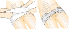
Control obvious source. Eg. apply external pressure to a bleeding wound, use a pelvic binder
What are the two main issues with circulation in trauma patients?
Haemorrhagic conditions
Cardiogenic issues
Common haemmorhagic conditions in trauma patients
Pelvic fractures
Massive haemothorax
Pelvic fractures
These fractures can result in massive blood loss into the pelvic cavity, which may not be immediately obvious. Using a pelvic binder can slow the internal bleeding.
Massive haemothorax
This occurs when 1500 mls of blood is lost into the pleural cavity. This prevents the lungs from inflating fully.
Treatment of massive haemothorax
Insert a chest drain, and provide blood products. Cardiothoracic surgery is then performed.
Common cardiogenic issue in trauma patients
Cardiac tamponade
Cardiac tamponade
Compression of the heart by an accumulation of fluid in the pericardial sac. This sac is ‘fixed’ (like the skull), meaning even a small pressure increase has a big effect. The heart then cannot pump during systole, or dilate to fill during diastole.
Treatment of cardiac tamponade
Pericardiocentesis - US guided needle enters the chest wall and punctures the pericardium to extract the blood gathered there.
Presentation of cardiac tamponade
History of penetrative trauma to thorax
Becks triad:
Hypotension: decreased stroke volume
Jugular venous distension: impaired venous return to the heart
Muffled heart noises: fluid inside the pericardium
What is the usual outcome of having any of these life threatening conditions?
The patient goes into shock.
Shock
End-organ dysfunction due to inadequate oxygen availability for tissues (perfusion).
What type of tissues are worst affected by shock?
Oxygen sensitive tissues like cardiac, gut, brain and renal tissues etc.
What are the two main causes of shock?
Reduced delivery of O2 (restricted blood vessels, inability for blood to pump, loss of blood…)
Increased demand for oxygen
Lethal triad of trauma
Acidosis
Hypothermia
Coagulopathy
How can trauma lead to acidosis?
If there is inadequate oxygen availability, the organs keep functioning in the absence of it using non-aerobic respiration. This leads to metabolic acidosis.
What is the most common type of shock in trauma patients?
Haemorrhagic shock
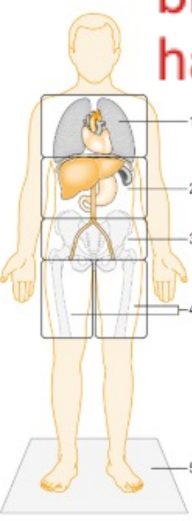
What are the 5 most common places for major blood loss in trauma patients?
Intrathoracic cavity
Intraperitoneal cavity
Retroperitoneal space
Thigh compartments
Floor
What are the 4 classes of haemorrhagic shock?
Class i - <15% (roughly <750ml), <100 bpm, normal BP, urine output = >30 ml/hr
Class ii - 15-30% (roughly 750-1500 ml), 100-120 bpm, normal BP, urine output = 20-30ml/hr
Class iii - 30-40% (roughly 1500-2000ml), 120-140 bpm, BP dropping, urine output = 5-15 ml/h
Class iv - >40% (roughly >2000 ml), >140 bpm, BP dropping, urine output = negligable
What happens to urine output as patients go into shock?
It begins to drop
What happens to heart rate as patients go into shock?
It increases
What will happen to the patients blood pressure when they go into the later stages of shock?
It will drop.
Why is a low blood pressure for shock patients good (permissive hypotension)?
It is just enough to keep cerebral perfusion, but not enough to start bleeding again. 80mmHg systolic pressure is the rough target.
Fluid resuscitation
First stop bleeding
Provide blood products: red blood cells, platelets and fresh frozen plasma
Tranexamic acid - promotes clotting
Permissive hypotension
Disability
Level of consciousness
What is the best way to assess disability?
The Glasgow Coma Scale (GCS) - Gives the patient a score out of 15. The higher the score, the more alert the patient is.
What is a quick way to assess disability (not GCS)?
Alert, Verbal, Pain, or Unresponsive (AVPU) scale
What are the two most common reversible reasons for unconsciousness?
Temperature and blood glucose.
Exposure
Full exam of patient whilst keeping them warm.
Why is it important to keep trauma patients warm?
Clotting is temperature dependant.
Common sites to miss injuries
Back of head
Back
Buttocks
Perineum
Axillae (armpits)
Skin folds
If anything changes during the ABCDE assessment, what should you do?
Go back and reassess!
Mnemonic to remember life threatening trauma conditions
ATOM FC
ATOM FC
Airway obstruction
Tension pneumothorax
Open pneumothorax
Massive haemothorax
Flail chest
Cardiac tamponade
Secondary survey
Systematic examination with log roll, usually done by an orthopaedic registrar.