Ch 4: Vascular Anatomy
 Call Kai
 Learn
 Practice Test
 Spaced Repetition
 Match
 Flashcards
 Knowt Play
1/132

Earn XP
Description and Tags
powerpoint
133 Terms
What are the layers of the vessel walls (both arteries and veins)
tunica intima, tunica media, and tunica adventitia

How do the vessel layers differ between arteries and veins
arteries have thicker tunica media (muscle layer) while veins have a thinner layer

What are traits of large arteries
elastic arteries with less smooth muscle
examples of large arteries
the aorta and the largest branches (ex: iliacs, renals, celiac)
Size of small/medium sized arteries
approx. 4mm in diameter
What is the smooth muscle layer like in small/medium sized arteries
its well-developed
examples of small/medium sized arteries
includes all arteries except aorta and major branches
What are arterioles
“stopcocks” of the vascular system
What does smooth muscle layer of arterioles do
circular smooth muscle layer controls contraction and resistance
How does the size/composition of walls of vein compare to arteries
the walls are thinner than arteries, and contain more elastic fibers and collagen than muscle fibers

What are venule walls mainly made of
mainly connective tissue
Examples of large veins
portal vein, superior and inferior vena cava, and main branches
What portion of the the large vein walls is thicker and what does it contain
they have thick adventitia layer with fibrous and elastic tissues

Size of small/medium sized veins
diameter of 1-10mm
Examples of small/medium veins
includes all veins expect portal vein and vena cava and their main branches
What portion of the the small/medium vein walls is thin
tunica media
What is a venous valve and where are they located
They are bicuspid inward projections of the tunica intima and are only in the veins (not arteries)

Function of venous valves
prevent retrograde movement of blood
What is a sinus in regards to venous valves
enlarged area behind leaflets
Where is there an abundance/absence of valves
Numerous in lower extremities
Absent in veins of thorax and abdomen
What are capillaries
smallest vessel in the body, which is the primary place where nutrient exchange occurs
What are capillary walls composed of
primarily of a layer of endothelial cells with a small amount of basement membrane
Where do the common carotid arteries (CCA) arise/ascend
left: directly from the aorta
right: originates from brachiocephalic artery
they ascend the neck laterally

Where does CCA bifurcate and what does it bifurcate into
it bifurcates at upper border of the thyroid cartilage into the internal and external carotid arteries

Where are the internal carotid artery (ICA) and external carotid artery (ECA) located in relation to each other
the ICA is lateral to the ECA
does the internal carotid artery have branches
no extracranial branches
3 portions of the intracranial segment if the ICA
petrous
cavernous
cerebral
What does the cerebral portion of ICA give rise to
the ophthalmic artery, which in turn gives rise to branches that anastomose with ECA branches
does the ECA have branches
yes, it has several extracranial branches
branches of the ECA
superior thyroid
lingual
facial
occipital
posterior auricular
ascending pharyngeal
maxillary
superficial temporal
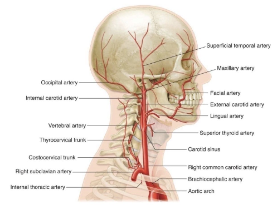
What artery normally does not supply blood tot he brain
the ECA
Where do the vertebral arteries arise/ascend
arise from subclavian artery
ascend the neck through transverse processes of cervical vertebrae
the vertebral arteries enter the brain through what
foramen magnum
vertebral arteries join where to form what
join intracranially to form basilar artery
What forms the circle of Willis
branches of ICA and vertebral arteries
ICA terminates into 4 branches (name the branches)
anterior cerebral
middle cerebral
posterior communicating
anterior choroidal
how are anterior cerebral arteries connected
through anterior communicating artery

How are posterior cerebral arteries (branches of basilar artery) connected to anterior circulation
through posterior communicating arteries

What does the external jugular vein (EJV) do
returns blood from portions of cranial cavity, face, and neck
Where does external jugular vein (EJV) empty into
subclavian vein

What does the internal jugular vein (IJV) do
returns blood from brain ad well as face and neck
How does IJV course in regards to ICA and CCA
it courses anterolateral to ICA and CCA
the IJV unites with what to form what
it unites with subclavian vein to form brachiocephalic vein

What is vertebral vein formed from and where does it emerge from
formed from numerous small tributaries
emerges from 6th cervical vertebra
where does vertebral vein empty into
brachiocephalic vein

Course/pathway of the aortic arch
begins at aortic valve, continues up and back crossing trachea, passes posteriorly on left of trachea, and curves downward and becomes the descending aorta
3 branches of the aortic arch
brachiocephalic (innominate) artery
left CCA
left subclavian artery

length of brachiocephalic (innominate) artery
4-5cm in length
What does brachiocephalic artery branch into and where does this happen
divides into right CCA and right subclavian at sternoclavicular joint

What does subclavian artery give rise to
gives rise to branches that supply the brain, neck, thoracic wall, and shoulder
What does the subclavian artery become and where
it becomes the axillary artery at outer border of the first rib
Where does axillary artery begin/continue through
outer border of first rib and continues through axilla

Where does brachial artery begin/continue through
begins at axilla and continues through upper arm to approx. 1cm below elbow joint

largest branch of brachial artery
the deep branchial artery (profunda brachii)
Where does the brachial artery terminate
in bifurcation into radial and ulnar arteries

is the radial or ulnar artery usually larger
ulnar artery

Where does ulnar artery course
courses along ulnar border of forearm

branches of ulnar artery
branches include ulnar recurrent and interosseous arteries and palmar and dorsal carpal branches
Where does ulnar artery terminate
in deep and superficial palmer arch in hand

Where does radial artery course
passes along radial aspect of forearm to wrist

branches of radial artery
branches include radial recurrent, muscular, palmar carpal, and superficial palmar arteries

Where does radial artery terminate
in superficial and deep palmar arches in the hand

What does palmar arch supply
digital arteries

What kind of vein is the cephalic vein
a superficial vein
Where does cephalic vein course
it winds around radial border of forearm and continues along lateral border of bicep muscle

Where does cephalic vein empty into
axillary vein

What kind of vein is the basilic vein
a superficial vein
Where does basilic vein course
courses along ulnar aspect of forearm and continues along medial border of biceps muscle

the basilic vein joins what to form what
joins brachial vein to form axillary vein

What do both cephalic and basilic veins communicate with
medial cubital vein

What are paired deep veins of the upper extremity
the radial and ulnar veins

How do the radial and ulnar veins course
they follow the path of the arteries

Where do radial and ulnar veins unite, and this forms what
they unite near antecubital fossa to form brachial veins

2 characteristics/traits of the brachial veins
usually paired
follow the course of artery

Where does axillary vein begin
its a single vein that begins at junction of brachial and basilic veins

What is subclavian vein a continuation of/where does it begin
its a continuation of the axillary vein and begins just past outer border of first rib

Where is brachiocephalic vein formed/located
at junction of internal jugular and subclavian veins, one at each side of the neck

Where is superior vena cava formed
by the junction of the 2 brachiocephalic veins

where does superior vena cava course
just behind the right side of the sternum

Where does the aorta terminate
at the level of the 4th lumbar vertebra

What does the aorta bifurcate into
the left and right common iliac arteries

what do the common iliac arteries bifurcate into
external and internal iliac arteries

What does the internal iliac arteries (hypogastric arteries) supply
pelvic organs
What does the external iliac arteries supply
they continue distally to supply lower extremities

the external iliac arteries become what, and where
they become common femoral artery at the inguinal ligament

What forms the common iliac veins
external iliac vein and internal iliac vein joining together

What do veins of the pelvis drain
legs and pelvic organs
Where does the left CIV and right CIA cross
left CIV passes beneath right CIA at level of aortic bifurcation
What forms the IVC
the left and right common iliac veins joining together

What is the common femoral artery (CFA) a continuation of
the external iliac artery below the inguinal ligament

What does the CFA divide into
superficial femoral and profunda femoris arteries

How does profunda femoris (deep femoral) artery course
posterior and lateral to superficial femoral artery

Where does profunda femoris (deep femoral) artery give off branches
into the thigh
How does superficial femora artery (SFA) course
distally, passing through the adductor canal

What does the SFA become
the popliteal artery

What is the popliteal artery a continuation of
the SFA

Where does popliteal artery course
behind the knee in popliteal fossa

branches of popliteal artery
branches include sural and genicular arteries around the knee

where does popliteal artery terminate
in anterior tibial, posterior tibial, and peroneal arteries

What does popliteal artery initially bifurcate into
anterior tibial artery and tibioperoneal trunk
