Musculoskeletal Disease Imaging
 Call Kai
 Learn
Learn Practice Test
 Spaced Repetition
 Match
Match Flashcards
 Knowt Play
1/140

There's no tags or description
Looks like no tags are added yet.
Name | Mastery | Learn | Test | Matching | Spaced | Call with Kai | Chat |
|---|
No analytics yet
Send a link to your students to track their progress
141 Terms
Osteochondrosis
developmental disorder seen in rapidly growing young animals when endochondral ossification fails at an articular surface, leaving cartilage that turns thick and prone to fracture or fragmentation
How does Osteochondrosis appear on radiographs?
as a defect in subchondral bone beneath the affected articular cartilage
Osteochondritis dissecans (OCD)
Osteochondrosis that has progressed to formation of a cartilage or mineralized flap/fragment
Radiographs will only show a Osteochondritis dissecans (OCD) cartilage flap if...
it contains enough mineralized subchondral bone
*A flap made entirely of cartilage may be invisible radiographically!
Four locations Osteochondrosis is commonly found in dogs:
1.) caudal humeral head
2.) medial humeral condyle
3.) femoral condyles
4.) talar trochlear ridges
Seven locations Osteochondrosis is commonly found in horses:
1.) medial femoral condyle
2.) lateral femoral trochlear ridge
3.) intermediate ridge of the tibia
4.) lateral talar trochlear ridge
5.) medial malleolus
6.) sagittal ridges of the distal third metacarpal/metatarsal bones
7.) cervical articular processes
Normal radiographic image of canine humeral head
smooth, continuously curved subchondral bone surface

Radiographic appearance of osteochondrosis of the canine humeral head
surface may become flattened or irregular, often with subchondral sclerosis underneath the lesion

If an osteochondrosis at the canine humeral head progresses to OCD, there will be a...
thin mineralized flap or separate mineral fragment visible

Radiographic appearance of osteochondrosis of the canine femoral condyle
produces an irregular, undulating, or concave lucent defect in the subchondral bone, often with surrounding subchondral sclerosis
One femoral condyle may remain smooth while the affected condyle has an irregular surface

Radiographic appearance of osteochondrosis of the canine talus
trochlear ridge of the talus may become flattened or blunted
tarsocrural joint space appears widened

Radiographic appearance of osteochondrosis of the equine medial femoral condyle
range from a smooth subchondral defect to a larger, expansile, cyst-like defect surrounded by sclerosis
commonly called osseous cyst-like lesions.

Radiographic appearance of osteochondrosis of the equine lateral trochlear ridge of the distal femur

Radiographic appearance of osteochondrosis of the equine intermediate ridge of the distal tibia
DMPLO view is particularly useful for highlighting this ridge!

Radiographic appearance of osteochondrosis of the equine lateral trochlear ridge of the talus
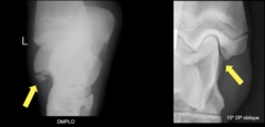
Three diseases under elbow dysplasia:
1.) ununited anconeal process (UAP)
2.) fragmented medial coronoid process (FCP)
3.) medial humeral OC (OCD)
elbow dysplasia results in joint ________
incongruity
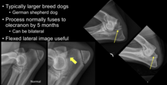
ununited anconeal process (UAP)
failure of the anconeal process to fuse to the olecranon
What positioning of the elbow is best for visualizing ununited anconeal process (UAP)?
flexed lateral elbow
What does ununited anconeal process (UAP) look like radiographically?
a radiolucent line between the anconeal process and olecranon
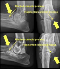
fragmented medial coronoid process (FCP)
most common cause of elbow dysplasia!!
What does fragmented medial coronoid process (FCP) look like radiographically?
blunting of the medial coronoid process or a fragment
medial humeral OC (OCD)
concave defect in the subchondral bone with surrounding sclerosis

medial humeral OC (OCD) can occur with what other elbow dysplasia?
fragmented medial coronoid process (FCP)
Hip dysplasia
developmental malformation of the coxofemoral joint where head of femur slips out of acetabulum
In a normal hip, ____% of the femoral head should be covered by the acetabulum
50%
Radiographic findings of hip dysplasia
subluxation/luxation of the femoral head from the acetabulum
shallow acetabula
enthesophytes on femoral neck
osteophytes on femoral head and acetabulum

Five Congenital Diseases
1.) Panosteitis
2.) Hypertrophic osteodystrophy
3.) Craniomandibular osteopathy
4.) Avascular necrosis of the femoral head
5.) Femoral neck metaphyseal osteopathy
Panosteitis
inflammation of an entire bone; self limiting and cause unknown
What breed of dog is classically affected by Panosteitis? How old?
5-14 month old Germand Shepherds (or other large breed dogs)
Clinical signs of Panosteitis
shifting leg lameness with and pain with deep palpation of the affected bones
Panosteitis radiographic findings
patchy medullary sclerosis, often centered near the nutrient foramen

nutrient foramen
small opening in the diaphysis where an artery enters the bone to provide nourishment
Hypertrophic osteodystrophy
inflammatory condition of long bones which affects rapidly growing puppies
What region is hypertrophic osteodystrophy most often found?
distal antebrachium
What dogs are classically affected by Panosteitis? How old?
large breed puppies 3-6 months old
Radiographic findings of hypertrophic osteodystrophy
radiolucent line within the metaphysis adjacent to the physis
sclerosis of the surrounding metaphyseal bone

Severe/chronic hypertrophic osteodystrophy can cause...
substantial metaphyseal remodeling and sclerosis
*radiolucent metaphyseal line may no longer be visible

Craniomandibular osteopathy
bony proliferation of the maxilla and mandible
What breeds is craniomandibular osteopathy common in? What age?
terriers, labs, and dobermans 3-10 months old
Clinical signs of craniomandibular osteopathy
pain and difficulty opening the mouth
Radiographic findings of craniomandibular osteopathy
periosteal proliferation along one or both hemimandibles
can progress and surround the tympanic bullae

Avascular necrosis of the femoral head
aka Legg-Calvé-Perthes disease; occurs when the blood supply to the femoral head and neck is disrupted
What breeds is Legg-Calvé-Perthes disease common in? What age?
small breed dogs, 4-12 months old
Radiographic findings of Legg-Calvé-Perthes disease
mottled lucency of the femoral head and neck, often mixed with areas of sclerosis
misshapen femoral head and heck; thinning of the neck
acetabular joint space widening

How can hip dysplasia and Legg-Calvé-Perthes disease be differentiated?
Hip dysplasia can produce severe remodeling and malformation, but it does not typically cause actual loss/resorption of the femoral head and neck. Avascular necrosis does.
Femoral neck metaphyseal osteopathy
abnormal bone remodeling/growth around the femoral neck, causing the neck to become thickened
What species and age is femoral neck metaphyseal osteopathy common in?
young male cats
Radiographic appearance of femoral neck metaphyseal osteopathy
mottled lucency and progressive thinning of the femoral neck
characteristic "apple core" appearance!

Laminitis
can result from inflammation, endocrine disease, or mechanical overload and causes separation of the laminae of the hoof
Radiographic findings of laminitis
sinking and rotation of P3
remodeling at the distant tip of P3
Periosteal proliferation along the dorsal surface of P3
Gas within the hoof wall

Three measurements made to assess severity of laminitis:
1.) Founder distance
2.) Hoof wall thickness
3.) Hoof wall to P3 length ratio
Founder distance
line is made at the level of the coronary band, and a vertical line is drawn down to the extensor process of P3

Normal founder distance length
4-15 mm
An increased founder distance indicates what?
that P3 has sunk distally within the hoof capsule
Hoof wall thickness
measured at both a proximal and distal location, perpendicular from the hoof wall to the surface of P3

Normal hoof wall thickness
14-22 mm
What would be an abnormal hoof wall thickness? What would this indicate?
if the distal line is longer than the proximal line, this indicates that P3 has rotated away from the dorsal hoof wall
Hoof wall to P3 length ratio
calculation: hoof-wall thickness divided by the length of P3
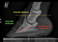
Why is a hoof wall to P3 length ratio calculated?
to correct for differences in horse size and radiographic magnification
Normal hoof wall to P3 length ratio
30%
An increased hoof wall to P3 length ratio indicates what?
abnormal separation between P3 and the hoof wall
Chronic laminitis radiographic appearance
progressively greater remodeling of the distal phalanx

Aggressive vs non-aggressive bone lesions
Aggressive bone lesions progress rapidly, have poorly defined margins, and cause greater bone destruction
Non-aggressive bone lesions are slow-growing and well-defined
Aggressive bone lesions are often due to...
malignancy or infection
Five major features to focus on to determine if a bone lesion is aggressive or non aggressive:
1.) location
2.) pattern of bone destruction
3.) zone of transition
4.) new bone formation
5.) rate of change
Lesion location
The distribution of a lesion, if it is one or several bones, and what part of the bone is involved
Three types of distribution of a lesion
solitary, multifocal, or generalized
Examples of a solitary bone lesion
primary bone tumor or localized infection
Examples of multifocal bone lesions
metastasis or disseminated infection
Examples of generalized bone lesions
suggestive of metabolic, nutritional, or congenital disease
If multiple adjacent bones across a joint are affected, think about ________ disease
If distant bones are involved, think about ________ disease processes
joint
disseminated
Examples of describing what part of the bone is involved
-epiphysis --> articular surface of bone
-physis --> growth plate between epiphysis and metaphysis (if present)
-metaphysis --> connect the epiphysis to the diaphysis
-diaphysis --> long shaft of bone
-periosteum --> outer membrane that covers the bone
-cortex --> hard outer portion of a bone
-medullary cavity --> hollow space inside bone
Three patterns of bone destruction
1.) geographic
2.) moth eaten lysis
3.) permeative
geographic lysis
relatively large area of bone loss with sharply defined margins

moth eaten lysis
multiple small, irregular, poorly defined areas of bone loss
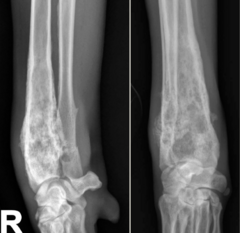
permeative lysis
contains numerous tiny, poorly defined lucencies, giving the bone a "sponge-like" appearance
Which patterns of bone destruction are aggressive?
Which patterns of bone destruction are non aggressive?
very aggressive: permeative
aggressive: moth eaten lysis
non aggressive: geographic lysis
*in order of bone margins becoming less defined; less defined = more aggressive
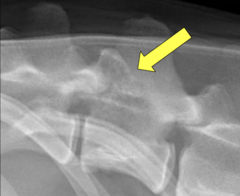
zone of transition
how quickly the radiograph changes from abnormal bone to normal bone.
Short zone of transition
you can easily identify where the lesion stops and normal bone begins; suggests a slower growing process
Long zone of transition
the lesion gradually fades into normal bone, making it difficult to determine exactly where the lesion ends

A ________ zone of transition is associated with a non aggressive bone lesion
A ________ zone of transition is associated with a aggressive bone lesion
short
long
Three types of new bone formation:
1.) smooth, lamellar
2.) palisading
3.) medullary
smooth, lamellar bone formation
forms organized layers of new bone along the cortex; usually indicates a slower process

palisading bone formation
consists of new bone extending perpendicular to the long axis of the bone

medullary bone formation
new bone formation within the medullary cavity
ex: panosteitis!

The different types of bone formation can all be either...
aggressive or non aggresive!
A _______ rate of change indicates non aggressive bone lesions
A _______ rate of change indicates aggressive bone lesions
slow
fast
Characteristics of a aggressive lesion
poorly defined, has moth-eaten or permeative lysis, has a long zone of transition, may have irregular periosteal proliferation, and changes rapidly over time
Three categories of aggressive lesions:
1.) neoplastic
2.) infectious
3.) immune mediated
Malignant neoplastic bone lesions
cancerous growths of bone
Malignant neoplasia can be divided into three categories:
1.) primary
2.) metastatic
3.) multicentric
primary malignant neoplasia
tumor that originates within the affected bone
examples of primary malignant neoplasia
-osteosarcoma
-chondrosarcoma
-fibrosarcoma
-hemangiosarcoma
Characteristics of primary malignant neoplasia
geographic, moth-eaten, and permeative bone lysis
cortical destruction
associated tissue mass

metastatic malignant neoplasia
neoplastic bone tumors in multiple bones
metastatic bone tumors typically arise from...
carcinomas (esp urogenital carcinomas)
metastatic bone tumors appearance
multiple areas of medullary sclerosis (though lesions can have variable appearances)
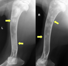
multicentric malignant neoplasia
originates at multiple locations rather than simply spreading from one primary bone tumor
multicentric bone tumors appearance
numerous relatively well-defined areas of bone lysis throughout the skeleton; "punched out" lesions
