622 11/3 GLA MHC and Antigen Presentation
 Call Kai
 Learn
 Practice Test
 Spaced Repetition
 Match
 Flashcards
 Knowt Play
1/71

There's no tags or description
Looks like no tags are added yet.
72 Terms
Antigen Presenting Cells
part of adaptive immunity, break down pathogens into little pieces called antigens and then present them to T cells so they know what the enemy looks like
T Cells
in adaptive immune system, has specific receptors to recognize small specific antigens
B Cell Receptors/Antibodies vs T Cell Responses
b cell receptors/antibodies can bind and respond to unprocessed, native antigens
t cell responses require antigen processing and presentation
Antigen Processing
the breakdown of the protein components of a pathogen into tiny pieces (protein fragments) called antigenic epitopes
Antigen Presentation
the ability of antigen presenting cells (APCs) to display, or present the small pieces of pathogen made by antigen processing on its surface as a signal to T cells that a foreign antigen is present
3 Professional APCs
dendritic cells
b cells
macrophages
Dendritic Cells
the best APCs, mostly reside where tissues are in contact with external environment (skin, GI, respiratory)
named so bc of their dendrite like projections
CD
cluster of differentiation
CD Markers
surface markers of cells, some of them have functions while others simply serve as identifying marker on that cell
CD Markers Table

Epitope
a small peptide of a foreign antigen
What is the function of the MHC in APCs?
in APCs, an epitope is loaded into MHC proteins which are then lifted to the surface, like a red flag, telling T cells that there is an infection present
MHC (Major Histocompatability Complex)
complex of cell surface proteins that help the adaptive immune system, specifically T cells, recognize self vs non self
Human Leukocyte Antigen Genes (HLA)
genes in humans that express MHCs
estimated to be more than 10,000 alleles in the human population
What determines MHC protein affinity for potential antigens?
its shape/conformation
this is important bc MHC genes have lots of variability, and having such variations results in a diversity of antigens that can be loaded onto the MHC proteins
2 MHC Classes
class i
class ii
MHC Class I
expressed by all nucleated cells and platelets in the body (not rbcs)
presents antigen to CD8+ cytotoxic T cells
MHC Class II
only produced on professional APCs
presents antigen to CD4+ helper T cells
Shape of MHC Class I
binding cleft and main structure made up of one chain (alphe chain)
a structural component called b2-microglobulin which holds the MHC

Shape of MHC Class II
two main chains (a and b) which together make up the binding cleft

What length of peptides to MHC Class I bind to, and how?
they bind to peptides 8-10 AA in length
binding cleft is closed at the ends, making MHC like a baseball glove with the peptide as the baseball
Anchor Residues
specific amino acids essential for binding peptide to a particular MHC Class I molecule
Why are APC so important?
APCs display both MHC class i and ii receptors, so they are able to present antigen to both cytotoxic and helper T cells
all other nucleated cells in the body display only the MHC i receptor and therefore are only able to present antigen to cytotoxic T cells
What length of peptides to MHC Class II bind to, and how?
they bind to peptides 10-30 AA in length
cleft is open at both ends, like a hot dog that is too long for the bun so the peptide sticks out at both ends
What is unique about the peptides that bind to MHC Class II?
they have internal conserved motifs, and lack anchor residues at the ends
2 Pathways Antigens can be Processed and Presented
MHC class i pathway
MHC class ii pathway
What types of pathogens are processed using the MHC Class I Pathway?
intracellular/endogenous pathogens (like viruses)
What types of pathogens are processed using the MHC Class II Pathway?
extracellular/exogenous pathogens (like most bacteria)
Common Features of the MHC I and MHC II Pathways
breaking down the antigen into small pieces and then association with an MHC protein complex and transport to the cell surface
How does our immune system respond to intracellular infections?
by having cytotoxic CD8+ cells recognize these infected cells and destroying them
process is mediated by the MHC class I pathway
MHC Class I Pathway Diagram
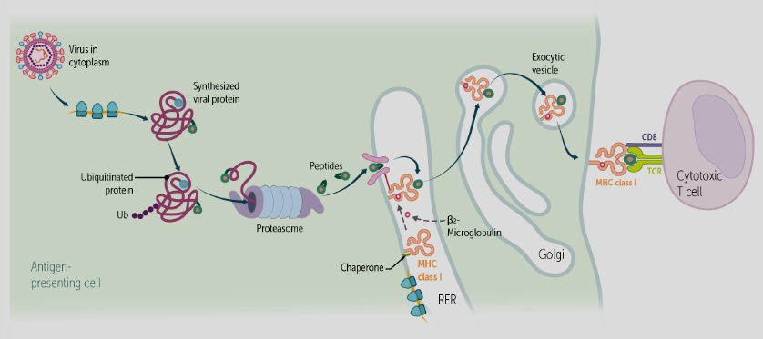
6 Steps in MHC Class I Pathway
Antigens in the cytoplasm are ubiquitinated and targeted for degradation by the proteasome
Proteosomes chop up the proteins into small peptides (8-10 amino acids in length)
Transporter associated with antigen presentation (TAP) uses ATP fuel to transport peptides into the lumen of the endoplasmic reticulum
Tapasin brings empty MHC Class I molecules into close proximity to TAP and the antigenic peptides
The peptide is fit into the binding cleft of the MHC Class I molecule (like a baseball glove) and Beta2-microglobulin holds the MHC in the correct shape
The MHC I/peptide complex is now stable and transported to the surface for display to CD8+ cytotoxic T cells
Steps 1-2 MHC Class I Pathway
antigens in cytoplasm are ubiquinated and targeted for degradation by the proteasome
proteasomes chop up the proteins into small peptides (8-10 aa in length)
Steps 3-4 MHC Class I Pathway
transporter associated with antigen presentation (TAP) uses ATP to fuel transport peptides into the lumen of the ER
tapasin brings empty MHC class i molecules into close proximity to TAP and the antigenic peptides
Steps 5-6 MHC Class I Pathway
the peptide is fit into the binding cleft of the MHC class i molecule like a baseball glove, and b2-microglobulin hold the MHC in the correct shape
the MHC i/peptide complex is now stable and transported to the surface for display to the CD8+ cytotoxic T cells
b2-microglobulin
part of MHC class i structure, holds MHC in the correct shape
associated with MHC i in the rough ER
How can our immune system attack cancer?
using the MHC class i pathway, endogenous cancer antigens are presented on the cell surface so the cells can be destroyed by the cytotoxic T cells
What cells perform the MHC Class II Pathway?
only professional APCs (dendritic cells, B cells, macrophages) because they are the only cells that express MHC II on their cell surface
MHC Class II Pathway Diagram

7 Steps of MHC Class II Pathway
Extracellular antigens or whole microbes are taken up by APCs and packaged into endosomes (like the phagolysosome after phagocytosis or the endolysosome after endocytosis)
Intracellular killing mechanisms cause the endosomal pH to drop. The acidified compartment triggers activity by enzymes called proteases (for example, lysozyme), that chop up the antigens within the endolysosomes
Empty MHC Class II molecules are constantly being synthesized in the ER. At this point, the empty binding cleft is held open by a peptide called the invariant chain
Vesicles containing these empty MHC Class II molecules fuse with the acidified endolysosomes
The activated enzymes in the end-some then chop up the invariant chain and leave only a piece of it in the binding cleft called CLIP (Class II invariant peptide)
A protein called HLA-DM replaces CLIP with the antigenic peptide
The MHC Class II/peptide complex is now stable and is transported to the surface for display to helper CD4+ T cells
Steps 1-2 MHC Class II Pathway
extracellular antigens or whole microbes are taken up by APCs and packaged into endosomes (such as the phaoglysosomes after phagocytosis or the endolyososome after endocytosis)
intracellular killing mechanisms cause the endosomal pH to drop, and acidification triggers activity of proteases that chop up the antigens within the endolysosomes
Steps 3-4 MHC Class II Pathway
empty MHC class ii molecules are constantly being synthesized in the ER, and the empty binding cleft is held open by a peptide called the invariant chain
vesicles containing the empty MHC class ii molecules fuse with the acidified endolyzozomes
Steps 5-7 MHC Class II Pathway
the activated enzymes in the end-some chop up the invariant chain and leave only a piece of it in the binding cleft called the CLIP
a protein called HLA-DM replaces CLIP with the antigenic peptide
the MHC class ii/peptide complex is now stable and is transported to the surface for display to helper CD4+ T cells
CLIP
class ii invariant peptide, the piece of the invariant chain left in the MHC class ii peptide after it has been chopped by the activated enzymes in the end-some
Where is the antigen loaded onto MHC II?
the endolysosome
What receptors do professional APCs have on their surface?
MHC type i and MHC type ii
If professional APCs have MHC I and MHC II receptors, what does helper T cell (MHC II) presentation accomplish?
cytokine secretion to activate a general immune response, which eventually rids the body of the antigens
4 Ways APCs are Involved with Different Types of T Cells
activation of naive T cells (whether CD4 or CD8) is done by dendritic cells, making these cells the key to early T-cell differentiation
stimulation of CD8+ effector cytotoxic T cells, causing them to kill infected cells, is done using the MHC class I pathway
activation of CD4+ effector T cells is done mostly by dendritic cells, with assistance from the other professional APCs (B cells and macrophages). These are the only cells that contain the MHC class II protein needed for this process
activation of memory T cells is mostly done by B cells and macrophages
Dendritic Cells (APC) Importance to T Cells
activation of naive T cells (whether CD4 or CD8) is done by dendritic cells, making these cells the key to early t-cell differentiation
APCs Impact on CD8+ Effector Cytotoxic T Cells
stimulation of CD8+ effector cytotoxic T cells by APCs causes them to kill infected cells is done using the MHC class i pathway
APCs Impact on CD4+ Effector Cytotoxic T Cells
activation of CD4+ effector T cells is done mostly by dendritic cells, with assistance from other processional APCs
these are the only cells that contain the MHC class ii protein needed for this process
Impact of APCs on Memory T Cells
activation of memory T cells is mostly done by B cells and macrophages
Cross-Presentation
ability of dendritic cells to load exogenous pathogens onto MHC i proteins for presentation of cytotoxic T cells, leading to destruction of the infected cell
this differs from the way exogenous pathogens are usually loaded via the MHC ii pathway
Cross-Presentation Diagram

Classes of MHC Genes
class i
class ii
class iii

Variability of MHC Genes
have the greatest diversity of alleles found in different individuals inside a population (aka high polymorphism), with over 10,000 unique alleles in the population
the most genetically diverse locus present in any mammalian genome
How many MHC genes do we have and how do we get them?
each person has 12 total MHC class i and ii genes
each parent donates 6
Co-dominance of MHC Genes
they are all expressed equally (not a blend)
3 Human Class I HLA Genes
HLA-A
HLA-B
HLA-C
3 Human Class II HLA Genes
HLA-DP
HLA-DQ
HLA-DR
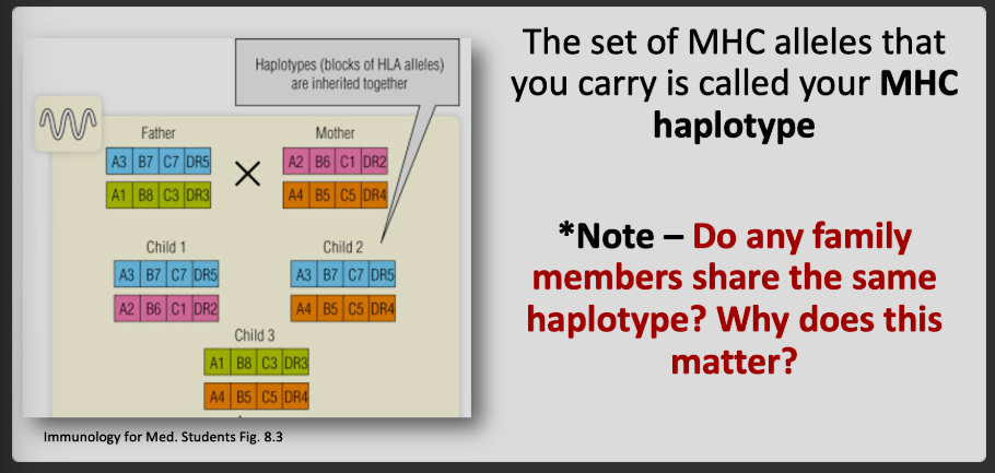
MHC Haplotype
made up of the set of MHC alleles on each chromosome
because each individual has two copies of each chromosome, everyone has two HLA haplotypes
each parent donates one haplotype
MHC Haplotypes Diagram
HLA-DR3 Allele
assocaited with an increased risk for certain autoimmune conditions including, type i diabetes, systemic lupus erythematesus, graves disease and myasthenia gravis
Autoimmunity
when your immune system attacks your own body
Why do some MHC alleles have a higher association with an autoimmunity?
for a peptide to be presented to a T cell it has to perfectly fit into the binding cleft of the MHC
often in autoimmunities, a self-peptide fits just perfectly into a patient’s MHC and is presented to T cells, starting an inappropriate immune response
Psoriasis as Example of Relationship Between MHC and Autoimmunity
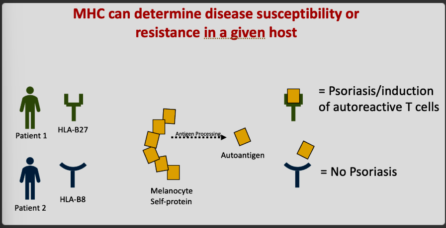
Chart of Diseases Associated with HLA Alleles

Advantage vs Disadvantage of Certain MHC Alleles
some alleles can present a peptide with high affinity to responding T cells, activating more of them, and enabling a better immune response to infection
some alleles can make a person more susceptible to disease if the allele cannot bind to or present peptides to T cells, limiting the immune response and making infection more severe (ex: COVID)
How does MHC variation help protect from disease?
since the immune system of each individual is different, perfect pathogens cannot evolve to spread through a population
possibly less likely for microbe to imitate MHC alleles if there is a large variety of MHC
MHC and Transplant Matching
the differences between MHC molecules of the donor and recipient are the major cause of tissue incompatability and transplant rejection, highlighting the importance of tissue typing before transplantation
name stems from its initial recognition as the site of genes that cause T cells to reject tissues transplanted from unrelated donors to recipients
Which MHC Classes have a direct role in antigen presentation?
class i to CD8
class ii to CD4
Where are the MHC genes located?
the short arm of chromosome 6
the do not participate in genetic recombination