Assessing Heart and Peripheral Vascular System
 Call Kai
 Learn
Learn Practice Test
 Spaced Repetition
 Match
Match Flashcards
 Knowt Play
1/83

There's no tags or description
Looks like no tags are added yet.
Name | Mastery | Learn | Test | Matching | Spaced | Call with Kai | Chat |
|---|
No analytics yet
Send a link to your students to track their progress
84 Terms

What is the green structure?
Apex of the heart.
What intercostal space is the base of the heart found in and which is the apex found in?
Base: Left 2nd intercostal space
Apex: Left 5th intercostal space

Identify each valve
1= Tricuspid valve
2 = Pulmonary valve
3 = Mitral valve
4 = Aortic valve
Path of electrical conduction
SA node → AV node → Bundle of His → Purkinje fibers
Role of SA node
Pace maker of the heart. Generates impulses of 60-100 BPM. Tells both atria to contract simultaneously.
Role of AV node
Backup pacemaker that will take over of AV node isn’t working. Generates impulses at 40-60 BPM, so pt will have this HR if AV node not working.
Role of bundle of his and purkinje fibers
Stimulates ventricular contraction/cardiac muscle. Generates 20-40 BPM, so pt will have very low HR if SA and AV node not working and body has to rely on these.
P-wave
Atrial depolarization (contraction). Accounts for 5-10% of cardiac output.
QRS complex
Ventricular depolarization (contraction)
T-wave
Ventricular repolarization (relaxation)
What happens during systole?
Contraction. Large amounts of blood in ventricles causes AV vales to close and semilunar valves to open as ventricles contract and send blood out.
What happens during diastole?
Relaxation and filling. AV valves are open as increased pressure in atria makes them open and blood from atria goes into ventricles. “Atrial kick” is when atria contract.
S1 heart sound
Produced by closing of the AV valves. “lub” sound. Signals beginning of systole. Loudest at apex.
S2 heart sound
Closure of semi-lunar valves. “dub” sound. From end of ventricle contraction + beginning of ventricle relaxation. Loudest at base.
S3 heart sound
Extra “dub” sound: “lub dub-dub”. Caused by blood hitting ventricle wall d/t rapid filling of ventricles. Best heard with the bell in the left lateral position. Considered an abnormal heart sound.
S4 heart sound
Extra “lub” sound: “lub-lub dub”. Caused by blood flowing rapidly into stiff ventricles after atria contract. Best heard with bell over apical area in supine/left lateral position. Considered an abnormal heart sound.
Pericardial friction rub
High-pitched, scratchy sound caused by inflammation of the pericardial sac. Best heard with diaphragm and pt leaning forward.
Description of murmurs
Intensity - levels I-VI with I being faint and VI being very loud
Timing - systolic or diastolic
Pitch - high, med, low
Pattern - increasing or decreasing volume
Quality
Location heard
Radiation of sound
Position of pt
Systolic vs diastolic murmurs
Systolic: heard when heart contracts (between S1 and S2)
Diastolic: heard when heart relaxes
SIgns and symptoms of murmurs
Client may have no signs or symptoms if murmur isn’t severe. Cyanosis (late sign), edema, JVD, chest pain, shortness of breath, dizziness, fainting.
Carotid artery
Located between the trachea and the R/L sternocleidomastoid muscle. Supplies blood to the brain (internal carotid) and superficial structures of head and neck (external carotid)
Carotid artery pulse
Since carotid artery is close to heart, it is good for assessing amplitude and contour. Its absence is very concerning.
Jugular veins
Internal and external. Are not routinely assessed for pulse/pressure but can be used for IV access (by provider).
History of present health concern for cardiac exam
Chest pain, tachycardia, heart skipping beats, tire easily, dyspnea, coughing mucous, dizziness, nocturia (frequent urination at night), swelling in lower extremities, frequent heartburn?
How do you know if heartburn is a GL problem or a cardiac problem?
GI = can be resolved by an antacid
Cardiac = can’t be resolved by antacid
Personal health history for cardiac exam
History of: heart murmur, rheumatic fever, heart surgery, ECG, blood pressure, lipid profile, medications.
Family history for cardiac exam
Hypertension, myocardial infarction, coronary artery disease, hyperlipidemia, diabetes
Lifestyle questions for cardiac exam
Smoking, alcohol, physical activity, stress, diet, ADLs, sexual activity (can reveal non adherence to meds), pillows used when sleeping (issues with laying flat), nocturia.
General routine screening in cardiac exam
Inspect jugular veins, auscultate/palpate carotid arteries, inspect pulsations on anterior chest, palpate apical pulse, palpate for abnormal pulsations, auscultate S1/S2 and for abnormal sounds and murmurs.
Observing jugular venous pulse
Stand on pts right side with pt in suping 30-45 degrees. Have pts turn head to left. Shine light on neck. Look for distension of the vein (JVD).
Note: Can be done on left side as well.
When do we assess the carotid artery?
In older adults and/or suspected cardiovascular disease
Auscultating the carotid artery
Auscultate BEFORE palpating. Place the bell over the artery and have client hold breath. Press lightly. Abnormal = bruit sounds, indicates atherosclerotic disease
Palpating the carotid artery
Alternates sides, never do both at once. Note amplitude, contour, trills, and elasticity. Normal: 2+ equal bilaterally w/ no trills
Anterior heart inspection
Pt in supine. Pt should have no visible lifts/heaves in chest. These would be pulsations if present.
Palpating apical pulse
Left anterior 5th intercostal at the midclavicular line. Should feel like a gentle tap and is small in amplitude.
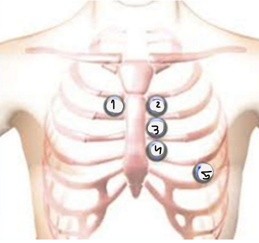
Name the 5 traditional areas for heart palaption/ausltation
1 = aortic - 2nd intercostal on right sternal boarder
2 = pulmonic - 2nd intercostal on left sternal boarder
3 = Erb point - 3rd intercostal on left sternal boarder
4 = tricuspid - 4th intercostal on left sternal boarder
5 = mitral - 5th intercostal on the left midclavicular line
Auscultation of heart
Use diaphragm. Should be 60-100 BPM and regular. Irregular is when there is different timing in between sounds.
What to do if rhythm is irregular upon auscultation
Palpate radial pulse while auscultating apical pulse. Assess for a full min. If regular, pulses will go at the same time. If they don’t assess for the pulse rate deficit.
Auscultating for extra heart sounds/murmurs
Start with diaphragm and bell. Auscultate all 5 traditional areas. Auscultate in various positions like left lateral.
Considerations when assessing heart of newborns
Formen ovale (allows flow b/t atria) should close within 1 hr after birth. Ductus arteriosus (shunts blood from pulmonary artery to aorta) should close within 10-15 hrs. May have systolic murmur that will resolve in 1-2 days. HR is 120-160. Listen at 4th intercostal margin.
Considerations when assessing heart of children/adolescents
Apical pulse at 4th intercostal. Heart rate will decrease by age 8. Some children will have a physiologic murmur. S3 may be heard at the apex of some children but should eventually dissipate with age.
Physiologic heart murmur
Complexly harmless heart murmur. Common in infants and children. May come and go depending on mood, exercise, etc.
Considerations when assessing heart of childbearing population
Increase in HR (10-15 BPM) expected. Increase in blood volume expected.
Considerations when assessing heart of older adults
Assess for orthostatic hypertension. Apical pulse may be difficult to palpate as chest diameter changes with age.
What is coronary heart disease
Includes coronary artery disease, heart rhythm abnormalities, and congenital defects.
Managing risk factors of coronary heart disease
BP monitoring, body weight, lipid management, smoking, diet, exercise, stress. Educate pts on preventative measures and lifestyle changes.
Function of arteries
Carry oxygenated blood away from heart and to tissues.
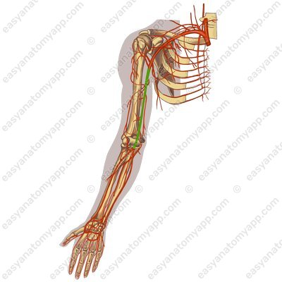
Name the indicated (green) vessel
Brachial artery

Name the indicated (green) vessel
Radial artery

Name the indicated (green) vessel
Ulnar artery

Name the indicated (green) vessel
Femoral arteries
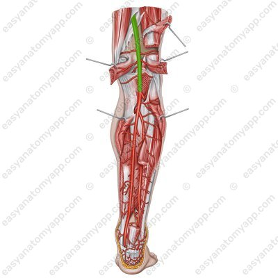
Name the indicated (green) vessel
Popliteal artery

Name the indicated (green) vessel
Dorsalis pedis artery

Name the indicated (green) vessel
Posterior tibial artery
Vein function
Carry deoxygenated blood back to the heart.
The 3 types of veins + their examples
Deep - femoral and popliteal
Superficial - small and great saphenous
Perforator - connect superficial to deep
Function(s) of lymphatic system
Drain excess fluid back to venous system. Carries immune cells + helps with immune response. Absorbs fat from SI for the body.
Lymph nodes
Healthy = small/can’t feel
Infection = large/inflamed/swollen upon inspection or palpation
History of present health concern in peripheral vascular assessment
Any color, temp, or texture changes in skin? Any pain or cramping in legs? Pain with walking or stairs? Any varicose veins? Wounds or sores? Swelling? Swollen glands or lymph nodes? Sexual dysfunction?
Personal history in peripheral vascular assessment
Previous circulatory issues? Heart or blood vessel surgury/treatment?
Family history in peripheral vascular assessment
Varicose veins, hypertension, coronary artery disease, elevated cholesterol/triglycerides?
Lifestyle in peripheral vascular assessment
Smoking, exercise, oral contraceptive use in females (clot risk), stress, ADLs, medications, compression sock/support hose use?
General routine screening in peripheral vascular assessment
Inspect arm size, color, edema. Palpate fingers, hands, arms for temp. Capillary refill. Upper and lower extremity pulses. Inspect legs for distribution of hair, temp, ulcers, edema. Inspect for varicosities and thrombophlebitis.
When do you use a doppler
Unpalpable pulses, determining patency of bypass graft, assessing tissue perfusion
Doppler sounds
We use to assess artery pulses. On an artery = whooshing sound with pulsing. On a vein = non-pulsating rush.
How to use a doppler
Apply lukewarm gel over the vessel. Lightly place the probe on gel. Listen to sound. Clean off skin. Mark site for easy reassessment. Document findings.
Assessing lymph nodes in extremities
Only assessed if there is a concern. Epitrochlear nodes found in groove between bicep and tricep. Inguinal lymph nodes found in crease between thigh and hip.
Considerations for peripheral vascular assessment in newborns
Should be pink and well perfused. Assess radial, brachial, and femoral pulses. Weak pulse indicates that something in system didn’t form/close properly.
Considerations for peripheral vascular assessment in childbearing population
Dependent edema expected, varicose veins due to extra pressure expected, watch for thrombophlebitis as clotting risk increases with pregnancy.
Considerations for peripheral vascular assessment in older adults
Evaluate for arterial and venous sufficiency. Be aware that symptoms may not be typical. Lymph nodes naturally smaller.
Peripheral artery disease (PAD)
Arteries slowly become narrowed or blocked for build up of plaque
PAD risk factors
Smoking, diabetes, and history of coronary arty disease or stroke are the biggest ones. Others are obesity, uncontrolled hypertension, high cholesterol, age (>50), and family history.
PAD symptoms
Intermittent claudication relived with rest (most common in calf), painful cramping, leg weakness/numbness, coldness in lower leg/foot, sores that don’t heal, hair loss, shiny skin, no pulse/weak pulse.
Late PAD symptoms
Severe ischemic rest pain that disrupts sleep and ADLs.
PAD education
Smoking cessation, blood sugar control, exercise, diet, healthy weight, cholesterol levels, BP.
What is arterial insufficiency?
Impeded blood flow in arterial system. Causes sharp, constant pain and diminished or absent pulses. Skin becomes dry and shiny, cool or cold, and has loss of hair.
Ulcers in arterial insufficiency
Seen on tips of toes, heels, or other pressure areas. Very painful and deep. Circular in shape. Minimal edema. Black to gangrene as tissues becomes necrotic.
Acute limb ischemia
Impeded blood flow to limb that can be due to exacerbation of arterial insufficiency. Classic presentation of six Ps: Pallor, Pain, Pulseless, Paralysis, Paresthesia, Poikilothermic (inability to regulate temp).
What to do when tissue becomes necrotic
Note line of demarcation to track growth so that we don’t amputate too early.
Venous insufficiency signs/symptoms
Aching and cramping pain as blood pools. Pulses are present but may need a doppler as edema blocks palpation. Skin becomes thick, tough, and reddish-blue-brown (hemosiderin).
Ulcers in venous insufficiency
Seen in medial malleolus or anterior tibial area. Minimal to severe pain depending on deepness. Superficial. Irregular boarder. Moderate to severe edema, may have leakage. Granulation tissue (beefy red to yellow in color).
Lymphedema vs edema
Lymphedema fluid is rich in protein and typically unilateral. Edema fluid contains very little protein and is usually bilateral.
Lyphedema
Caused by abnormal or blocked lymph vessels preventing proper drainage. Assessment shows non-pitting edema, usually unilateral, no skin ulceration or pigmentation.
Peripheral edema
Accumulation of fluid causing swelling due to fluid imbalance. Assessment shows usually bilateral but can be unilateral, skin ulceration and pigmentation may be present.