Lower Leg, Knee, and Patella Positioning (Condensed) (RAD 1101)
 Call Kai
 Learn
Learn Practice Test
 Spaced Repetition
 Match
Match Flashcards
 Knowt Play
1/167

There's no tags or description
Looks like no tags are added yet.
Name | Mastery | Learn | Test | Matching | Spaced | Call with Kai | Chat |
|---|
No analytics yet
Send a link to your students to track their progress
168 Terms

Leg - AP Projection
Explain how the patient should be positioned for an AP projection of the leg.
1. Supine with leg extended
Explain how the patient's body part should be positioned for an AP projection of the leg.
1. Leg in true AP position (toes up)
2. Foot is dorsiflexed to 90 degrees
3. Femoral condyles parallel to the IR
When taking an AP projection of the leg, the CR is perpendicular to the _________ of the leg
midpoint
NOTE: When taking an AP projection of the leg, the IR is typically ______________ to the person's leg in order to get the entire leg
diagonal (or caddy cornered)

NOTE: When taking an AP projection of the leg, the SID is typically increased to about ________ rather than kept at 40" SID otherwise you may clip your ankle and knee joints
50"
The structures shown in an AP projection of the leg are the....
- entire tibia and fibula (fibula is posterior and lateral to the tibia)
- both knee and ankle joints included

When meeting criteria for an AP projection of the leg, what should be met?
- The long axis of the leg should be in alignment with the long axis of the IR
- Ankle and knee joint open
- Distal fibula superimposed over tibia
- Slight overlap of proximal head of fibula over tibia by 1/4 inch


Leg - Lateral Projection
Explain how the patient should be positioned for a lateral projection of the leg.
1. Lateral recumbent
Explain how the patient's body part should be positioned for a lateral projection of the leg.
1. Turned toward affected side
2. Patella perpendicular to the IR
3. Femoral condyles superimposed
4. Malleoli superimposed
5. Knee flexed 20-30 degrees
How much should a child's knee be flexed for a lateral projection of the leg?
15 degrees
When taking a lateral projection of the leg, the CR is _________ to the IR and midpoint of lower leg
perpendicular
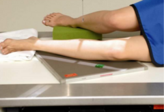
NOTE: If the patient is unable to turn for a lateral projection of the leg like a trauma patient need to be left in AP, what are you able to do?
obtain by doing a cross-table lateral a with horizontal beam which will require 2 separate XTL's (cross-table laterals) that overlap each other by about an inch
The structures shown in a lateral projection of the leg are the....
- entire tibia and fibula superimposed
- both knee and ankle joints included
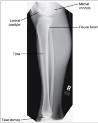
When meeting criteria for a lateral projection of the leg, what should be met?
- The long axis of the leg should be in alignment with the long axis of the IR
- Ankle and knee joint open
- Distal fibula lying over posterior half of tibia
- Slight overlap of the proximal head of fibula over tibia


What is the error in this lateral leg X-ray? How should it be corrected?
Leg is rotated. The head of the fibula is separated from the tibia and the distal fibula is too posterior. To correct, roll patella away to the IR (backward rotation of the leg).
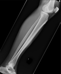
What is the error in this lateral leg X-ray? How should it be corrected?
Leg is rotated. The head of the fibula is too superimposed over the tibia and the distal fibula is too anterior. To correct, roll patella toward the IR (forward rotation of the leg)

Knee - AP Projection
Explain how the patient should be positioned for an AP projection of the knee.
1. Supine; leg extended
For the AP projection of the knee, the femoral condyles are ___________ to the IR
parallel
To demonstrate the knee joint space in the AP projection, the CR is _______ (1 cm) ________ to what?
1/2 inch distal (below) to the patellar apex
Angle of tube in an AP projection of the knee depends on what?
patient size
For an asthenic (thin, frail build) patient, you should place the CR 3-5 degrees _________ if the ASIS to tabletop is < _______ cm in an AP projection of the knee
caudad; 19

For a sthenic (common physical build and body shape) patient, the CR is angled __________ if the ASIS to tabletop is _______ cm in an AP projection of the knee
perpendicular; 19-24
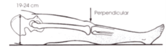
For a hypersthenic (massive, heavy build) patient, you should place the CR 3-5 degrees _________ if the ASIS to tabletop is > _______ cm in an AP projection of the knee
cephalad; 24

NOTE: The CR is parallel to the ________ ________ in an AP projection of the knee
tibial plateau
The structures shown in an AP projection of the knee are the....
- knee joint
- distal femur
- proximal tibia and fibula

When meeting criteria for an AP projection of the knee, what should be met?
- The femoral condyles should be symmetrical
- The intercondylar eminences should be centered within the intercondyloid fossa
- The head of the fibula should overlap the tibia by approximately 1/4 inch
- The patella should be seen in the center of the femur
- The knee joint should be opened
- The intercondyloid fossa should be partially seen with the knee fully extended


What is the error in this AP knee X-ray? How should it be corrected?
Medial or internally rotated which causes the lateral condyle to appear larger, intercondyloid fossa will be distorted, and there will be less overlapping of the fibular head and the tibia. To correct, externally rotate the leg.

What is the error in this AP knee X-ray? How should it be corrected?
Knee is laterally or externally rotated which this will cause the medial condyle to appear larger, the intercondyloid fossa will be distorted, and there will be more superimposition of the fibula and tibia. To correct, internally rotate the leg.

What is the error in this AP knee X-ray? How should it be corrected?
Knee not fully extended which causes intercondyloid fossa to be opened too much and the femur is distorted. Knee is internally rotated as the fibula is too lateral. To correct, extend the entire leg and rotate the knee externally.

Knee - Lateral Projection
Explain how the patient should be positioned for a lateral projection of the knee.
1. Lateral recumbent
Explain how the patient's body part should be positioned for a lateral projection of the knee.
1. Knee flexed 20-30 degrees
2. Patella is perpendicular to the IR
3. Femoral condyles are perpendicular to the IR

When taking a lateral projection of the knee, the CR is _______ degrees ________ through the knee joint
5-7 degrees cephalad

For the lateral projection of the knee, why is the CR angled 5-7 degrees cephalad?
Due to a magnified medial femoral condyle, plus it is at a lower level than the lateral femoral condyle
When taking a lateral projection of the knee, the CR is centered _______ _______ to the medial femoral epicondyle
1" distal
For a lateral projection of the knee, the CR angle is determined based on what?
width of pelvis
For a lateral projection of the knee, the CR angle is at what when the pelvis is wide?
5-7 degrees cephalad
For a lateral projection of the knee, the CR angle is at what when the pelvis is narrow?
less of an angle
NOTE: If the patient is unable to turn into the lateral position for a lateral knee projection like in cases of trauma, a _______ ________ ________ may be obtained
cross table lateral (XTL)

The structures shown in a lateral projection of the knee are the....
- knee joint
- femoropatellar joint
- patella in profile
- superimposed femoral condyles
- proximal tibia and fibula
- shows posterior and anterior displacement of fractures

When meeting criteria for a lateral projection of the knee, what should be met?
- The distal articulating surfaces of the medial and lateral femoral condyles should be superimposed, and the knee joint space should be open
- Knee flexed 20-30 degrees
- The patella should be situated proximal to the femur
- The patellofemoral joint should be open
- The tibia should be partially superimposed over the fibular head
- Patella seen in profile

Lateral Pediatric Knee - A pediatric patient's knee should be flexed _____ - ____ degrees
10-15 degrees


What is the error in this lateral knee X-ray? How should it be corrected?
Knee is flexed too much where the patellofemoral (between the patella and femur) joint will not be opened. To correct, flex the knee less.

What is the error in this lateral knee X-ray? How should it be corrected?
Knee is not flexed enough where the patellofemoral joint will not be opened. To correct, flex the knee more.

What is the error in this lateral knee X-ray? How should it be corrected?
The patella was situated too far from the IR (under-rotated knee). The head of the fibula is more superimposed on the tibia than normal. To correct, roll the patella closer (forward) to the IR.

If the lateral knee is positioned like this, what is the error in this lateral knee projection? How should it be corrected?
The patella was situated too far from the IR (under-rotated knee). To correct, roll the patella closer (forward) to the IR.

What is the error in this lateral knee X-ray? How should it be corrected?
The patella was situated too close to the IR (over-rotated knee). There is increased separation of the tibia and fibula. To correct, roll the patella farther away (backward) from the IR and/or angle the tube less.

If the lateral knee is positioned like this, what is the error in this lateral knee projection? How should it be corrected?
The patella was situated too close to the IR (over-rotated knee). To correct, roll the patella farther away (backward) from the IR and/or angle the tube less.

What is the error in this lateral knee X-ray? How should it be corrected?
CR was not angle cephalad. Note that medial femoral condyle obscures the knee joint space. To correct, angle the tube 5-7 degrees cephalad.

What is the error in this lateral knee X-ray? How should it be corrected?
Slight rotation as patella is too close to the IR, and clothing artifact is in the way. To correct, roll the patella away from the IR and have patient remove patients.
Case Study - Tibial Plateau Fx (Fracture)
Serious injury as it is an unstable fracture and want to make sure patient is not standing at all


Weightbearing Knees - AP Projection

Weightbearing Knees - AP Projection X-ray
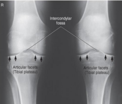
Weightbearing Knees - AP Projection X-ray Diagram

Weightbearing Knees - Lateral Projection
Weight-bearing knees in the AP and lateral projections is meant for what?
For arthritis studies to show narrowing of joint space
For the weight-bearing knees in the AP and lateral projections, the CR is __________ to the IR
perpendicular
Rosenberg Method, Camp-Coventry Method, Homblad Method, and Beclere Method are projections that show what?
Intercondylar Fossa - "Tunnel Views"
What is the purpose of the Intercondylar Fossa "Tunnel Views" exams?
For arthritis studies to show narrowing of the knee joint space and ICF
For Intercondylar Fossa "Tunnel Views", one thing it is looking at is for "joint mice" in which this is when what has broken off in the joint space?
tibial spines (eminences)

Intercondylar Fossa - PA Bilateral Weightbearing Knees Projection (Rosenburg Method)

Intercondylar Fossa - PA Axial Bilateral Weightbearing Knees Projection (Rosenburg Method)
Explain how the patient should be positioned for a PA or PA Axial Bilateral Weightbearing Knees projection (Rosenburg Method) for the intercondylar fossa.
1. Upright PA with knees touching the IR
2. Patient bends (flex) their knees so that the femoral area are at a 45 degree angle

For a PA Bilateral Weightbearing Knees projection (Rosenburg Method), the CR is perpendicular to what?
perpendicular to the center of the IR and to the tibia and fibula
For a PA Axial Bilateral Weightbearing Knees projection (Rosenburg Method), the CR is angled _______ ____ degrees
caudad 10 degrees
When meeting criteria for a PA or PA Axial Bilateral Weightbearing Knees projection (Rosenburg Method) for the intercondylar fossa, what should be met?
- Both knees presented without rotation
- Knee joints centered to exposure area
- Tibial plateaus in profile
- Intercondylar fossae visible
- Soft tissue and bony trabecular

Intercondylar Fossa - PA Axial Projection (Camp-Coventry Method)
Explain how the patient should be positioned for a PA Axial projection (Camp-Coventry Method) of the intercondylar fossa.
1. Prone
Explain how the patient's body part should be positioned for a PA Axial projection (Camp-Coventry Method) of the intercondylar fossa.
1. Knee flexed 40-50 degrees
2. No rotation of the leg
3. Foot resting on support

When taking a PA Axial projection (Camp-Coventry Method) of the intercondylar fossa, the CR is ________ to the lower leg
perpendicular
When taking a PA Axial projection (Camp-Coventry Method) of the intercondylar fossa, the CR is angled ________ through the posterior knee joint (popliteal fossa)
caudally
When taking a PA Axial projection (Camp-Coventry Method) of the intercondylar fossa, how is the tube angled in relation to the knee?
angled at the same amount that the knee is flexed
When meeting criteria for a PA Axial projection (Camp-Coventry Method) of the intercondylar fossa, what should be met?
- Open intercondylar fossa
- Knee joint space open
- Apex of patella above ICF
- Symmetrical appearance of femoral condyles
- No rotation demonstrated by slight overlap and centered intercondylar eminence
- Soft tissue and bony trabecular detail

PA Axial Projection - Homblad Method
Explain how the patient should be positioned for a PA Axial projection (Homblad Method) of the intercondylar fossa.
1. Kneeling or standing
Explain how the patient's body part should be positioned for a PA Axial projection (Homblad Method) of the intercondylar fossa.
1. Knee flexed
2. Knee joint centered to the IR
3. Femoral shafts 70 degrees to the IR

When taking a PA Axial projection (Homblad Method) of the intercondylar fossa, the CR is perpendicular to the lower leg through the _______ _________ (knee joint)
popliteal fossa
Homblad Method Modifications

Homblad Method Modifications


AP Axial Projection - Beclere Method
Explain how the patient should be positioned for an AP Axial projection (Beclere Method) of the intercondylar fossa.
1. Supine
Explain how the patient's body part should be positioned for an AP Axial projection (Beclere Method) of the intercondylar fossa.
1. Knee flexed 40-45 degrees
2. IR placed under the knee
3. No rotation of the leg

When taking an AP Axial projection (Beclere Method) of the ICF, the CR is ___________ to the lower leg
perpendicular
When taking an AP Axial projection (Beclere Method) of the ICF, the CR is angled ___________ through the knee joint
cephalad
When taking an AP Axial projection (Beclere Method) of the ICF, the CR enters _______" _______ to the apex of the patella
1/2" distal
In all 4 methods for the ICF or "tunnel views", the structures shown and criteria for evaluation is/are the....
- intercondylar fossa
- intercondylar eminences in profile
- tibial plateaus
- loose bodies in joint space


Patella - PA Projection
Explain how the patient should be positioned for an PA projection of the patella.
1. Prone, leg extended
2. Supports under thigh and leg
Explain how the patient's body part should be positioned for an PA projection of the patella.
1. Center IR to patella
2. Rotate heel laterally 5-10 degrees to make patella parallel to the IR (or 5 degrees internal rotation of the anterior knee)
When taking an PA projection of the patella, the CR is ________ to the IR
perpendicular
When taking an PA projection of the patella, the CR enters the _________ ________ and exits the ________
popliteal fossa; midpatella
The structures shown in a PA projection of the patella are the....
- patella
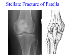
- stellate fractures, if present

NOTE: A patella may be done AP, but what is shown better when done PA?
details
When meeting criteria for a PA projection of the patella, what should be met?
- Clear visualization of the patella through the superimposing femur
- No rotation
- Soft tissue and bony trabecular detail

Stellate Fracture of Patella

Patella - Lateral Projection
Explain how the patient should be positioned for a lateral projection of the patella.
1. Lateral recumbent