A&P Tissue Types, Skin Layers & Skeletal System
 Call Kai
 Learn
 Practice Test
 Spaced Repetition
 Match
 Flashcards
 Knowt Play
1/138

There's no tags or description
Looks like no tags are added yet.
139 Terms
Simple Squamous Epithelium
A single layer of flat, scale-like cells specialized for rapid diffusion and filtration, found in lung alveoli, capillary walls, and linings of blood and lymph vessels.


Stratified Squamous Epithelium
Multiple layers of flat cells designed to protect underlying tissues against abrasion, found in the epidermis, mouth, esophagus, and vagina.
Simple Cuboidal Epithelium
A single layer of cube-shaped cells with large central nuclei specialized for secretion and absorption, found in kidney tubules and small gland ducts.

Stratified Cuboidal Epithelium
Multi-layered cube-shaped cells providing protection, found in sweat and salivary gland ducts.

Simple Columnar Epithelium
A single layer of tall, column-like cells specialized for absorption and secretion, often ciliated, found in the digestive tract and uterus.

Stratified Columnar Epithelium
Multi-layered tall cells providing protection and secretion, found in rare areas like parts of the pharynx and male urethra.


Pseudostratified Epithelium
A single-layer epithelium whose nuclei sit at varying heights, giving the false appearance of multiple layers.
Endocrine Glands
Ductless glands that secrete hormones directly into the bloodstream or interstitial fluid.

Exocrine Glands
Glands that secrete their products through ducts onto body surfaces or into body cavities, such as sweat and sebaceous glands.

Bone Connective Tissue
Hard, rigid connective tissue composed of a calcified extracellular matrix containing calcium salts and collagen fibers, providing structural support and protection.

Hyaline Cartilage
The most abundant avascular cartilage featuring a frosted-glass matrix with fine collagen fibers that supports, cushions, and resists compressive stress in joint cavities, costal cartilages, nose, trachea, and larynx.


Elastic Cartilage
Flexible avascular cartilage containing a high density of elastic fibers that maintains shape while allowing flexibility, found in the external ear and epiglottis.
Fibrocartilage
Tough avascular cartilage packed with thick collagen fibers that absorbs heavy compressive shock, found in intervertebral discs, the pubic symphysis, and knee menisci.

Dense Regular Connective Tissue
Tissue composed of parallel collagen fibers that provides strong tensile strength and resists tension in one direction, found in tendons and ligaments.

Dense Irregular Connective Tissue
Tissue with irregularly arranged collagen fibers able to withstand tension exerted in many directions, found in the dermis of the skin and fibrous joint capsules.

Areolar Connective Tissue
A gel-like loose connective tissue containing all three fiber types that wraps and cushions organs, holds tissue fluid, and underlies epithelia.

Adipose Tissue
Loose connective tissue packed closely with fat cells (adipocytes) that stores energy, insulates against heat loss, and cushions organs.

Reticular Connective Tissue
A delicate network of reticular fibers forming a soft internal skeleton (stroma) that supports immune cells in lymphoid organs like lymph nodes, bone marrow, and the spleen.

Skeletal Muscle Tissue
Voluntary, striated, multinucleated muscle tissue attached to bones responsible for body movement.

Cardiac Muscle Tissue
Involuntary, striated muscle tissue found in the heart wall, featuring intercalated discs and single central nuclei.

Smooth Muscle Tissue
Involuntary, non-striated muscle tissue found in the walls of hollow internal organs like the stomach, intestines, and blood vessels.

Mature Red Blood Cells (RBCs)
Specialized blood cells that lack a nucleus when mature to maximize oxygen transport.
Epidermis
The outermost superficial layer of the skin composed of keratinized stratified squamous epithelium.
Dermis
The thick, fibrous connective tissue layer located beneath the epidermis that provides strength, flexibility, and houses glands, hair follicles, and blood vessels.
Papillary Layer of the Dermis
The superficial layer of the dermis composed of loose areolar connective tissue featuring dermal papillae that form fingerprints and house capillary loops and touch receptors.

Reticular Layer of the Dermis
The deeper, thicker layer of the dermis composed of dense irregular connective tissue that provides strength and elasticity and houses glands and hair follicles.

Hypodermis (Subcutaneous Layer)
The layer of adipose and areolar connective tissue beneath the dermis that anchors the skin to underlying muscles and stores fat.
Stratum Basale
The deepest epidermal layer where continuous mitotic cell division occurs, containing keratinocytes, melanocytes, and tactile cells.
Stratum Spinosum
The "spiny" epidermal layer above the basale where keratinocytes begin producing keratin fibers and dendritic cells provide immune defense.
Stratum Granulosum
The granular layer where cells accumulate keratohyalin granules, flatten, and plasma membranes thicken as they begin to die.
Stratum Lucidum
A clear, thin, dead epidermal layer found exclusively in thick skin such as the palms of the hands and soles of the feet.
Stratum Corneum
The outermost epidermal layer consisting of 20-30 layers of dead, keratin-filled cells that provide waterproofing and protection.
Arrector Pili Muscle
Smooth muscle attached to hair follicles that contracts to pull hair upright, producing "goosebumps" in response to cold or fear.

Eccrine Sweat Glands
Merocrine sweat glands widely distributed across the skin surface responsible for thermoregulation through evaporative cooling of water-based sweat.
Sebaceous Glands
Holocrine glands connected to hair follicles that secrete oily sebum to lubricate and waterproof skin and hair.
Adult Bone Count
206 bones in the average adult human skeleton.
Functions of the Skeletal System
Provides structural support, protects internal organs, enables movement, stores minerals (calcium and phosphorus), and produces blood cells (hematopoiesis).
Axial Skeleton
Comprises 80 bones including the skull, vertebral column, and thoracic cage (ribs and sternum); provides central support and protects vital organs.
Appendicular Skeleton
Comprises 126 bones including the upper and lower limbs and pectoral and pelvic girdles; enables movement and locomotion.
Compact Bone
Dense, hard outer bone tissue composed of repeating osteons (Haversian systems) that provides strength and protection.
Spongy Bone
Porous inner bone tissue composed of trabeculae that reduces skeletal weight and houses red bone marrow, located in epiphyses.

Long Bones
Bones significantly longer than they are wide, featuring a shaft and two expanded ends built for leverage and movement, such as the femur, humerus, radius, ulna, tibia, and fibula.
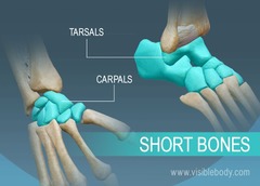
Short Bones
Cube-shaped bones roughly equal in length and width that provide stability and limited gliding motion, such as the carpal bones and tarsal bones.

Flat Bones
Thin, flattened, curved bones consisting of two compact bone layers sandwiching spongy bone that protect organs and offer muscle attachment, such as the sternum, ribs, scapulae, and cranial bones.
Irregular Bones
Bones with complex, unique shapes that do not fit into other categories, such as the vertebrae, hip (coxal) bones, sphenoid, and ethmoid bones.
Sesamoid Bones
Small, sesame-seed-shaped bones that develop within tendons to reduce friction and mechanical stress across joints, such as the patella and pisiform.
Diaphysis
The tubular, hollow shaft of a long bone composed of compact bone that runs between the proximal and distal ends and encloses the medullary cavity.
Epiphysis
The wider proximal and distal ends of a long bone filled with spongy bone and covered by articular cartilage.
Articular Cartilage
Smooth hyaline cartilage covering the joint ends of bones at the epiphyses to reduce friction and absorb shock.
Periosteum
A dense fibrous connective tissue membrane covering the outer surface of bone, serving as a site for ligament and tendon attachment and appositional growth.
Endosteum
A delicate membranous lining inside the medullary cavity and trabeculae of bones containing osteogenic cells.
Medullary Cavity
The central hollow cavity inside the diaphysis of a long bone that contains yellow bone marrow in adults.
Red Marrow
Hematopoietic tissue found in the spaces of spongy bone that produces red and white blood cells (hematopoiesis).
Yellow Marrow
Inactive fat stored inside the medullary cavity of adult long bones.
Endochondral Ossification
The process of bone development where bone forms by replacing a hyaline cartilage model, forming most bones of the skeleton.
Intramembranous Ossification
The process where bone develops directly from sheets of mesenchymal connective tissue, forming flat skull bones, clavicles, and the mandible.
Bone Growth in Length
Occurs at the epiphyseal (growth) plate via interstitial growth, where cartilage is continuously formed and replaced by bone.
Bone Growth in Width (Appositional Growth)
Occurs beneath the periosteum, where osteoblasts deposit new bone tissue on the outer surface while osteoclasts reabsorb bone in the medullary cavity.
Wolff's Law of Bone
The principle that bone grows or remodels in response to the mechanical stress and physical forces placed upon it.
Vitamin D in Bone Health
Essential for promoting calcium and phosphorus absorption in the small intestine, ensuring proper bone mineralization and strength.
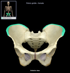
Crest
A narrow, usually prominent ridge of bone.
Epicondyle
A bony projection situated above a condyle.


Tuberosity
A large, rounded bone marking that serves as a point of attachment for muscles, tendons, and ligaments.
Frontal Bone
The single cranial bone forming the forehead, the anterior cranium, and the roof of the eye orbits.

Parietal Bones
The paired cranial bones forming the superior and lateral walls of the cranium, joined at the sagittal suture.


Temporal Bones
The paired cranial bones forming the lower lateral walls and floor of the cranium, housing hearing structures and the smallest bones in the body (auditory ossicles).
Occipital Bone
The single cranial bone forming the posterior base and walls of the skull, featuring the foramen magnum.
Sphenoid Bone
The bat-shaped "keystone" bone of the cranium that spans the width of the middle cranial floor and articulates with all other cranial bones.


Ethmoid Bone
The complex, deep cranial bone located between the nasal cavity and orbits that helps form the nasal septum and roof of the nasal cavity.
External Acoustic Meatus
The canal opening in the temporal bone that leads inward to the eardrum and inner ear structures.

Mastoid Process
A prominent round projection on the temporal bone located just behind the ear, serving as a muscle attachment site.

Styloid Process
A thin, needle-like projection extending downward from the temporal bone that serves as an anchor point for muscles and ligaments of the tongue and neck.

Coronal Suture
The fibrous joint connecting the frontal bone to the parietal bones.
Sagittal Suture
The fibrous joint extending posteriorly from the coronal suture to unite the two parietal bones along the midline of the skull.
Lambdoid Suture
The fibrous joint connecting the parietal bones to the occipital bone.
Squamous Suture
The fibrous joint connecting the parietal bone to the temporal bone.
Zygomatic Bone
The cheekbone; forms the prominence of the cheek and part of the lateral wall and floor of the orbit.

Maxilla
The upper jaw bone; features the infraorbital foramen and articulates with all other facial bones except the mandible.
Mandible
The lower jaw bone; features the ramus and mental foramen and is the largest, strongest, and only freely movable bone of the face.
Lacrimal Bone
A small, delicate facial bone located in the medial wall of each orbit containing the tear duct groove.

Vomer
A plow-shaped facial bone that forms the inferior and posterior portion of the nasal septum.

Infraorbital Foramen
An opening in the maxilla located directly below the eye orbit that transmits nerves and blood vessels to the upper lip and cheek.

Mental Foramen
A small opening on the anterior surface of the mandible that allows blood vessels and nerves to pass to the chin and lower lip.

Ramus of Mandible
The vertical portion of the mandible that articulates with the temporal bone at the temporomandibular joint (TMJ).


Cervical Vertebrae
The 7 vertebrae of the neck region (C1-C7) characterized by transverse foramina that serve as passageways for vertebral arteries to the brain.
Thoracic Vertebrae
The 12 vertebrae of the chest region (T1-T12) that feature costal facets to articulate with the ribs.

Lumbar Vertebrae
The 5 large, heavy vertebrae of the lower back region (L1-L5) built to support upper body weight.

Typical Parts of a Vertebra
Includes the weight-bearing vertebral body, vertebral arch (formed by pedicles and laminae enclosing the vertebral foramen), and processes (spinous, transverse, and articular).
Atlas (C1)
The 1st cervical vertebra; lacks a vertebral body and spinous process and articulates with the occipital condyles to allow nodding ("yes").

Axis (C2)
The 2nd cervical vertebra; features the dens (odontoid process) projecting superiorly to act as a pivot point for head rotation ("no").


Sternum
The flat breastbone located in the anterior chest that belongs to the axial skeleton and articulates with the clavicles and costal cartilages.
True Ribs (Vertebrosternal Ribs)
The first 7 pairs of ribs (1-7) that attach directly to the sternum via their own costal cartilage.

False Ribs (Vertebrochondral Ribs)
Rib pairs 8, 9, and 10 that attach to the cartilage of the ribs above them and thus indirectly to the sternum (broadly includes pairs 8-12).

Floating Ribs (Vertebral Ribs)
Rib pairs 11 and 12 that have no anterior connection to the sternum, attaching only to the vertebrae and body wall muscles.

Scapula
The anatomical name for the shoulder blade, a flat triangular bone of the pectoral girdle.

Humerus
The single long bone of the upper arm belonging to the appendicular skeleton.

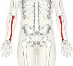
Ulna
The medial bone of the forearm (in anatomical position).
Carpal Bones Mnemonic
"Steve Left The Party To Take Carol Home" stands for Scaphoid, Lunate, Triquetrum, Pisiform, Trapezium, Trapezoid, Capitate, and Hamate.
Scaphoid Bone
The largest, boat-shaped carpal bone in the proximal row on the lateral (thumb) side; most frequently fractured wrist bone.


Lunate Bone
A crescent- or moon-shaped carpal bone located in the middle of the proximal row that articulates with the radius and ulna (via the triangular fibrocartilage complex).