AnaPhy Prelims - Skeletal
 Call Kai
 Learn
 Practice Test
 Spaced Repetition
 Match
 Flashcards
 Knowt Play
1/60

There's no tags or description
Looks like no tags are added yet.
61 Terms
Osseous Tissue
a complex and dynamic living tissue, continually engages in a process called remodeling
Remodeling
the construction of new bone tissue and breaking down of old bone tissue.

diaphysis
bone’s shaft or body
long, cylindrical, main portion of the bone

Epiphyses
the proximal and distal ends of the bone

Metaphyses
region between the diaphysis and epiphyses
epiphyseal plate
layer of hyaline cartilage that allows diaphysis of the bone to grow length
epiphyseal line
when a bone ceases to grow in length at about ages 14–24, the cartilage in the epiphyseal plate is replaced by bone
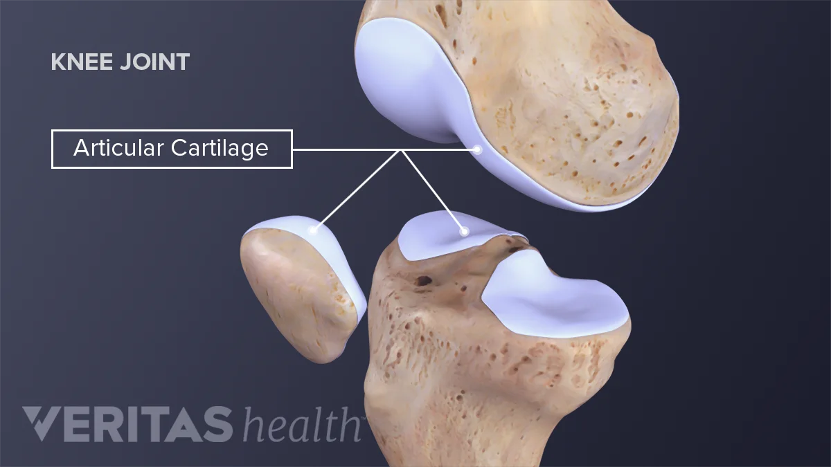
articular cartilage
thin layer of hyaline cartilage covering the part of the epiphysis where bone forms articulation with another bone
reduces friction and absorbs shock at freely movable joints

periosteum
tough connective tissue sheath and its associated blood supply that surrounds the bone surface wherever it is not covered by articular cartilage
Sharpey’s fibers
Thick collagen fibers that connect the periosteum firmly to the bone underneath. Think: “anchors the periosteum to the bone.”
Medullary cavity / Marrow cavity
A hollow space inside the shaft (diaphysis) of a long bone. It contains yellow bone marrow and blood vessels in adults.
Yellow bone marrow
Fatty tissue found inside the medullary cavity of adults. It mainly stores fat.
Endosteum
A thin membrane that lines the inside of the medullary cavity. It contains bone-forming cells and a small amount of connective tissue.
hydroxyapatite
Hard mineral crystals made mainly from calcium and phosphate.
They give bone its hardness.
Osteoprogenitor cells
unspecialized bone stem cells that can develop into osteoblast.
only bone cells to divide
found along the inner portion of the periosteum, in the endosteum, and in the canals within bone that contain blood vessels
osteoblasts
bone building cells that produce materials needed to make bone matrix and start calcification - osteocytes
osteocytes
mature bone cells
osteoclasts
huge cells derived from the fusion of as many as 50 monocytes
concentrated in the endosteum
resorption
breakdown of bone extracellular matrix
compact bone tissue
strongest form of bone tissue
found beneath periosteum
Osteon / Haversian system
The basic structural unit of compact bone. It looks like a small cylinder made of several layers.
Concentric lamellae
Circular layers of mineralized bone matrix that surround the central canal. Think of tree rings.
Central canal / Haversian canal
A small canal in the center of an osteon that contains blood vessels and nerves.
Lacunae
Small spaces between the lamellae that contain osteocytes. Think: “little lakes where bone cells live.”
Osteocytes
Mature bone cells that live inside the lacunae and maintain the bone.
Canaliculi
tiny channels connecting the lacunae to each other and to the central canal. Allows nutrients and waste to move through the bone
Extracellular fluid
fluid inside the canaliculi that helps carry nutrients and remove wastes
Gap junctions
connection between neighboring osteocytes that allow them to communicate with each other
Spongy bone tissue
bone tissue found inside bones
lightweight and made of a network of thin bony structures
Trabecular bone
Another name for spongy bone tissue.
Trabeculae
Thin, beam-like structures of spongy bone that support the bone and are arranged along lines of stress.
Red bone marrow
Soft tissue that produces blood cells. It is found in the spaces between trabeculae in certain bones.
Yellow bone marrow
Fat-rich tissue that stores energy as fat. It can fill the spaces between trabeculae in certain bones.
Hemopoiesis
The process of producing blood cells. It occurs in red bone marrow.
Sesamoid bones
Bones that develop within tendons, such as the patella (kneecap).
Intramembranous ossification
bone forms directly within mesenchyme
arranged in sheetlike, resembles membrnaes
endochondral ossification
bone forms within hyaline cartilage that develops from mesenchyme
Ossification center
the site of cluster
Mesenchyme → osteoprogenitor cells → osteoblasts
Development of ossification center
Osteoblasts secrete matrix → osteocytes form → calcium/minerals harden matrix
Calcification
Matrix forms → trabeculae fuse → spongy bone → red bone marrow develops
Formation of Trabeculae
Mesenchyme at the outside condenses → periosteum forms
Development of periosteum
Bone is destroyed and rebuilt → reaches adult size and shape
Remodeling
Mesenchymal cells cluster and differentiate into chondroblasts
development of cartilage model
Cartilage ECM
Chondroblasts produce______, forming a hyaline cartilage model.
The cartilage model grows in length through interstitial growth.
It grows in thickness through appositional growth.
growth of cartilage model
Hypertrophy
When chondrocytes in the middle enlarge
Interstitial growth
endogenous growth
cartilage model grows in length
Appositional growth
exogenous growth
cartilage model grows in thickness
nutrient artery enters the diaphysis
blood vessels and osteoprogenitor cells enter the developing region
perichondrium becomes periosteum
Spongy bone trabeculae form.
Ossification progresses toward both ends of the shaft.
Development of primary ossification center
Osteoclasts resorb some of the newly formed bone.
A medullary cavity develops in the diaphysis.
The shaft wall develops into compact bone.
Development of the medullary (marrow) cavity
Blood vessels enter the epiphyses.
Secondary ossification centers develop, usually around birth.
Bone replaces the cartilage in the epiphyses.
Spongy bone remains inside the epiphyses; a medullary cavity does not normally form there.
Development of the secondary ossification center
hyaline cartilage becomes articular cartilage
hyaline cartilage remains between the diaphysis and epiphysis
Formation of articular cartilage and the epiphyseal (growth) plate.
Chondroblast
cells that produces cartilage ECM
Chondrocyte
mature cartilage cell
Hyaline cartilage model
cartilage structure that serves as the template for the developing bone
Periosteum
connective tissue covering the cartilage model
periosteum
connective tissue covering the outer surface of bone
Bone remodeling
ongoing replacement of old bone tissue by new bone tissue
bone resorption
removal of minerals and collagen fibers from bone by osteoclasts
bone deposition
addition of minerals and collagen fibers to bone by osteoblasts.