PHS 3507 - Nervous Tissue (Chapter 12)
 Call Kai
 Learn
Learn Practice Test
 Spaced Repetition
 Match
Match Flashcards
 Knowt Play
1/73

There's no tags or description
Looks like no tags are added yet.
Name | Mastery | Learn | Test | Matching | Spaced | Call with Kai | Chat |
|---|
No analytics yet
Send a link to your students to track their progress
74 Terms
Nervous System Overview
The nervous system detects changes in the environment that affect the body
It works together with the endocrine system to help the body respond to these changes
The nervous system controls:
Behavior
Memories
Movement
Nervous tissue is excitable, meaning it can respond to stimulation and produce electrical signals
These electrical signals are called nerve impulses (action potentials)
3 Steps of the Nervous System
1. Sensory function = Detect
Detects internal and external stimuli (changes in/outside the body)
2. Interpretation = Analyze
The nervous system interprets/analyzes the information as detected
3. Motor response = React
The nervous system causes the body to respond or react
Nervous System Overview
Sensory Neuron → Interneuron → Motor Neuron
Interneuron connects the sensory with motor neuron

Nervous System Organization
The nervous system contains over 100 billion neurons
It has 10-50 times more support cells, neuroglia (gilal cells)
These cells are organized into 2 main divisions:
Central Nervous System (CNS)
Peripheral Nervous System (PNS)
Neurons vs. Neuroglia
Neurons = the “thinking” cells
They recieve, process, and transmit information
They do this by controlling the flow of electrical charge across their cell membrane
Basically, each neuron does a small version of what the whole nervous system does
Neuroglia (glial cells) = support cells
They provide support and nutrition for the brain
They do not process/manipulate information like neurons
They maintain the internal environment so neurons can work properly
Divisions of the Nervous System
Central Nervous System (CNS)
Consists of the brain and spinal cord
Peripheral Nervous System (PNS)
Consists of all nervous tissue outside of the brain and spinal cord
Nerves
Ganglia
Enteric plexuses
Sensory receptors

Further Division of the Nervous System
Most signals that tell muscles to contract and glands to secrete start in the CNS
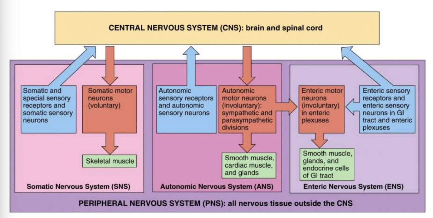
The PNS is further divided into 3 parts:
Somatic Nervous System (SNS)
Autonomic Nervous System (ANS)
Enteric Nervous System (ENS)

Somatic Nervous System (SNS)
Somatic Sensory (Afferent) Neurons
Carry information from sensory receptors in the head, body wall, and limbs toward the CNS
Somatic Motor (Efferent) Neurons
Carry nerve impulses away from the CNS into the skeletal muscles of the body
Controls voluntary movement
Interneurons
Conduct nerve impulses between the afferent and efferent neurons within the CNS
Autonomic Nervous System (ANS)
Sensory Neurons
Carry information from autonomic sensory receptors, mainly in the visceral organs like the stomach or lungs towards CNS
Motor Neurons
Carry nerve impulses from the CNS to the smooth muscle, cardiac muscle (heart), and glands
These are under involuntary control
The motor part of the ANS has 2 branches that usually have opposite actions:
Sympathetic division
Parasympathetic division
Enteric Nervous System (ENS)
The ENS is called the “brain of the gut”
The ENS was once considered part of the ANS
It involuntarily controls GI propulsion, acid secretion, and hormone secretion
It contains over 100 million neurons
These neurons are located in enteric plexuses (networks of neurons) that extend through most of the GI tract
SNS, ANS, ENS
Ganglia
Ganglia are small groups of neuron cell bodies located outside the brain and spinal cord
Closely associated with cranial & spinal nerves
There are different types of ganglia depending on the neurons they contain:
Somatic ganglia → somatic neurons
Autonomic ganglia → autonomic neurons
Enteric ganglia → enteric neurons
Neurons and Neuroglia
Neurons and neuroglia work together in different parts of the nervous system
Neurons
Are the functional units of the nervous system → they do the actual receiving, processing, and sending of information
Form complex networks in the brain and spinal cord that allow the CNS to control different regions of the body
Neuroglia (Glial Cells)
Are smaller than neurons, but greatly outnumber them
Act like the “glue” of the nervous sytem
They support and maintain neuronal networks, helping neurons work properly
Neurons
Several different types of neurons, most have:
Cell body, axon, dendrites, and axon terminals

Neurons Overview
Neurons gather information at dendrites and process it in the dendritic tree and cell body
Then they transmit the information down their axon to the axon terminals

Dendrites
Dendrites mean “little trees” because they look like small branches
They are the receiving end of the neuron—they receive signals/information from other cells
They are short, highly branched structures that conduct impulses toward the cell body
Lots of branches allow them to receive many signals
Dendrites also contain organelles
Cell Body
The cell body contains a nucleus surrounded by cytoplasm
Like other cells, neurons contain organelles such as:
Lysosomes
Mitochondria
Golgi complexes
Rough ER (RER) → protein production
In neurons, the rough ER is called the Nissl bodies
Nissl bodies give the neuron a striped “tiger appearance”
Neurons have no mitotic apparatus
This means mature neurons generally cannot undergo mitosis (cell division)

Axons
Axons carries nerve impulses away from the cell body towards:
Another neuron
An effector cell (muscle or gland)
Three important areas near the beginning of the axon
Axon hillock: where the axon connects to the cell body
Initial segment: the beginning part of the axon
Trigger zone: the junction between the axon hillock and inital segment

Axon Terminals
The axon and its branches (collaterals) eventually split into many small endings called axon terminals (telodendria)
Telodendria can be highly branched, similar to dendrites
They interact with the dendrites of the “downstream” neurons
Downstream neuron = the next neuron that will receive the information
The tops of some axon terminals become enlarged into bulb-shaped structures called synaptic end bulbs

Synapse
A synapse is the place where communication happens between:
Two neurons, or
A neuron and an effector cell
Presynaptic Cell
The cell sending the signal
Postsynaptic Cell
The cell receiving the signal
Synaptic Cleft
The tiny gap between the presynaptic and postsynaptic cells

Synaptic Vesicles and Neurotransmitters
Synaptic end bulbs and other swollen areas (varicosities) on the axon terminals of the presynaptic neuron contain many synaptic vesicles
Synaptic vesicles = tiny membrane-covered sacs that store neurotransmitters
Neurotransmitters = chemicals that help send signals to the postsynaptic cell
Many neurons can contain 2 or even 3 different types of neurotransmitters
Each neurotransmitter can have a different effect on the postsynaptic cell

Action Potentials at a Synapse
An action potential (electrical signal) travels down the presynaptic neuron
But the action potential cannot physically cross the synaptic cleft because there is a gap between the cells
So the neuron uses neurotransmitters (chemical messengers) to carry the message across the gap
The neurotransmitter binds to the postsynaptic cell
This can cause a new action potential to be generated in the postsynaptic cell

Axonal Transport
Materials are made or recycled in the neuron cell body
Some of these materials are needed in the axon and axon terminals
Axonal transport moves these materials between the cell body and axon terminals
Neurons have 2 transport systems that move these materials
Slow axonal transport
Moves axoplasm (material inside the axon)
Moves in one direction only:
Cell body → axon terminals
Fast axonal transport
Moves material in both directions:
Cell body → axon terminals
Axon terminals → cell body
Axonal Transport Cont.
Slow axonal transport
Supplies new axoplasm (cytoplasm in axons) to developing or regenerating axons and replenishes axoplasm in growing and mature axons
Fast axonal transport
Anterograde (foward)
Cell body → axon terminals
Moves organelles and synaptic vesicles (new supplies)
Retrograde (backward)
Axon terminal → cell body
Move membrane vesicles and other cellular materials back to the cell body (old supplies)
Once back, these materials are degraded/broken down or recycled
Fast Retrograde Transport
Substances that enter a neuron at the axon terminals can be carried back toward the cell body
This is called fast retrograde transport
It can carry:
Helpful substances
Trophic chemicals (Nerve Growth Factor; NGF)
Harmful substances
Tetanus toxin
Rabies virus
Polio virus
These harmful substances can use the neuron’s retrograde transport to travel toward the CNS
A deep cut or puncture wound in the head or neck can be serious than a similar injury in the leg
There is a shorter distance to the brain, so a harmful substance traveling through neurons can reach the brain faster than a similar injury in the leg
Classifying Neurons
Neurons can have many different sizes and shapes
Some neurons are extremely long → almost as long as a person is tall, extending toward the lowest part of the brain
Dendrites branch differently depending on the type of neuron and where it is located in the nervous system
Some neurons have:
Very short axons
No axons at all
Neurons can be classified based on 2 things:
Structure → What the neuron looks like
Function → What the neuron does
Structural Classification
Structural classification = classifying neurons based on how many processes extend from the cell body
Processes = the axons and dendrites coming off the cell body

Multipolar Neurons
Multipolar neurons have:
Many dendrites
One axon
They are found throughout the brain and spinal cord
The vast majority of neurons in the human body are multipolar

Bipolar neurons
Bipolar neurons have:
One dendrite
One axon
They help carry information for the special senses:
Sight, Smell, Hearing, Balance
Because of this bipolar neurons are found in:
Retina of the eye (sight)
Inner ear (hearing and balance)
Olfactory area (smell)

Unipolar (Pseudounipolar) Neurons
Unipolar neurons have one process extending from the cell body
That one process splits into branches that function in receiving and carrying sensory information
They are commonly sensory neurons
They carry information about:
Touch
Stretching
This information comes from the extremities → arms, hands, legs, and feet

Functional Classification of Neurons
Functional classification = classifying neurons based on what they do
It is based on:
Whether their effect is excitatory or inhibitory
Which direction the action potential (AP) travels compared with the CNS
1. Sensory (Afferent) Neurons
Carry action potential into the CNS through cranial or spinal nerves
Most sensory neurons are unipolar
2. Motor (Efferent) Neurons
Carry action potential away from the CNS
Carry signals to effectors in the periphery (muscles, glands) through cranial or spinal nerves
Most motor neurons are multipolar
Interneurons (Association Neurons)
Interneurons are mainly located inside the CNS (brain and spinal cord)
They are found between sensory and motor neurons
Their job is to integrate (process) sensory information coming from sensory neurons
After processing the information, they activate the appropriate motor neuron to produce a response
Most interneurons are multipolar in structure
Neurogila
Neuroglia (glial cells) are support cells of the nervous system
Unlike neurons, they do not generate or conduct nerve impulses
They support neurons by:
Forming the Blood-Brain Barrier (BBB)
Forming the myelin sheath → nerve insulation
Making cerebrospinal fluid (CSF) → circulates around the brain and spinal cord
Participating in phagocytosis
Types of Neuroglia
4 types of neuroglia in the CNS
Astrocytes → support neurons in the CNS
Maintain the chemical environment (Ca2+ & K+)
Oligodendrocytes → produce myelin in CNS
Microglia → participate in phagocytosis
Ependymal cells → form and circulate CSF
2 types of neuroglia in the PNS
Satellite cell → support neurons in the PNS
Schwann cell —> produce myelin in PNS
Myelination
Myelination = the process of forming a myelin sheath around an axon
Myelin sheath acts like insulation around the axon
Allows nerve impulses (action potentials) to travel faster
Different cells make myelin depending on the location
CNS → Oligodendrocytes
PNS → Schwann cells
Nodes of Raniver
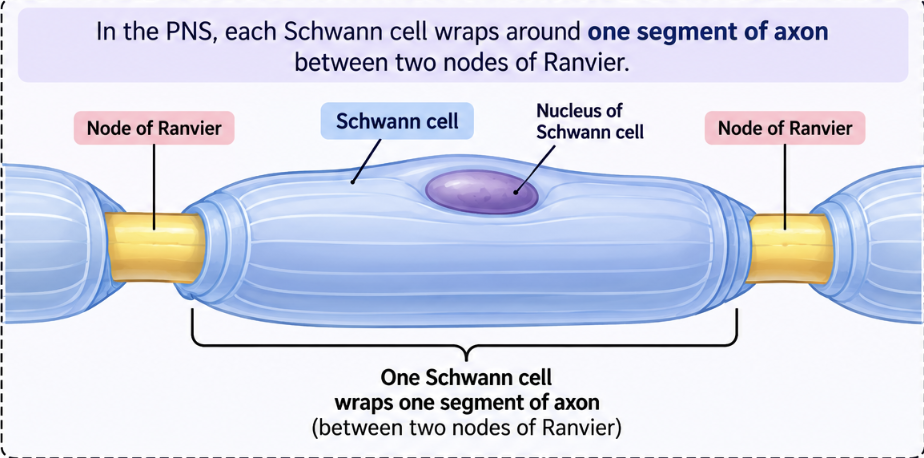
Nodes of ranvier = small gaps between sections of the myelin sheath where the axon is exposed
In the PNS, each Schwann cell wraps around one segment of axon between two nodes of ranvier
Each myelinated segment is about 1 mm long and can have up to 100 layers of myelin
The amount of myelin increases from birth to maturity
More myelin allows nerve impulses to travel much faster
Multiple Sclerosis involves the immune system attacking and destroying myelin, which interferes with normal nerve conduction
Neuron Regeneration
Around birth, most neurons lose their ability to undergo mitosis
This means if a neuron is injured, it usually cannot make daughter cells to replace itself
This is different from epithelial cells, which can divide and replace damaged cells
Instead, damaged neurons may sometimes repair/regenerate damaged parts
PNS
Some nerve regeneration can occur
Schwann cells play an important role in helping damaged nerve fibers regenerate
CNS
Regeneration essentially doesn’t occur at all
Astrocytes tend to form scar tissue around the damaged area instead of helping the nerve regenerate
Neurolemma
The neurolemma (sheath of Schwann) is the outer layer of a Schwann cell
It contains the Schwann’s cell’s nucleus and cytoplasm
It surrounds the myelin sheath
When an axon in the PNS is injured:
The neurolemma helps the axon regenerate
It forms a regeneration tube
This tube guides and stimulates the damaged axon to regrow in the correct direction

Neuron Regeneration & Demylination
For a neuron to regenerate after injury, it must:
It must be in the PNS
Its cell body must still be intact/alive
It must have functional Schwann cells with a neurolemma around the myelinated axon
The neurolemma helps guide axon regrowth
Demyelination
Demyelination = loss or destruction of the myelin sheath around axons
It can result from:
Disease, radiation therapy, chemotherapy
Even one episode of demyelination can cause the affected nerves to deteriorate
Gray and White Matter
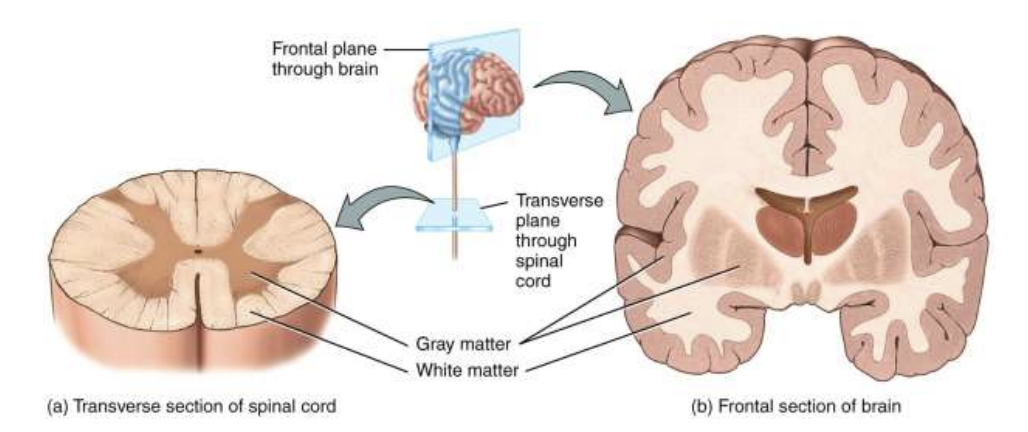
White Matter
Made of mostly groups of myelinated axons from many neurons
It looks white because myelin contains a lot of lipid (fat)
Gray Matter
Made mostly of neuron cell bodies and dendrites
Looks more gray because these areas lack large amount of myelin
Electrical Signals in Neurons
Like muscle cells, neurons are electrically excitable
They can respond to stimulation by changing their electrical charge
Neurons use 2 types of electrical signals:
Graded potentials
Used for short-distance communication
Action potential
Used for long-distance communication
Resting Membrane Potential (RMP)
Neurons need a resting membrane potential (RMP) before they can produce electrical signals
RMP = there is a difference in electrical charge across the neuron’s membrane
Similar to a battery having a difference in electrical charge between its two ends
The RMP is created by:
Ion gradients → different amount of ions (like Na+ and K+) inside vs. outside the neuron
Ion channels → allow ions to move across the membrane
Some ion channels open or close in response to specific stimuli
The lipid bilayer does not allow ions to easily pass directly through it because it acts as an insulator
Therefore, ions such as Na+ and K+ must travel through ion channels to cross the membrane

Ion Channels & Negative Charge
Ion channels are found in the plasma membrane of all cells
They are especially important in neurons because neurons use ion movement to create electrical signals
Cells use a lot of energy to maintain a difference in electrical charge across their membrane
At rest, the inside of a neuron is more negative than the outside
Electrochemical Gradient
When an ion channel opens, specific ions can move across the cell membrane according to their electrochemical gradient
Electrochemical gradient = 2 forces working together
Chemical (concentration) gradient
Ions move from high concentration → low concentration
Electrical gradient
Positive ions (cations) are attracted towards negative areas
Negative ions (anions) are attracted toward positive areas
Opposite charges attract
Types of Ion Channels
Active (gated) channels open in response to a specific stimulus
Ligand-gated channels
Open when a chemical (ligand), such as a neurotransmitter, binds to them
Mainly found at synapses
Voltage-gated channels
Open/close when the electrical potential across the membrane changes
Mainly found along the axon
Important for action potentials
Mechanically-gated channels
Open in response to physical/mechanical deformation, such as pressure on a receptor
Leakage Channels
Leakage channels are also gated, but not active
They open and close randomly which allow ions to “leak” across the membrane
Resting Membrane Potential (RMP)
RMP is measured when the neuron is resting → it is not currently sending a nerve impulse
At rest:
A small amount of negative charge builds up in cytosol along the inside of membrane
An equal amount of positive charge builds up in the extracellular fluid along the outside surface of the membrane
This difference in charge across the membrane creates the resting membrane potential
The rest of the cytosol is electrically neutral
Why the RMP is Negative
At rest, the inside of the neuron is slightly negative compared with the outside
This happens mainly because:
More K+ leaks out of the neuron than Na+ leaks in
This is because there are more K+ leakage channels than Na+ leakage channels
There are also large negatively charged proteins that stay trapped inside the cytosol
Why do we need the Na+/K+ pump?
Na+ is constantly leaking in
K+ is constantly leaking out
If this continued without being corrected, the ion gradients would gradually disappear and the RMP would eventually be lost
The Na+/K+ ATPase (sodium-potassium pump) fixes this:
Pumps Na+ back out as it leaks in
Pumps K+ back in as it leaks out
This maintains the ion gradients needed for the RMP
Resting Membrane Potential (RMP) Cont.
A typical neuron has an RMP of about -70 mV
The negative sign means the inside of the neuron is more negative than the outside
Polarization & Graded Potentials
A neuron with a resting membrane potential (RMP) is called polarized
Polarized = there is a difference in charge across the membrane
Inside is more negative than outside
When polarized, the neuron is “primed” or ready to potentially produce an action potential
But an action potential doesn’t happen immediately
1. Graded potentials happen first
2. They depolarize the neuron → make the inside less negative
3. If the neuron reaches threshold, an action potential starts
Where do graded potentials come from?
A stimulus opens:
Ligand-gated channels → opened by chemicals/neurotransmitters
Mechanically gated channels → opened by pressure/stretch
Ions flow through these channels → producing a small, localized electrical current
This is a graded potential
It spreads to nearby parts of the membrane for only a short distance
It gets weaker as it travels and dies out within a few millimeters
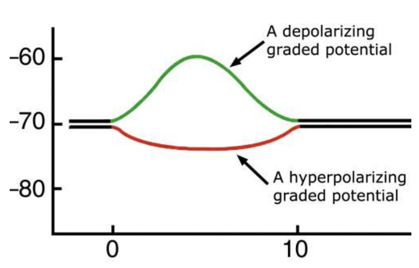
Depolarizing vs. Hyperpolarizing Graded Potentials
Start at the neuron’s RMP, usually around -70 mV
Depolarizing graded potential
Inside becomes less negative
Example: -70 mV → -60 mV
Moves the neuron closer to firing an action potential
Hyperpolarizing graded potential
Inside becomes more negative
Example: -70 mV → -80 mV
Moves the neuron farther away from firing an action potential
Graded Potentials
Graded potentials can have different names depending on:
The type of stimulus
Where in the neuron they occur
They have variable amplitudes (strengths)
Some graded potentials are small
Others are larger
Multiple graded potentials can interact:
Summate (add together) → make a larger graded potential
Cancel each other out → make a smaller graded potential
Graded potentials occur mainly in the dendrites and cell body
They do not travel down the axon
Action Potential (AP)
Unlike a graded potential, an action potential (AP) travels down the entire axon to the axon terminals
During an action potential:
The membrane potential reverses → inside temporarily becomes positive
Then it returns to its resting membrane potential (RMP)
Threshold
The stimulus must be strong enough to reach threshold
Threshold (liminal) stimulus reached → a full-strength action potential occurs and travels down the axon
Subthreshold (subliminal) stimulus → not strong enough → no action potential
Action Potential Phases
An AP has two main phases:
Depolarizing phase
Repolarizing phase

Beginning of an Action Potential
The neuron starts at its RMP of about -70 mV
A depolarizing graded potential makes the inside less negative
If it reaches threshold (about -55 mV) → an action potential begins
Then:
Voltage-gated Na+ channel open
Na+ rapidly rushes into the neuron
Since Na+ is positive, the inside becomes more and more positive
This is called the depolarization phase of the action potential

Depolarization → Repolarization
Depolarization
Only about 20,000 Na+ ions enter each small area of the membrane
Even this small amount causes a large change in membrane potential
The membrane potential can reach about +30 mV
So the inside becomes positive
Repolarization
K+ channels open
K+ rushes out of the neuron
Since K+ is leaving, the inside becomes more negative again
The membrane potential moves back toward the RMP of about -70 mV
After-Hyperpolarization
During repolarization, voltage-gated K+ channels are open, so K+ moves out of the neuron
K+ flow out of the neuron, making the inside more negative
Sometimes the K+ channels stay open longer than needed
Because they are still open, more K+ continues to leave
This makes the membrane potential more negative than the normal RMP, reaching about -90 mV
This phase is called after-hyperpolarization
When the voltage-gated K+ channels finally close, the membrane potential returns to the normal RMP of about -70 mV
All-or-None Principle
If the neuron does not reach threshold → no action potential
If a neuron reaches threshold → a full action potential occurs
A stronger stimulus does not make a bigger action potential
Absolute Refractory Period
After an action potential starts, there is a short period called the absolute refractory period
During this time, the neuron cannot start another action potential, even if the stimulus is extremely strong
This happens because they voltage Na+ channels are activated and then inactivated
The inactivated Na+ channels must reset back to their resting state before another action potential can occur
Relative Refractory Period
The relative refractory period happens after the absolute refractory period
During this time, the neuron can produce another action potential, but it requires a stronger-than-normal stimulus
This happens because:
Na+ channels have reset → so another AP is still possible
But voltage-gated K+ channels are still open
K+ is still leaving the neuron, the inside if more negative than normal
Therefore, it’s harder to reach threshold
Action Potential Propagation
The action potential (AP) starts at the trigger zone near the axon hillock
It then propagates (travels) down the axon
The electrical current spreads forward and triggers the next part of the neuron
There are 2 ways this happens:
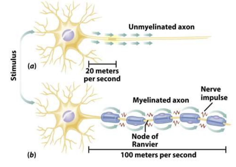
Continuous conduction → unmyelinated axons
The AP is regenerated along each neighboring section of the axon
This is slower
Saltatory conduction → myelinated axons
Myelin covers most of the axon
The AP is regenerated mainly at the nodes of Raniver
It appears to jump from node to node
This is much faster
What Affects Action Potentials
What affects how fast an action potential travels?
Axon diameter
Amount of myelin
Temperature
Nodes of Ranvier
What tells the brain how strong a stimulus is?
Frequency code → how fast/often neurons fire
Recruitment → how many neurons fire
Fiber Types
Axons are classified into A, B, and C fibers based on their size, speed, and myelination
A Fibers
Largest and fastest → 130 m/sec
Myelinated
Carry and touch pressure sensations
Many motor neurons are also A fibers
B Fibers
Medium size and speed → 15 m/sec
Myelinated
Include:
Visceral sensory neurons
Autonomic preganglionic neurons
C Fibers
Smallest and slowest → about 2 m/sec
Unmyelinated
Include:
Sensory neurons
Autonomic motor neurons
Signal Transmission at a Synapse
Signal transmission goes one way:
Presynaptic neuron (sender) → Postsynaptic neuron (receiver)
Steps:
1. Action potential reaches the synaptic end bulb of the presynaptic neuron
2. Voltage-gated Ca2+ channels open
3. Ca2+ enters the presynaptic neuron
4. Ca2+ triggers the release of neurotransmitters
5. Neurotransmitters travel across the synaptic cleft
6. Neurotransmitters bind to ligand-gated receptors on the postsynaptic cell
7. This produces graded potentials in the postsynaptic cell

Electrical → Chemical → Electrical
As a chemical synapse, the signal changes forms:
Presynaptic neuron: electrical → chemical
Action potential arrives = electrical signal
Causes neurotransmitter release = chemical signal
Postsynaptic neuron: chemical → electrical
Neurotransmitter binds to receptors
This creates a postsynaptic potential (graded potential) = electrical signal
Synaptic Delay
These steps take a tiny amount of time
This creates a synaptic delay of about 0.5 milliseconds

Excitatory vs. Inhibitory Neurotransmitters
Both excitatory and inhibitory neurotransmitters effects occur in the CNS and PNS
Excitatory → makes the postsynaptic cell more likely to fire an action potential
Inhibitory → makes the postsynaptic cell less likely to fire an action potential
Example: Acetylcholine
ACh is commonly released by neurons in the PNS and by some neurons in the CNS
At the neuromuscular junction (NMJ) → ACh is excitatory → helps skeletal muscles contract
At some other synapses → ACh can be inhibitory
Amino Acid Neurotransmitters
Many amino acids can act as neurotransmitters in the brain
Glutamate → mainly excitatory
Released by nearly all excitatory neurons in the brain
Makes the next neuron more likely to fire
GABA → inhibitory
Used at about 1/3 of brain synapes
Makes the next neuron less likely to fire
Valium (diazepam) enhances GABA’s inhibitory effects
This decreases neuronal activity → causes sedation
Modifying Neurotransmitter Effects
The effects of neurotransmitters can be changed at different steps:
Synthesis → the body can make more or less neurotransmitter
Release → neurotransmitter release can be increased or blocked
Removal → neurotransmitter can be removed faster or prevented from being removed
Receptors → receptors can be activated or block
Agonist vs. Antagonist
Agonist → activates/stimulates a receptor
Increases or mimics the normal effect
Antagonist → blocks a receptor
Decreases/prevents the normal effect
EPSPs vs. IPSPs
A neurotransmitter can cause 2 types of graded potentials in the postsynaptic neuron:
EPSP = Excitatory Postsynaptic Potential
Causes depolarization
Makes the inside less negative
Moves the neuron closer to threshold
Makes the neuro more likely to fire an action potential
Usually, one EPSP alone is not enough to reach a threshold
IPSP = Inhibitory Postsynaptic Potential
Causes hyperpolarization
Makes the inside more negative
Moves the neuron farther from threshold
Makes the neuron less likely to fire an action potential
Spatial vs. Temporal Summation
Summation = graded potentials (EPSPs and IPSPs) added together
Spatial summation = where
Postsynaptic potentials come from different locations/neurons around the same time
Their effects combine
Temporal summation = when
Postsynaptic potentials arrive every close together in time
Usually, the same input fires repeatedly before the previous graded potential disappears
Their effects add together
What happens after summation?
The neuron adds up all EPSPs and IPSPs:
ESPS → toward the threshold
IPSPs → away from threshold
Net effect determine whether action potential is started

Neurotransmitter Removal
After a neurotransmitter sends its message, it must be removed from the synaptic cleft
If it stayed there, it would keep affecting the postsynaptic cell continuously
This could keep stimulating or inhibiting a neuron, muscle, or gland when it shouldn’t
There are 3 ways neurotransmitters are removed:
Diffusion → neurotransmitters moves away from the synaptic cleft
Enzymatic degradation → an enzyme breaks it down
Reuptake → cells take the neurotransmitters back up
Example: Acetylcholine (ACh)
ACh is released into the synaptic cleft
After it sends its signal, the enzyme acetylcholinesterase (AChE) breaks it down
How Neurons Process Information
A neuron receives many ESPSs and IPSPs from other neurons
These signals are added together (summated)
Their net effect is evaluated at the trigger zone
Integration
Integration = when a postsynaptic neuron combines all EPSPs and IPSPs it receives and decides how to respond
Reaches threshold → action potential fires
Doesn’t reach threshold → no action potential
This happens over and over between many neurons, especially interneurons in higher brain regions, including the thalamus and cerebral cortex
Neural Circuits
A neuronal network = a group of interconnected neurons that communicate and process information together
One network can contain thousands or even millions of neurons
There are different ways neurons can be connected, called neural circuits
Diverging
Converging
Reverberating
Parallel after-discharge

Neural Circuits
Diverging circuit = few → many
A small number of neurons stimulate a much larger number of neurons
Converging circuit = many → few
The opposite of diverging
Many neurons send information toward a smaller number of neurons
Reverberating circuit = loop
Signals travel through a circuit and then feed back through it repeatedly
Used for repetitive/ongoing activites such as:
Breathing, coordinated muscle activity, waking up, short-term memory
Parallel after-discharge = one → many paths → one
One presynaptic neuron stimulates multiple neurons/pathways
Those pathways eventually synapse with the same postsynaptic neuron
Used for precise activities, such as mathematical calculations