Muscle Tissue and Muscle Fiber Structure
 Call Kai
 Learn
Learn Practice Test
 Spaced Repetition
 Match
Match Flashcards
 Knowt Play
1/20

There's no tags or description
Looks like no tags are added yet.
Name | Mastery | Learn | Test | Matching | Spaced | Call with Kai | Chat |
|---|
No analytics yet
Send a link to your students to track their progress
21 Terms
Generation of force by muscle tissue
creates movement, maintains posture, stabilizes joints, generates heat, and regulates the flow of materials through hollow organs
Three types of muscle tissue
Skeletal muscle, cardiac muscle, and smooth muscle

Skeletal muscle tissue
Long, thin, striated, multinucleated cells arranged parallel to each other
mostly attached to the skeleton
requires nervous system stimulation and is voluntary
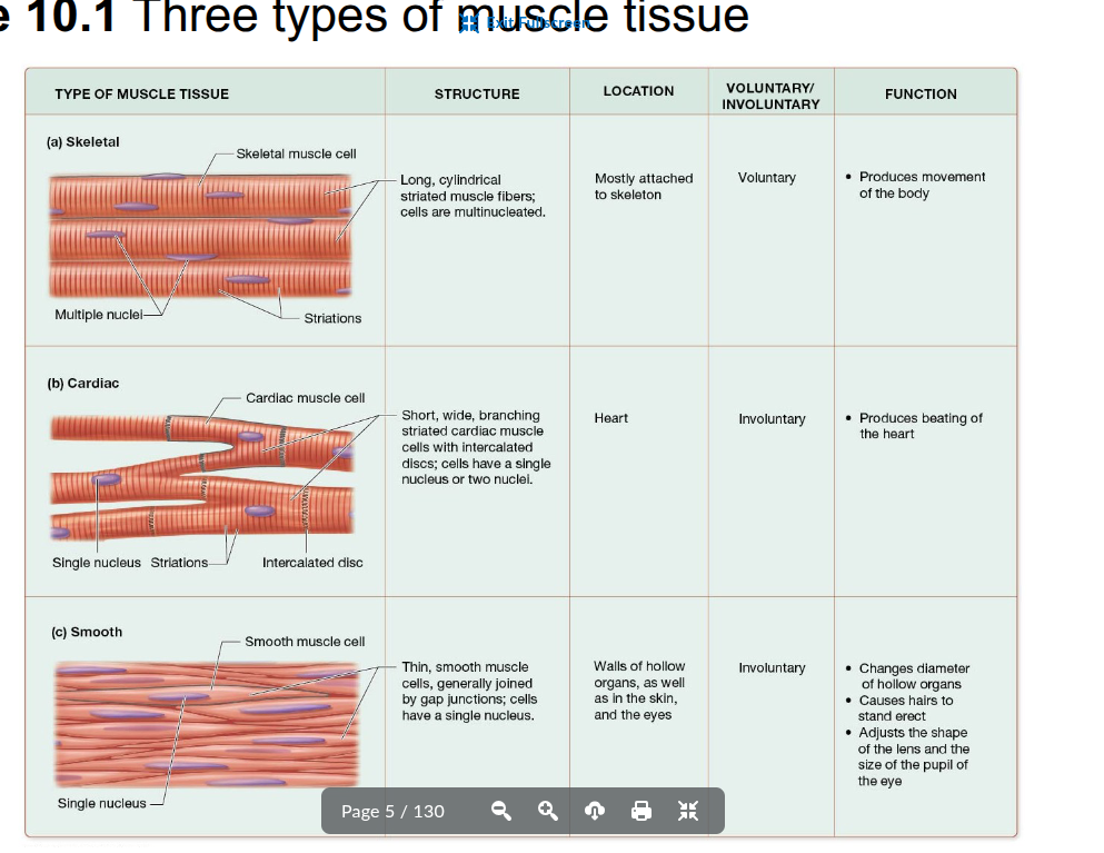
Cardiac muscle tissue
Shorter, wider, branching, striated cells usually containing a single nucleus
found only in the heart and is involuntary
Cells are connected by intercalated discs that help them contract as a unit

Smooth muscle tissue
Long, flattened cells with pointed ends and one centrally located oval nucleus
nonstriated and involuntary
Found in hollow organs, eyes, skin, and ducts of certain glands

Muscle tissue with alternating light and dark bands
skeletal and cardiac muscle are striated, while smooth muscle is not
Ability of a muscle cell to contract as proteins within the cell draw closer together
Ability of a muscle cell to respond to a chemical, mechanical, or electrical stimulus
Ability to conduct electrical charges across the plasma membrane
Ability of a muscle cell to be stretched without rupturing
Ability of a muscle cell to return to its original length after being stretched
Sarcoplasm
The cytoplasm of a muscle cell

Sarcolemma
The plasma membrane of a muscle cell
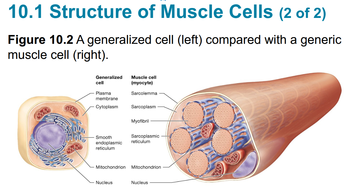
Myofibrils
Bundles of specialized proteins within a muscle cell, including proteins involved in contraction
make up about 50–80% of the cell's volume
Myofibrils are the most abundant organelle in the sarcoplasm and are surrounded by the sarcoplasmic reticulum, which stores and releases calcium ions

Sarcoplasmic reticulum (SR)
Modified smooth endoplasmic reticulum that forms a network around each myofibril
stores and releases calcium ions
Skeletal muscle fiber structure
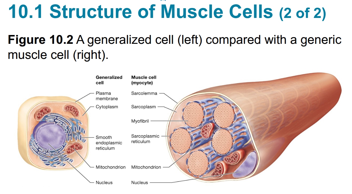
Long, cylindrical, striated cell that contains multiple nuclei and many myofibrils
Multiple nuclei—Muscle fibers arise from multiple embryonic Myoblasts, which fuse to form the muscle fibers

Multiple embryonic myoblasts fuse together to form a skeletal muscle fiber, resulting in a cell with multiple nuclei
T-tubules
tranverse tubules
Inward extensions of the sarcolemma that extend deep into the muscle fiber and surround the myofibrils
their tunnels contain extracellular fluid

Terminal cisternae
Enlarged portions of the sarcoplasmic reticulum located on each side of a T-tubule
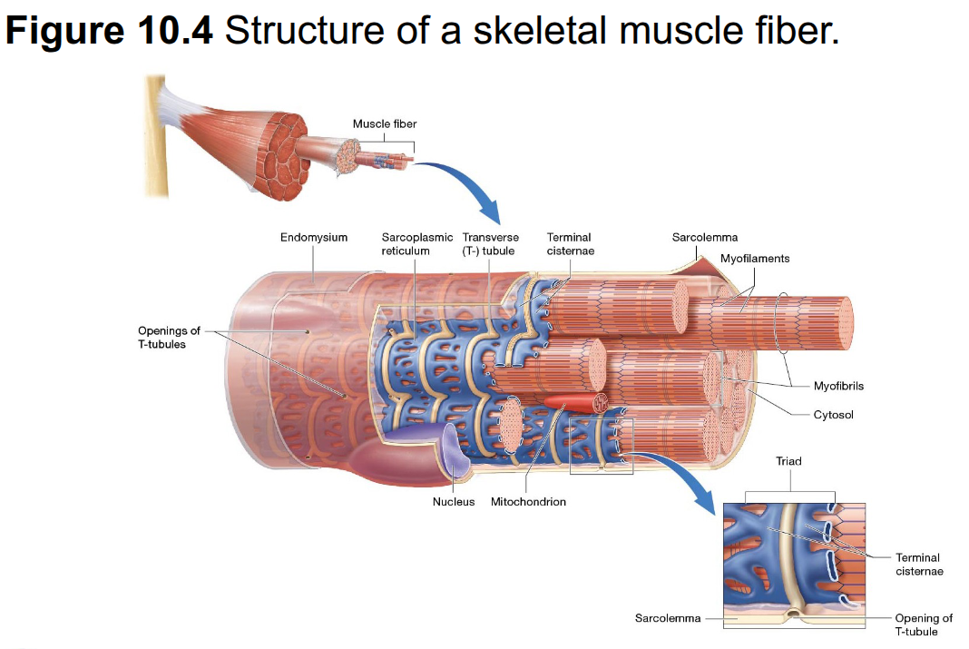
One T-tubule plus the two terminal cisternae of the sarcoplasmic reticulum located on either side of it
Skeletal muscle fiber organization
Sarcolemma surrounds the muscle fiber →
muscle fiber contains myofibrils →
myofibrils are surrounded by sarcoplasmic reticulum →
T-tubules extend inward from the sarcolemma →
terminal cisternae of the SR flank each T-tubule
