Anaemia
 Call Kai
 Learn
 Practice Test
 Spaced Repetition
 Match
 Flashcards
 Knowt Play
1/140

There's no tags or description
Looks like no tags are added yet.
141 Terms
anaemia
decreased amt of rbc or haemoglobin (dcrs pct/ hct)
why low hb?
bc carrys o2
consequence of anaemia→ poor o2 carrying
how is anaemia measured by
cats can form rouleaux naturally so this needs to be overcome.
haematocrit
calculated value from mean cell volume and RBC count, and haemoglobin conc, which provided by automated analysers. HCT can be affected by machine errors
HCT=(MCV x RBC count) + 10→ dont mem
PCV
directly measured value measured as percentage of packed RBCs in blood volume.
PCV can be affected by how RBCs pack together.
signs of anaemia
inadequate o2/ perfusion
pale mm
lethargy
exercise intolerance
CRT delays, cold extremities
compensatory mechs
tachypnoea
tachycardia
-poor pulse quality
-haemic/flowheart murmur (esp if lost contents of blood, more watery)
-signs relating to underlying pathology
underlying pathology signs of anaemia
-splenomegaly
-lymphadenopathy
-pain
-pica
-icterus
-melaena
physiologic anaemia causes→ normally mild
-young animals
-anaesthesia→ rbc sequestered in spleen
-overhydration→ iatrogenic dilution of cells
look for any reason why it might be pathological
how to categorise anaemia
-severity
-RBC indices
-regeneration
-other haematology clues
severity of anaemia
-does not relate to clinical condition
-can be useful for diagnosing
Grade of anaemia | Canine | Feline |
None (reference interval) | 41–58 | 31–48 |
Mild | 30–40 | 25–30 |
Moderate | 20–30 | 15–25 |
Severe | <20 | <15 |
RBC indices
-categorise RBCs by size, colour, shape and cellular contents
MCV (mean cell vol)
average volume of RBCs will change if large/small RBCs present
MCHC (mean corpuscular hb conc)
amount of haemoglobin in RBCs will change dependent on absolute amount of Hb but also affected by cell volume
rdw (red cell distribution width)
Variation in size→ but doesnt tell us how big/ small or if mix
normocytic
MCV within reference interval
-volume of cytoplasm
microcytic
small cells so MCV below reference interval
-size of cytoplasm
hyperchromic
-more/darker colour
-MCHC above reference interval and should always be artefactual e.g. haemolysis (too much Hb relative to intact RBCs, lipaemia (interference)
polychromasia
variable RBC colours (immature RBCs)
normochromic
MCHC within reference interval
hypochromic
less/lighter colour (MCHC below reference interval-immature RBCs)
what does the fact that rbc indices are means
A mean requires substantial change to move out of range
RDW – big or small cells or both
Importance of the blood smears
signs of regenerative anaemia
-reticulocyytes
-anisocytosis
anisocytosis
variable size RBCs
how does size of rbc change as matures
gets smaller
lose nucleus
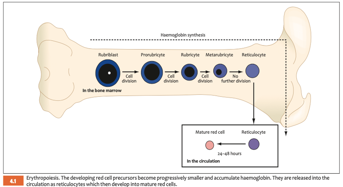
non specific RBC indicators common in regen anaemia (normal in some species low no)
-nucleated RBCs
-basophilic stippling
-howell jolly bodies (norm cats)
-heinz bodies (norm cats)
howell-jolly body
remnants of DNA
heinz bodies
denatured, damaged haemoglobin from oxidative damage
regenerative anaemia likely due to
haemorrhage or haemolysis
non regenerative anaemia
-decreased bone marrow production of RBCs
-This can be mild or severe, temporary/reversible or catastrophic
-chronic haemorrhage or haemolysis that was regen will eventually become non regen
pre regen anaemia
bone marrow takes 3-7 days to respond to anaemia so acute conditions may initially appear non regen
other clues of anaemia
Other abnormal RBCs?
What is the WBC count doing and what do they look like?→ systemic disease? anaemia of inflam disease?
Thrombocytopenia?→ see in haemorrhage bc lost/used too
large number of spherocytes with anaemia mean
immune mediated haemolysis
schistocytes with anaemia
intravascular red cell injury
keratocytes with anaemia
intravascular red cell injury
heinz bodies with anaemia
heinz body haemolysis, oxidant injury
dacrocytes (tear drop) with anaemia
-iron deficiency
-myelofibrosis
echinocytes with anaemia
-renal azotaemia
-chemo
acanthocytes with anaemia
-splenic neoplasia
-liver disease
acanthocytes
-irregular, blunt tipped spicules
-fragmentation injury
what causes acanthocytes
fragmentation injury
-DIC
-fragile RBCs
-liver disease
-abnormal vessels
how can we cause echinocytes
used edta
overfilled edta
echinocytes
-sharp or blunt projections, tend to be uniform and evenly spaced
causes of echinocytes
-artefact
-drug exposure e,f, furosemide
-electrolyte disturbance
-renal disease
-snake evomation
schistocytes
RBC fragments
causes of schistocytes
-mechanical injury often some level of vascular damage e.g. DIC, glomerulonephritis, vaculitis
spherocytes
-sphere shaped
-no central pallor
-smaller and denser
-MCV normal
moderate to severe spherocytosis suspect
IMHA
describe the smear

increased zone of central pallor
microcytic hypochromic anaemia
anisocytosis
poikilocytosis
mostly associated with fe deficiency

How would you characterise this anaemia?
nucleated rbc (LHS) (metarubricyte)
howell jolly body (RHS)
variety in size
bigger and lacking central pallor in dog →polychromatophil, macrocyte, reticulocyte
anisocytosis
regenerative
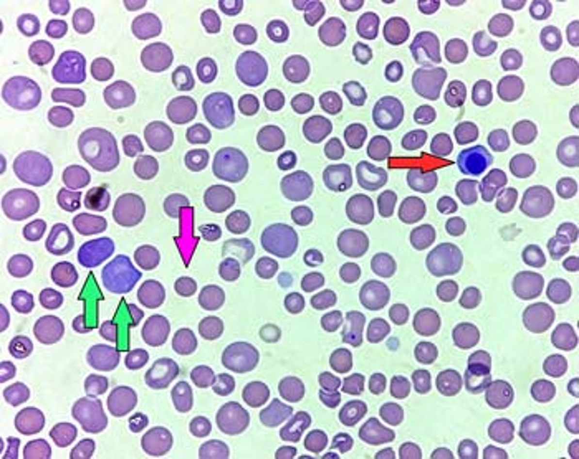
Blood smear from a dog with immune-mediated haemolytic anaemia (IMHA).
anisocytosis (variable size of RBCs)
polychromasia (presence of polychromatophils,
immature RBCs that are larger and more basophilic; green arrows).
A nucleated red blood cell is present (red arrow), consistent with red cell regeneration.
Spherocytes are present (small red cells lacking central pallor; pink arrow).
presence of schistocytes on a blood smear is most likely to indicate
imha
imtp
renal disease
intravascular rbc injury
infectious disease
intravascular rbc injury
severe renal disease too
which findings would most likely see in 3yo springer spaniel with imha
numerous echinocytes
microcytic hypochromic anaemia
numerus spherocytes
intracellular parasites in rbc
low rbc
numerus spherocytes
causes of regen anaemia
-blood loss
-haemolysis
non regen anaemia causes
suppression of erythropoiesis
-intra marrow or extra marrow
RBC loss causes- regen anaemia
haemorrhage due to trauma or coagulopathy
internal RBC losses causes
-GIT
-parasites
-pulmonary
-abdo cavity
external blood loss causes
-wounds
-epistaxis
-ectoparasites (young kittens with high flea burden)
RBC destruction- regen anaemia
haemolysis
causes of haemolysis
-toxicity
-infection
-immune mediated
-mechanical damage
-others
causes of reduced RBC production
-genetic
-immune mediated
-infectious/chronic disease/inflam
-nutritional deficiencies (iron deficiency anaemia)
-myelodysplasia
acute and spectacular blood loss
traumatic wounds,
blunt force (intracavitary bleeding),
haemorrhagic diarrhoea,
haematemesis,
epistaxis,
DIC…
mild and chronic blood loss and haemorrhage
-melaena
-chronic or multiple small wounds
-GI or blood borne parasitism
will blood loss cause an immediate drop in pcv
no→ all is non regenerative to start (comp mechs)
splenic rupture can be in hours tho
if blood loss prolonged, waht will result
iron deficiency
IMHA
-immune system dysfunction and loss of of tolerance (primary)
-antigen exposed due to cell damage
-similarity between self antigens and foreign antigens
-reaction to infectious agents bound to cell surface or reaction to non biologic Ag bound to cell surface

clinical signs of IMHA
-pyrexia
-pallor/jaundice
-CV abnormalties
-spherocytes
-leukocytosis
-thrombocytopaenia
-RBC parasites

what is intravascular haemolysis
= blood cells lyse in circulation
Serum will be pink/red, urine from red to brown due to haemoglobin
Example: extreme hypophosphataemia
can intravascular and intravascular haemolysis happen at the same time
In many conditions, either or both of these can happen (and at the same time!)
In IMHA, extravascular is more common
what is extravascular haemolysis
= blood cells lyse outside of circulation
Example: IMHA (macrophages in spleen attack RBCs with self Ag or intracellular infection)
what can both extravascular and intravascular haemolysis lead to
Both can lead to jaundice (haemoglobin breakdown → bilirubin)
Intravascular may be slower to develop jaundice as some haemoglobin will be excreted via kidneys
what is extra marrow non regen anaemia
influence from outside the bone marrow
what is intra marrow non regen anaemia
problem originates within the bone marrow
intra marrow non regen anaemia causes
-infection
-chronic damage
-neoplasia
-lack of raw materials (iron deficiency)
extra marrow non regen anaemia causes
-CKD→ bc stops producing EPO
-excess oestrogen (ferrets)
-some types of FeLV
what is most common form of non regen anaemia
Anaemia of inflammation (a.k.a. anaemia of chronic disease)
anaemia of inflammation presents on parameters (anaemia of chronic disease)
normocytic and normochromic
absolute (true) iron deficiency
-chronic haemorrhage (loss of iron rich Hb)
-can be regen or non regen
how does iron deficiency present on parameters
microcytic and hypochromic
chronic renal failure in non regen anaemia
EPO production reduced in advanced renal disease
what is PIMA
Precursor-targeted immune-mediated anaemia (PIMA)
non regen anaemia
Rare form of IMHA directed at RBC precursors in bone marrow
Diagnosis usually requires bone marrow aspirate
neoplasia causes of non regen anaemia (dont mem)
-haemorrhage
-haemolysis
-decreased or ineffective erythropoiesis
endocrinopathies causes of non regen anaemia (dont mem)
-hypothyroidism
-diabetes mellitus
-addisons
myelodysplastic syndrome of non regen anaemia (dont mem)
erythropoiesis ineffective
liver disease causes of non regen anaemia (dont mem)
-anaemia of inflam
-abnormal nutrient metabolism
-defective iron handling
-dogs with portosystemic shunt often microcytic even if not anaemic
infection of erythropoietic cells causes of non regen anaemia (dont mem)
-FeLV can infect precursors so direct suppression of early stage erythropoeitic cells
-anaemia of inflam
-immune mediated haemolysis
toxic insult to bone marrow causing non regen anaemia (dont mem)
chemo
space occupying disease in bone marrow in non regen anaemia (dont mem)
fibrosis and neoplasia
nutritional deficiencies causing non regen anaemia (dont mem)
-copper
-folate
-cobalamin
diagnostic tests for anaemia
Complete blood count (haematology)
Manual PCV
Blood smear
more specific tests for anaemia
Saline agglutination test/Coombs test (IMHA)
Infectious agent testing (e.g. 4Dx blood parasite screen, PCR)
Imaging (neoplasia)
Bone marrow aspirate (neoplasia, PIMA)

presented with a 5yo lab with anaemia (PCV 21%) you perform a blood smear what sus is the cause of anaemia
IMHA → spherocytes
which of following findings are consistent with acutely bleeding splenic hemangiosarcoma
pcv 25% numerus schistocytes and acanthocytes
pcv 40 polychromasia and many reticulocytes
pcv 15 numerus spherocytes
pcv 12 with heinz bodies in the rbc
pcv 25% numerus schistocytes and acanthocytes → fragmentation damage
clinical signs of anaemia
-pallor
-icterus
-tachycardia
-tachypnoea, dyspnoea
-pyrexia if infectious or IMHA
-weak, thready, bounding or absent peripheral pulses
-cold extremities
collapse coma death
traumatic wound
GI blood loss signs
-haematemesis (spitting up blood)
-melaena
-haemorrhagic diarrhoea
melaena
black, tarry stools
if anaemia/ o2 problem during anaethesia what pulses should you feel
pedal pulses → extremity
signs of coagulopathy
-internal bleeding
-epistaxis
-bleeding wounds
-sites of blood draw/cannulation
internal bleeding signs
-dyspnoea/tachypnoea
-abdo distension
-fluid thrull
-bruising
acute vs chronic anaemia
-more severe signs than chronic anaemia of same PCV
-compensatory mechanisms in chronic vs. sudden loss of circulating volume/oxygen-carrying capacity in acute disease
-Hypovolaemia, trauma, pain, pyrexia in acute
severe anaemia signs
-CV signs, lethargy/weakness