ANSC Lec. 13 Heart conduction, blood vessels, blood pressure
 Call Kai
 Learn
 Practice Test
 Spaced Repetition
 Match
 Flashcards
 Knowt Play
1/26

There's no tags or description
Looks like no tags are added yet.
27 Terms
what is Sinoatrial (SA) Node responsible for? what side of heart is it on?
= primary pacemaker cell cluster of the heart that spontaneously depolarizes to set the heart rate (~70 bpm).
General Location: Right atrium wall.

what is Atrioventricular (AV) Node responsible for? what side of heart is it on? what is AV Node Delay
=electrical gateway tissue that routes signals down to the ventricles and delays them slightly so the atria can finish contracting.
General Location: At the junction floor between the right atrium and right ventricle
NODE DELAY: deliberate slowing down of the electrical impulse to let the atria contract and empty completely before ventricles squeeze.
General Location: Within the AV Node cells.

what is Atrioventricular (AV) Bundle of His responsible for? what side of heart is it on? What about bundle branches?
BUNDLE OF HIS: sole electrical connection link passing signals between the atria and the ventricles through the fibrous cardiac skeleton.
General Location: Upper region of the interventricular septum.
Analogy: An insulated extension cord running through a thick wall to carry power into the next room.
BUNDLE BRANCHES: Two split pathways stemming off the AV bundle that guide electrical currents rapidly down toward the heart's apex.
General Location: Running down the left and right sides of the interventricular septum.
Analogy: A fork in the road splitti

what is Purkinje Fibers responsible for? what side of heart is it on?
Large, specialized conduction cells that distribute electrical signals upward into the ventricular walls for upward contraction.
General Location: Spreading throughout the ventricular apex and walls.
Analogy: Fingers of a glove sending warmth (electricity) to every individual finger simultaneously.

what is cardiac cycle: diastole, systole, isovolumic Ventricular Contraction, Ventricular Ejection, Isovolumic Ventricular Relaxation
= complete sequence of mechanical events spanning exactly one full heartbeat (comprising contraction and relaxation).
Diastole
What & Function: The phase of the cardiac cycle where the heart muscle relaxes to let its chambers fill back up with blood.
General Location: Atria and ventricles.
Analogy: Releasing a squish toy under water so it pulls fluid back inside its chamber.
Systole
What & Function: The active phase of the cycle where the cardiac muscle contracts to aggressively pump blood forward.
General Location: Atria and ventricles.
Analogy: Clenching your fist around a water balloon to force the liquid out.
Isovolumic Ventricular Contraction
What & Function: The early stage of ventricular contraction where all valves are shut tight; pressure skyrockets, but blood volume does not change.
General Location: Ventricles.
Ventricular Ejection
What & Function: The phase where high ventricular pressure forces semilunar valves wide open, shooting blood out into major arteries.
General Location: Ventricle chambers outflowing to the Aorta / Pulmonary Trunk.
Isovolumic Ventricular Relaxation
What & Function: Early diastole where ventricular pressure falls rapidly as muscles relax, all valves close, and volume stays static.
General Location: Ventricles.

what is EDV VS ESV and what does it represent?
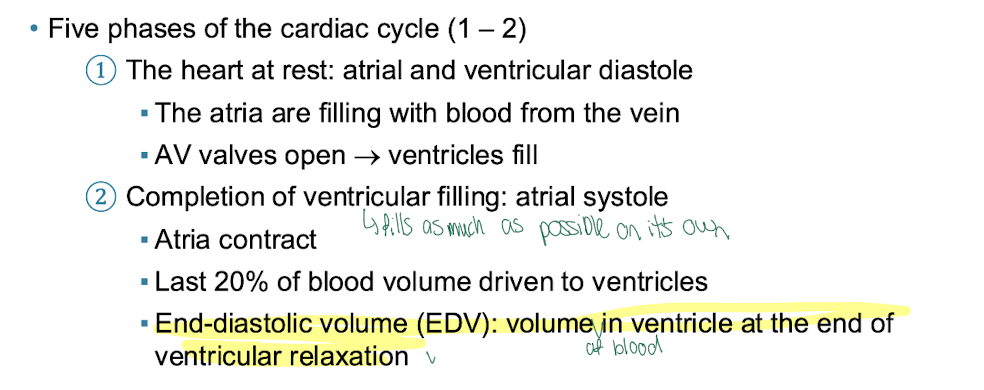
End-Diastolic Volume (EDV)
What & Function: The maximum total volume of blood contained within a ventricle right at the very end of its relaxation/filling phase.
General Location: Inside the left and right ventricles.
End-Systolic Volume (ESV)
What & Function: The leftover volume of blood remaining inside a ventricle immediately after it finishes contracting.
General Location: Inside the ventricles.

what is Cardiac Output (CO)
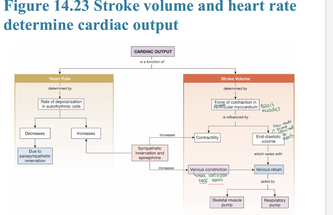
= The total volume of blood pumped out by a single ventricle per minute = (HR x Stroke Volume)
General Location: Systemic output from the heart.

how is stroke volume calculated?

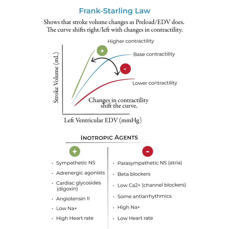
what is Frank-Starling Law of the Heart
intrinsic law stating that the heart will automatically contract harder if it is stretched more by extra incoming blood volume
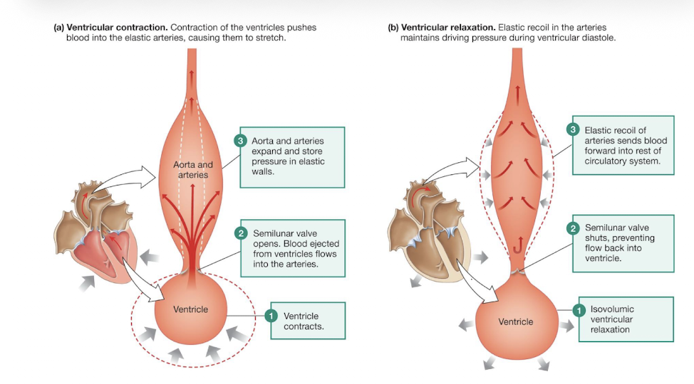
what is perfusion and elastic recoil? what does pressure reservoir refer to?
Perfusion
What & Function: The steady, physiological passage of blood through the circulatory system to successfully deliver nutrients/oxygen to tissues.
General Location: Capillary networks within all living organs.
Elastic Recoil
What & Function: The passive snap-back property of major arteries that keeps blood squeezing forward even while the heart is resting.
General Location: Aorta and large systemic arteries.
Analogy: Stretching a thick rubber fitness band; when your muscles relax, the band snaps back to its original size.
PRESSURE RESERVOIR= functional title for arteries because their elastic walls expand to store pressure during systole and release it during diastole.
General Location: Systemic Arteries.
whatare Arterioles

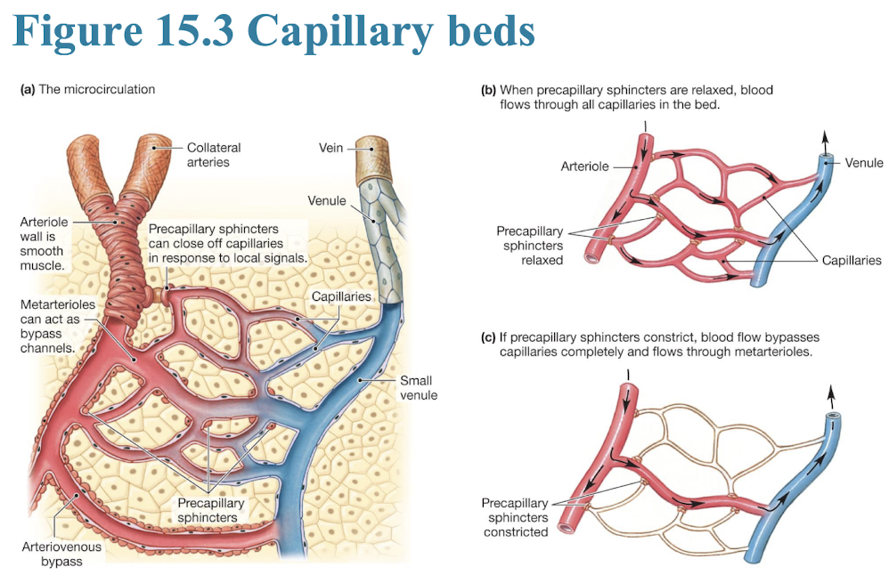
= Small, highly muscular branching vessels that act as the main sites of adjustable resistance to blood flow.
what are arteries
known as pressure reservoir, their elastic walls expand to store pressure during systole and release it during diastole.
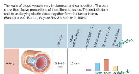
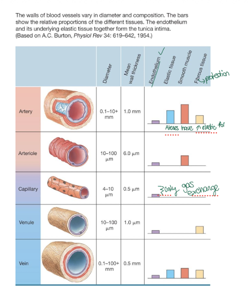
veins vs venules vs capillaries, arterioles, vs artery tissue makeup comparison
which type of blood vessel holds most of bodies blood?
veins, highly distensible walls allow them to safely hold the majority of the body's blood (~60%).
what is vascular smooth muscle, what is Muscle Tone
Muscle arranged in circular layers around vessels that changes the inner vessel diameter via contraction or relaxation.
General Location: Inside walls of all blood vessels except true capillaries.
MUSCLE TONE: baseline state of resting partial contraction maintained by blood vessels so they can adjust in either direction.
General Location: Vascular smooth muscle walls.
systolic vs diastolic pressure?
Systolic Pressure= highest peak blood pressure reached in systemic arteries during active ventricular contraction (~120 mmHg).
General Location: Large systemic arteries (e.g., Brachial Artery).
Diastolic Pressure= lowest baseline blood pressure remaining in arteries while the ventricles are relaxing (~80 mmHg).
General Location: Large systemic arteries.

what is pulse, pulse pressure
pulse: the palpable, traveling pressure wave created by the rapid ejection of blood from the left ventricle into the aorta.
General Location: Accessible superficial arteries (Radial, Carotid).
Pulse Pressure= calculated numerical strength or amplitude of the pressure wave(Systolic Pressure) - (Diastolic Pressure).
General Location:
Arterial systemic circulation branches.
what is venous return and where does it come from?
crucial volume of blood flowing back from the body's veins into the right atrium per minute.
General Location: Venae Cavae entering the right side of the heart.
what determines stroke volume vs cardiac output?
arteries vs arterioles vs metarterioles?

what are pericytes and walls of capilaries and key compnents ?

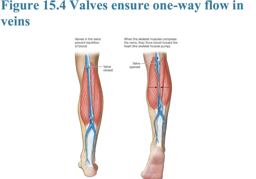
what is Skeletal Muscle Pump, where is it and where does it pump blood? wh
= peripheral vein compression caused by surrounding skeletal muscle movement that pushes venous blood upward past one-way valves.
General Location: Deep veins embedded in limbs (especially calves).
Analogy: Squeezing the bottom of a tube of toothpaste to force the paste out the top opening.
what is Respiratory Pump?
= Pressure changes created within the thorax and abdomen during breathing that draw venous blood upward toward the heart.
General Location: Thoracic and abdominal cavities.
Analogy: Using a drinking straw; creating low pressure at the top pulls liquid upward.
what is mean arterial pressure MAP, what is Myogenic Autoregulation
The calculated average driving pressure pushing blood through the body (Diastolic+ (1/3)*Pulse Pressure).
General Location: Entire systemic arterial tree.
Myogenic Autoregulation
What & Function: An intrinsic vascular feedback loop where smooth muscle contracts automatically when stretched by high pressure to keep blood flow stable.
General Location: Arterioles supplying organs (like kidneys or brain).
how is cardiovascular system controlled differently in autonomic system: PARASYMPA vs SYMPA, and where does this controll occur? what is tonic control
in medulla!


what are 5 phases of cardiac cycle


what are the 4 parts of electrocardiogram and what do they represent: reorder them: QRS complex, T wave , P wave, P-R segment. How many peaks vs waves?

