Anatomy Lab Practical 2: Bone
 Call Kai
 Learn
 Practice Test
 Spaced Repetition
 Match
 Flashcards
 Knowt Play
1/235

There's no tags or description
Looks like no tags are added yet.
236 Terms
Hyaline cartilage in bone features
Firm matrix (amorphous), chondrocytes, and collagen fibers
Hyaline cartilage in bone function
Resists compression
Hyaline cartilage in bone location
Covers ends of bones at moveable joints, costal (rib) cartilage
Where does growth in long bones occur?
Epiphyseal plates
4 zones for growth
Proliferation, hypertrophic, calcification, and ossification
Bone shapes
Long bones, short bones, flat bones, and irregular bones
Long bone examples
tibia, fibula, ula, radius, humerus, femur

Short bone examples
carpals, tarsals, metacarpals, metatarsals, phalanges

Flat bone examples
scapula, sternum

Irregular bone examples
vertebra, pelvis

Proximal
Towards point of attachment of a limb to the trunk
Distal
Away from point of attachment of a limb to the trunk
Medial
Toward midline in horizontal direction
lateral
Away from midline in horizontal direction
Cranial / cephalic
Toward the head
Caudal
Toward the tail (coccyx)
Anterior / ventral
The front of body
Posterior /dorsal
The back of the body
Axial skeleton
Cranium, facial, thoracic cage, and vertebral column bones

Shapes of bones in axial skeleton
Flat (cranium, sternum, and ribs) and irregular ( bones in vertebrae)
Bone markings
Projections that are sites of muscle and ligament attachment
Bone marking: tuberosity
Large rounded projection; may be roughened
Bone marking: crest
Narrow ridge of bone; usually prominent
Bone marking: trochanter
Very large, blunt, irregularly shaped process (only examples are in femur)
Bone marking: line
Narrow ridge of bone; less prominent than a crest
Bone marking: tubercle
Small rounded projection or process
Bone marking: epicondyle
Raised area on or above a condyle
Bone marking: spine
Sharp, slender, often pointed projection
Bone marking: process
Bony prominence
Bone marking: head
Bony expansion carried on a narrow neck
Bone marking: facet
Smooth, nearly flat particular surface
Bone marking: condyle
Rounded articular projection
Bone marking: ramus
Armlike bar of bone
Depressions and openings difference
depressions: indentations that accommodate other structures
Openings: passages through bones for nerves and blood vessels
Fissure
Narrow slit like opening for passage of vessels and nerves
Foreman
Round or oval opening through a bone for passage of vessels and nerves
Notch
Indentation at the edge of a structure for passage of vessels and nerves
Meatus
Canal-like passageway
Sinus
Bone cavity, filled with air and lined with mucous membrane
Fossa
Shallow basinlike depression in a bone, often serving as an articulate surface
Frontal bone
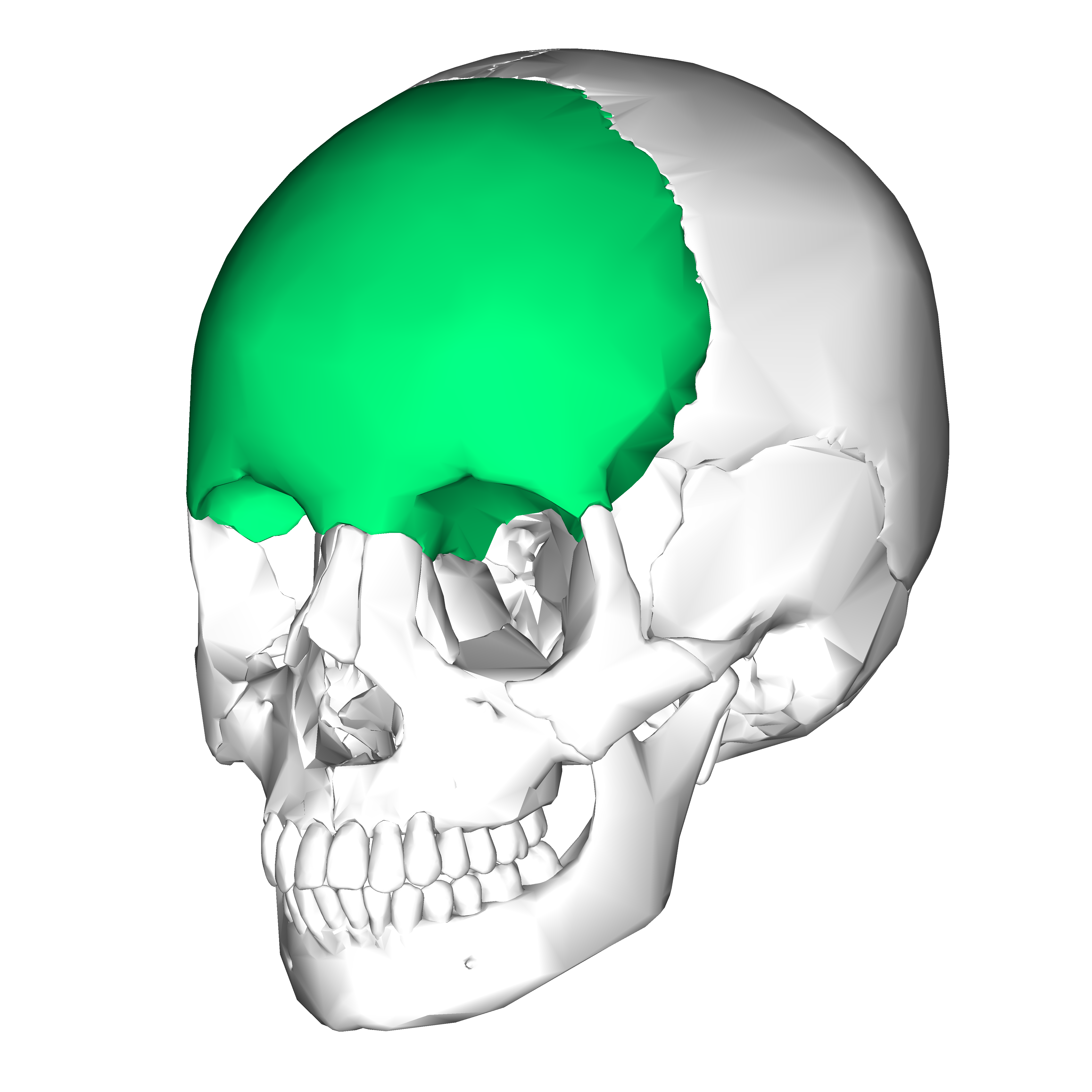
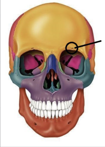
Supraorbital foreamen
Parietal bone

Temporal bone

Squamous suture

zygomatic process
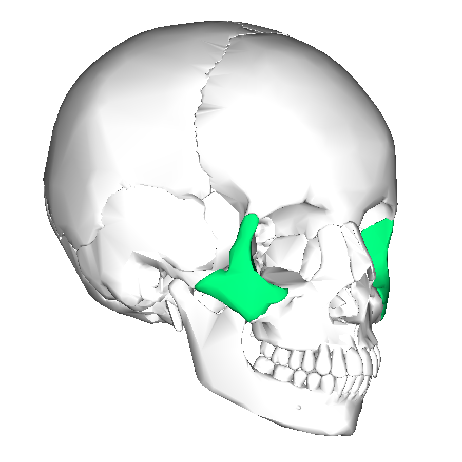
external acoustic meatus

Occipital bone

foreamen magnum

Sphenoid bone

optic canals

Ethmoid bone

Crista galli

cribiform plate

Coronal suture

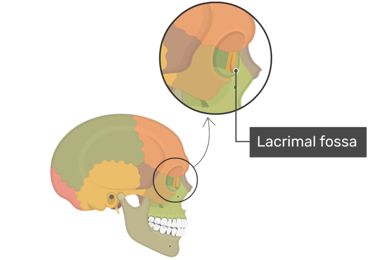
Lacrimal bone
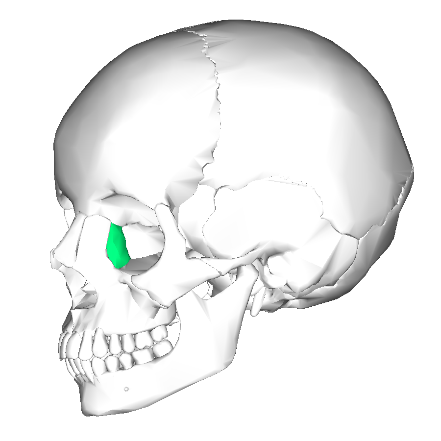
Lacrimal fossa
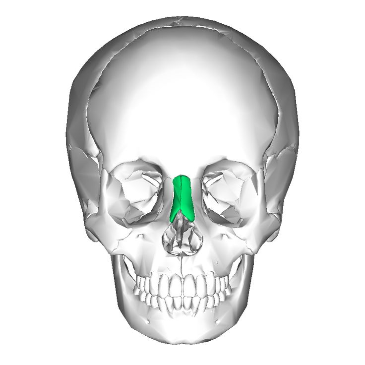
Nasal bone
Maxilla
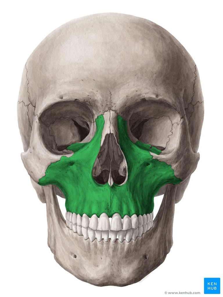
Alveolar processes

Mental foreamen

Coronoid process

Mandibular notch

Mandibular ramus

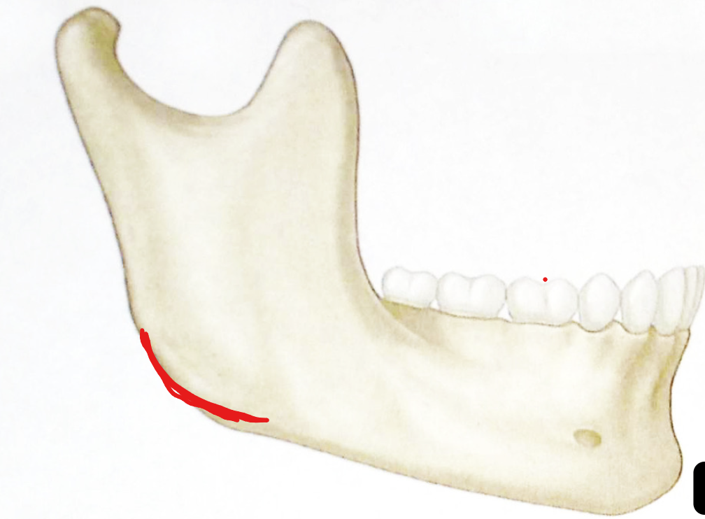
Mandibular angle
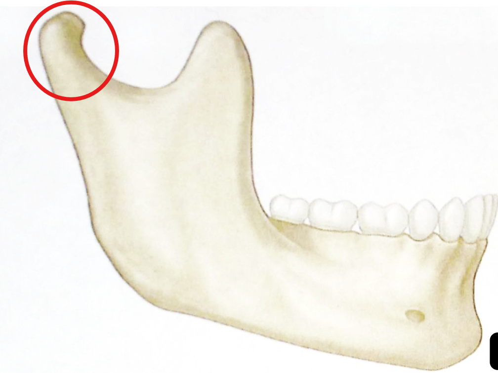
Condylar process
Styoid process

Mastoid process

Occipitomastoid suture

lambdoidal suture

Glabella

Supraorbital margin

Superior orbital fissure

Inferior orbital fissure

Middle nasal concha ( ethmoid bone)

Perpendicular plate (ethmoid bone)

Inferior nasal concha

Vomer

Sagital suture

Bregma
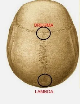
Sphenofrontal suture

Sphenoparietal suture

Sphenosquamous suture

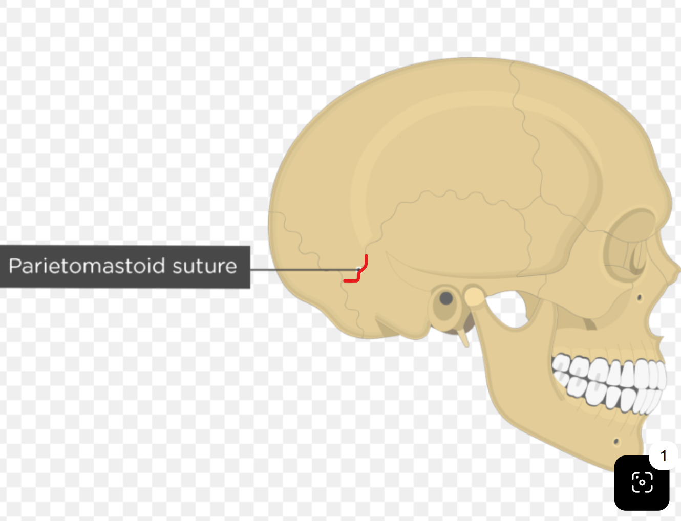
Parietomastoid suture
Hyoid bone
Located in throat above larynx
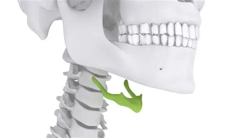
Greater horn (hyoid)
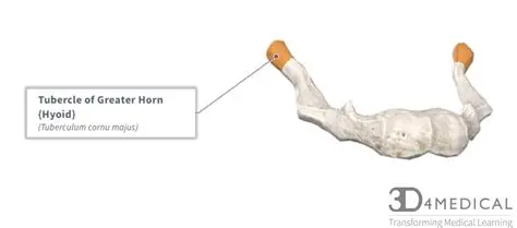
Lesser horn (hyoid)

Body of hyoid

Hyoid function
Serves as a point of attachment for many tongue and neck muscles
fontanelle
Holes in fetal skull

Posterior fontanelle

Ossification center ( in fetal skull)

Sphenoidal fontanelle

Anterior fontanelle

Mastoid fontanelle

Fetal skull V.S. Adult skull
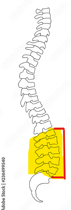
5 curvatures in vertebral column
Cervical 2. Thoracic 3. Lumbar 4. Sacral 5. Coccyx

Cervical curvature (concave)
7 vertebrae (c1-c7)

Thoracic curvature (convex)
12 vertebrae (T1- T12)

Lumbar curvature (concave)
5 vertebrae ( L1- L5)