(A&P 1) Lab 6: Physiology of the Muscular System
 Call Kai
 Learn
 Practice Test
 Spaced Repetition
 Match
 Flashcards
 Knowt Play
1/79

There's no tags or description
Looks like no tags are added yet.
80 Terms
The 6 main functions of the Muscular System
1. Produce skeletal movement
2. Maintain body position
3. Support soft tissues
4. Guard openings
5. Maintain body temperature
6. Store nutrient reserves
What is excitability in skeletal muscle?
The ability to respond to a stimulus by changing electrical membrane potential.
What is contractility in skeletal muscle?
It is exhibited when filaments slide past each other, which enables the muscle to cause movement.
What is elasticity in skeletal muscle?
The ability to return to its original length following a lengthening or shortening.
How is skeletal muscle defined?
Defined as voluntary (subject to subconscious control) striated muscle that is usually attached to one or more bones.
What are striations?
Alternating light and dark transverse bands visible in the muscle tissue. (In Skeletal and Cardiac muscle)
What is another term for a skeletal muscle cell?
Myofiber
Describe the cellular characteristics of skeletal muscle.
They are long, cylindrical, multinucleate cells with obvious striations.
What are the primary functions of skeletal muscle?
Voluntary movement
locomotion
manipulation of the environment
facial expression.
Where is skeletal muscle located in the body?
In skeletal muscles attached to bones, or occasionally to the skin

Name the three layers of connective tissue in skeletal muscle, from outermost to innermost.
Epimysium, Perimysium (which wraps fascicles), and Endomysium (which is between fibers).

Label the structures listed A-L in the image
Check correct answers.

Epimysium
Surrounds the entire muscle and connect to tendons.

Perimysium
surrounds bundles of muscle fibers separating them into fascicles

Endomysium
surrounds each muscle fiber

Sarcolemma
is the plasma membrane of a muscle fiber

Sarcoplasm
The cytoplasm which is occupied mainly bylong protein cords called myofibrils

What are the three main contents of the sarcoplasm and their functions?
Myofibrils: Long protein cords.
Glycogen: Stored energy.
Myoglobin: Red pigment that stores oxygen for muscular activity.
What is the specific name for the smooth endoplasmic reticulum in a muscle fiber?
sarcoplasmic reticulum (SR)

Label the structures listed A-Q in the image
Check Correct answers.

What are transverse (T) tubules?
Tubular infoldings of the sarcolemma

Label the structures A-F.
Check Correct Answers.

What is the primary function of the sarcoplasmic reticulum (SR)?
It acts as a reservoir of calcium ions, which activate the muscle contraction process.

What three structures make up a triad in a muscle fiber?
One T tubule and two terminal cisterns of the SR.

What is a myofibril?
A bundle of parallel protein calles myofilaments.

What protein makes up thick filaments?
Myosin

What protein makes up thin filaments
Actin
What is the function of the active site on an actin molecule?
allows actin to bind to a myosin molecule.
What is the function of tropomyosin during muscle relaxation?
blocks the active sites on actin, preventing myosin from binding to them.
What is troponin?
calcium-binding protein attached to tropomyosin molecules

Label Structures A-O (Skeletal muscle fiber + Myofibril)
Check for correct answers.

Label structures P-V as well as the rest of the blank labels.
Check for correct answers.

What protein makes up Elastic filaments?
Titin

What is the function of titin (elastic filaments)?
It anchors thick filaments to the Z disc at one end and the M line at the other.
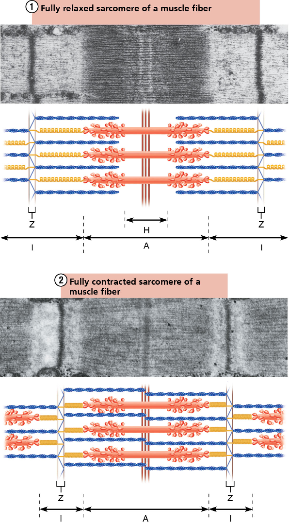
What is a helpful mnemonic for remembering the appearance of A bands and I bands?
Think "dArk" for A bands and "lIght" for I bands.

What filaments make up the dark A band?
Thick Myosin Filaments

What filaments make up the light I band?
Thin Actin Filaments

H Band
The lighter region in the middle of the A band where actin filaments do not reach.

Sarcomere
the functional contractile unit of the muscle fiber, goes from one Z disc to the next
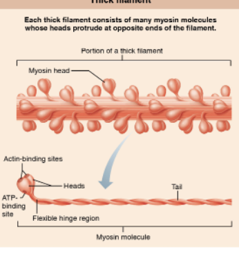
Thick Filaments (Myosin)
Made of multiple myosin molecules with protruding heads at opposite ends. The center of the sarcomere lacks these heads.
Myosin Heads
Contain specific binding sites for both Actin and ATP.

Thin Filaments (Actin)
Consist of two twisted strands of actin subunits, along with the regulatory proteins
troponin
tropomyosin.
Actin Subunits
Each subunit features a myosin-binding site.

What is a Motor Neuron?
specialized nerve cells that transmit signals from the brain and spinal cord to muscles and glands, allowing both voluntary and involuntary body movements
How do the motor neuron axons reach the muscle?
They extend from the spinal cord and travel together through a nerve.
What structures are formed where the branching axon terminals connect with the muscle fibers?
Neuromuscular Junctions
What makes up a motor unit (e.g., Motor unit 1 or Motor unit 2) as shown in the diagram?A
A single motor neuron and all the scattered muscle fibers its axon terminals connect to.
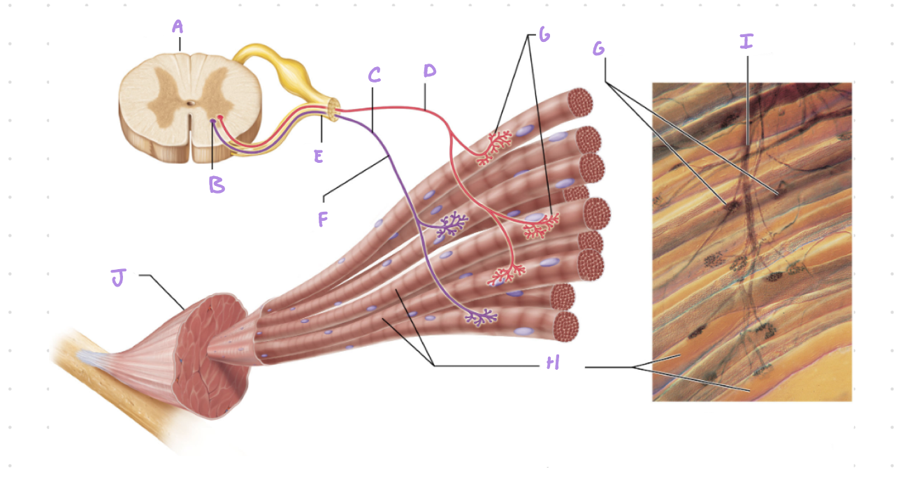
Label all structures
Check for correct answers.
Somatic Motor Neurons
The specific nerve cells that serve skeletal muscles; their cell bodies are located in the brainstem and spinal cord.
Somatic Motor Fibers
The axons of these motor neurons that carry signals out to the skeletal muscles.
Motor Unit
Defined as one single nerve fiber combined with all the individual muscle fibers it innervates.
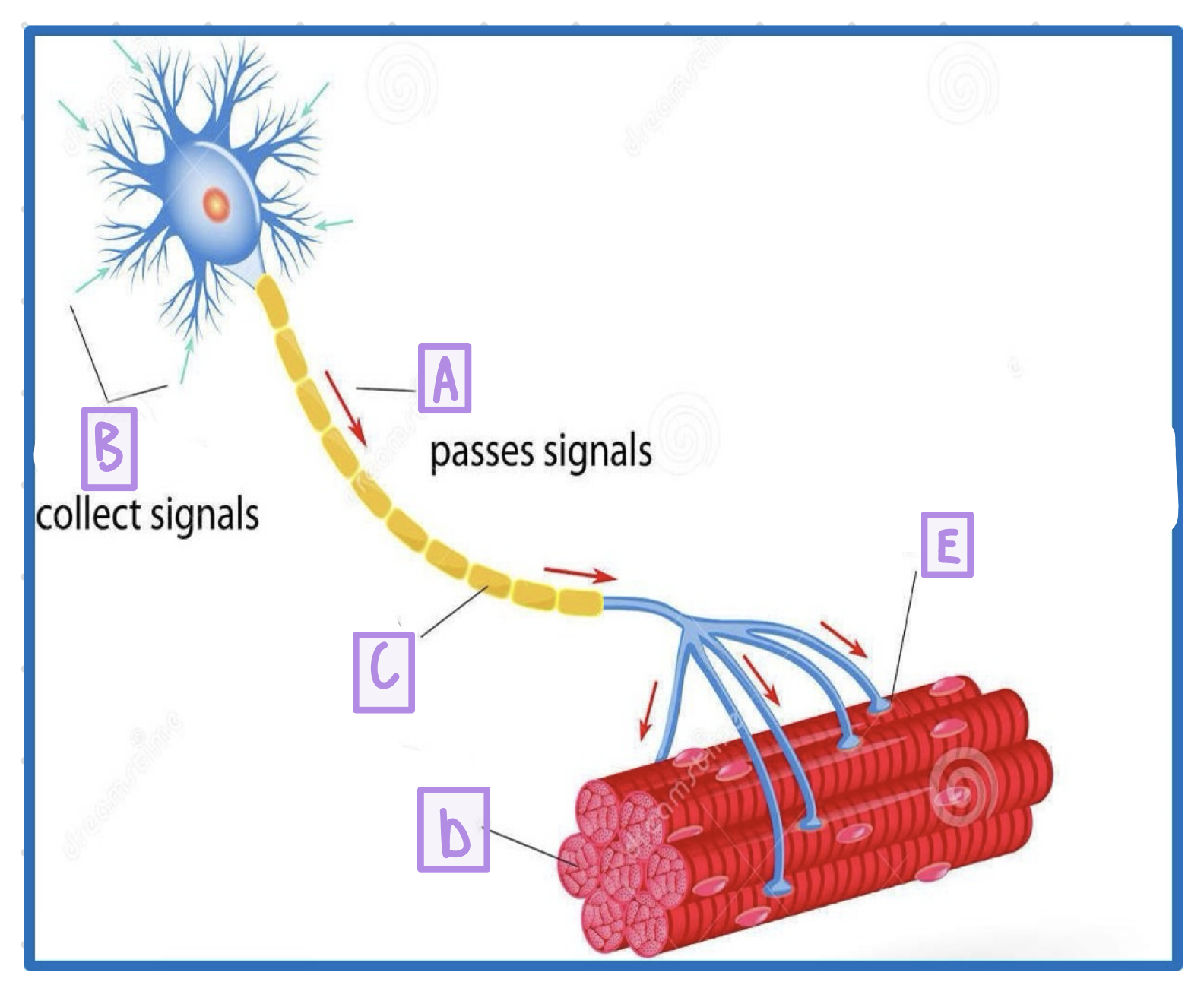
Label A-E
Check for correct answers.

A neuromuscular junction (NMJ) or motor end plate
two terms for the specific type of synapse where the target cell is a muscle fiber

Synapse
The point where a nerve fiber meets its target

What is the bulbous swelling at the end of a nerve fiber at a synapse called?
The synaptic knob

What is the narrow space separating the synaptic knob from the target cell called?
The synaptic cleft

What structures within the synaptic knob contain neurotransmitters?
Synaptic vesicles

What specific chemical neurotransmitter fills the synaptic vesicles at the neuromuscular junction?
Acetylcholine

Label 1-3
Check correct answers,
What causes synaptic vesicles to undergo exocytosis at the neuromuscular junction?
An electrical signal (nerve impulse) traveling down the nerve fiber.
What is released into the synaptic cleft when synaptic vesicles undergo exocytosis?
ACh (Acetylcholine)
What is the specific function of ACh at the neuromuscular junction?
It acts as a chemical messenger passing the signal from the nerve cell to the muscle cell.

Label 1-7
Check for correct answers.

What initiates the excitation process at the neuromuscular junction?
An action potential arriving at the terminal

After the action potential arrives, what neurotransmitter is released and where does it bind?
ACh is released and binds to receptors on the sarcolemma.

What happens to the sarcolemma immediately after ACh binds to its receptors?
It’s ions permeability changes

What is the local change in membrane (mb) voltage called during excitation?
Local deporilization, also known as the end plate potential.

What is the ultimate result of the local depolarization (end plate potential)?
It ignites an action potential in the sarcolemma

During the generation and propagation of the action potential, what is the status of the ion channels?
The Na+ channel is open and the K+ channel is closed.

During repolarization, what is the status of the ion channels?
The Na+ channel is closed and the K+ channel is open.

What are the 7 key stages of Excitation-Contraction Coupling?
ACh release: ACh binds to receptors, triggering an Action Potential (AP).
Propagation: The AP travels across the sarcolemma and down T-tubules.
Calcium release: The AP causes the Sarcoplasmic Reticulum (SR) to release Ca2+.
Sites exposed: Ca2+ binds to troponin, moving tropomyosin to expose myosin-binding sites.
Contraction: Myosin cross-bridges form and pull the thin filaments (using ATP).
Calcium removal: Ca2+ is actively pumped back into the SR.
Relaxation: Tropomyosin re-blocks the binding sites, and the muscle relaxes.
(ALWAYS PACK CALCIUM SO CELLS CAN RETURN RELAXED)

What happens to the specific regions of a sarcomere when a skeletal muscle fully contracts?
Z discs: Move closer together.
I bands: Narrow significantly.
H zone: Disappears.
A band: Width remains unchanged.
What are the sequential steps of muscle fiber relaxation?
STOP EVERY CRAZY TWITCHING TISSUE
Stop ➔ Signals stop (nerve stops releasing ACh)
Every ➔ Esterase (ACh Esterase breaks down remaining ACh)
Crazy ➔ Calsequestrin (Ca2+ is pumped back into the SR and stored by calsequestrin)
Twitching ➔ Troponin (Calcium dissociates from troponin)
Tissue ➔ Tropomyosin (Moves back to block actin, stopping the muscle tension)

What is denervation atrophy?
The shrinkage of a paralyzed muscle that occurs if its nerve connection is not restored.

Rigor mortis
uThe hardening of the muscles and stiffening of the body that begins 3 to 4 hours after death
Deteriorating membranes (sarcoplasmic reticulum and sarcolemma) leak calcium into the cytosol.
Because a dead body produces no ATP, myosin cannot release actin, leaving the filaments rigidly cross-linked.

What is a muscle twitch and what are the three distinct phases?
a motor unit’s response to a single action potential of its motor neuron
Latent Period
Period of Muscle Contraction
Period of Relaxation
What is one primary way to achieve a graded muscle response?
By increasing the firing rate of neurons.

What is temporal summation?
When muscles maintain a prolonged contraction by adding numerous individual contractions back-to-back
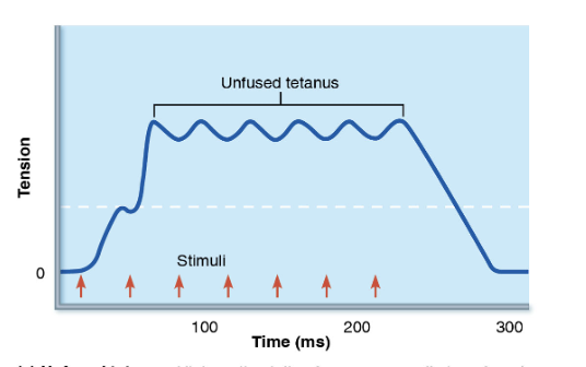
What happens during incomplete (unfused) tetanus?
Each new stimulus initiates a contraction when the muscle has only partly relaxed from the previous one.

What happens during complete (fused) tetanus?
Stimuli are repeated so rapidly that no decrease in tension or relaxation can be detected between them.