Other Lab Stuff
 Call Kai
 Learn
 Practice Test
 Spaced Repetition
 Match
 Flashcards
 Knowt Play
1/209

Earn XP
Description and Tags
G5, G6, G7
210 Terms

what kind of plate is it?
Blood Agar Plates
what does BAP stand for
blood agar plates
blood plates are made with 5% — —
sheep blood
what is the use of BAP’s
Can help to differentiate different organisms based on the organism's ability to create hemolysis (clearness) around the colony.
BAP aka
a differential medium.

what kind if plate is it
Tryptic Soy Agar
define Tryptic Soy Agar
A non-selective, nutritional medium that has the required nutrients to grow a variety of organisms.
Selective mediums
plates that contain specific materials and/or antibiotics to encourage or discourage certain colony growth
how to collect a liquid/broth sample (4)
Remove the lid of the container without putting the lid down, keep it held in your dominant hand and the container in your non-dominant hand
Flame the rim of the container for 1-2 seconds
Submerge your loop tip into the sample without touching the lid of the container
Loop should have a bubble in it (think bubbles you buy in the store with the wand), be careful not to break the bubble
how to collect a solid/plated sample (3)
Remove the lid of the container without putting the lid down
keep the lid held in your dominant hand and the container in your non-dominant hand
Gently touch the loop to the colony of bacteria or object to obtain a small amount of sample, careful not to disrupt the plating contents
how to streak a plate (6)
Using inoculating loop, transfer culture or specimen.
Sterilize loop in between inoculations by holding it in a Bunsen burner flame until hot.
Sample should be spread in a back and forth pattern that is separated into 4 different quadrants
Once you are finished with one quadrant, you will reflame your loop to sterilize it and then starting from where you left off with your previous quadrant you will start the same back and forth pattern with the next quadrant. This ensures you are diluting the sample as you move throughout your 4 quadrants and are also creating 4 separate quadrants
You will then place your plate back onto the lid inverted (upside down) and place in the incubator. This is to prevent condensation from affecting the colonies
Check growth every 24-48 hours and once growth is observed a macromorphology of the colonies can be done

what bacterial form
punctiform

what bacterial form
circular

what bacterial form
filamentous

what bacterial form
irregular

what bacterial form
rhizoid

what bacterial form (2)
spindle (lens)

what bacterial elevation
flat

what bacterial elevation
raised

what bacterial elevation
convex

what bacterial elevation
pulvinate

what bacterial elevation
umbonate

what bacterial margin (2)
entire (even)

what bacterial margin
undulate (wavy)

what bacterial margin
filamentous

what bacterial margin (2)
lobate (lobes)

what bacterial margin (2)
Erose (serrated)

what bacterial margin
curled
gram positive = — (color)
purple
gram negative = — (color)
pink

this is gram —
positive

this is gram —
negative

what bacterial arrangements
coccus

what bacterial arrangements
diplococcus

what bacterial arrangements
steptococcus

what bacterial arrangements
tetrad

what bacterial arrangements
staphylococcus

what bacterial arrangements
sarcina
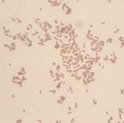
what bacterial shape (2)
rods/bacilli

what bacterial shape
cocci

what bacterial shape
vibrio
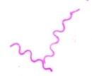
what bacterial shape
Spirillum / Spirochete

what bacterial shape
flagella
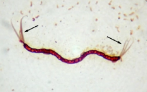
what bacterial shape
flagella

what bacterial shape
flagella

what bacterial shape
pili
how to Prepare slide with bacteria (8)
On the slide, with a grease pencil draw a circle the size of a dime on the bottom side.
Then turn it over and place a drop of saline in the center of the slide, the size of a dime
Touch a colony from the agar plate with an inoculating loop. Place the bacteria at the edge of the saline drop and gently mix.
Do not mix too hard.
Mixture should look creamy and not too thick or it will take too long to dry
Label slide with your initials and date (use a pencil)
Allow slide to air dry till smear looks somewhat dry
Pass slide through flame two to three times specimen side up. Do not overheat the slide.
what is the gram stain, stains and order (4)
crystal violet
iodine
decolorization
safranin
How to perform the catalase test
Using a sterile inoculating loop, touch an isolated colony of the bacteria and smear it onto the glass slide in an even layer.
Add two to three drops of 3% hydrogen peroxide onto each bacterial smear. Do not mix the sample
Watch for a bubbling effect. Bubbles indicate a catalase-positive result (such as Staphylococcus), while no bubbles indicate a catalase-negative result (such as Streptococcus)
bubbles in a catalase test indicates
catalase positive
no bubbles in a catalase test indicates
catalase-negative
culture answers the question : …
what kind of bacteria are we dealing with?
sensitivity answers the question : …
what antimicrobial will best get rid of this infection?
— of amino acids produces ammonia
Deamination
Ammonia is toxic and needs to be converted to — to to be excreted as waste
urea
Urea filtered by the —
kidneys
— - —% of urea reabsorbed to help create renal medullary concentration gradient
40 - 60 %
define Azotemia
Increased levels of nitrogenous waste products in the blood
define Uremia
Greatly increased BUN/Creatinine
clinical signs of uremia (5)
anorexia
nausea
vomiting
depression
ulceration of GIT mucosa
— — is the Most common reason for azotemia
pre renal
Pre-renal Azotemia Causes (6)
Decreased GFR
Increased protein catabolism
High protein diets
Electrolyte disturbance
acid-base disturbance
High USG
decreased GFR causes (4)
Decreased perfusion of the kidney
Shock
dehydration
CV disease
Increased protein catabolism causes (7)
Necrosis
starvation
infection
fever
corticosteroid admin
BUN increases
CREA normal
post renal causes (4)
Increased BUN
increased Creatinine
Animal unable to void normal amounts of urine
Urine SG – isosthenuric
Electrolyte abnormalities!
define isosthenuric
USG is the same as protein free plasma, indicates damage to the kidneys
protein malnutrition is caused by
low BUN
define protein malnutrition (2)
Severely restricted diet in protein
Disease preventing amino acid absorption
— is not affected by protein intake
Creatinine
what organ produces urea
liver
what are some abnormalities caused from increased levels (3)
Protein Malnutrition
Hepatic Disease
Decreased Muscle Mass
Ammonia also formed in GIT by bacteria …
acting on dietary proteins
creatine is produced in and released from — —
muscle cells
creatine is not reabsorbed in the —
kidneys
what does SDMA stand for
Symmetric dimethylarginine
what are the normal levels of SDMA
<15 ug/dL
what does BUN stand for
Blood Urea Nitrogen
what are the units of measurement for BUN (2) (SI unit and conventional unit)
SI Units (mmol/L)
Conventional units (mg/dL)
Carnivores tend to have — BUN normal ranges than herbivores
higher
why do carnivores tend to have higher normal ranges than herbivores
Because of their diets being higher in protein
CREA stands for
creatine
what are the normal creatine values (2) (range mg/dL and mmol/L)
1-2 mg/dL
90-180 mmol/L
true or false : creatine levels don’t vary in different species
true
what are the 3 sources of water
Water as fluid consumed
Water in food
Metabolic water
how is water lost (5)
Urine
Feces
Sweat
Exhaled air
Body secretions
what are the 2 types of fluid for the body
Intracellular fluid
Extracellular fluid
what does ICF stand for
Intracellular fluid
define Intracellular fluid
fluid within the cells
intracellular fluid is — - —% of body weight
35 - 45%
what does ECF stand for
Extracellular fluid
define Extracellular fluid
fluid outside of the cells
intracellular fluid is —% of body weight
15%
where is ECF divided to in the body (3)
Intravascular (plasma)
Interstitial fluid
Lymphatic
where is Intravascular (plasma)
within blood/circulatory system
where is Interstitial fluid
in the tissue, between cells
where is lymphatic
within the lymphatic system
what contributes to Osmotic Pressure in Plasma (6)
albumin
Plasma proteins
glucose
urea
Na
Cl
what does Osmotic Pressure in Plasma manage (4)
water balance regulation
Movement of water across the semipermeable cell membrane
Equalize concentrations between ICF and ECF
Pressure due to solutes dissolved
balance of water is regulated by (2)
thirst
kidney
what does the Hypothalamus do
monitors plasma osmotic pressure