Head & Neck (Temporal and Infratemporal Fossa) - Video 6
 Call Kai
 Learn
Learn Practice Test
 Spaced Repetition
 Match
Match Flashcards
 Knowt Play
1/46

Earn XP
Description and Tags
Head & Neck Exam 1
Name | Mastery | Learn | Test | Matching | Spaced | Call with Kai | Chat |
|---|
No analytics yet
Send a link to your students to track their progress
47 Terms
Infratemporal fossa:
Home of CN V3 and everything mandibular (plus some)
Pterygopalatine fossa:
Home of CN V2 and everything maxillary (plus some)
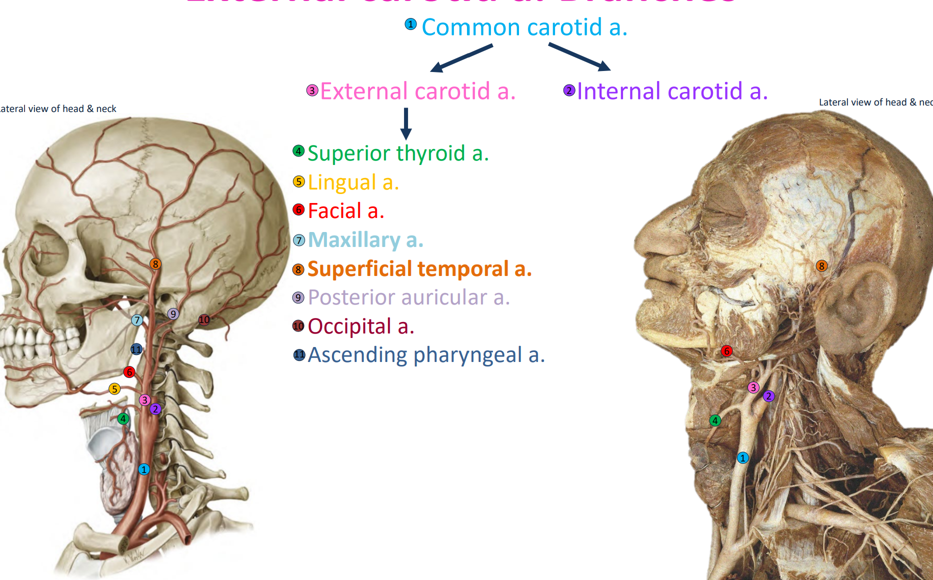
Arteries of the Head & Neck → External carotid a. Branches

Arteries of the Head & Neck - External carotid a. Branches
Temporal Fossa: General Location

Temporal Fossa (Muscles → 1)
1 muscle of mastication: Temporalis m.

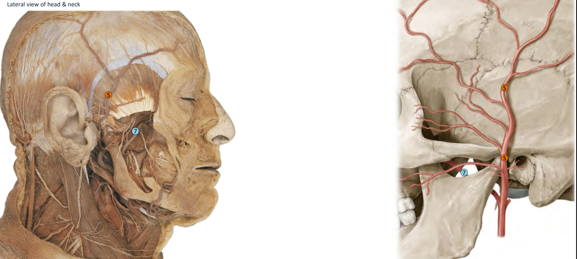
Temporal Fossa (Arteries→ 2)
External carotid artery → Superficial temporal artery and its branches
Anterior temporal artery
Middle temporal artery
Posterior temporal artery
Maxillary artery and its branches
Deep temporal arteries
Temporal Fossa (Nerves→ 2)
Trigeminal nerve (CN V3) branches:
Deep temporal nerves – motor to temporalis muscle
Auriculotemporal nerve – general sensory
.
Trigeminal nerve (CN V2) branches:
Zygomaticotemporal nerve – general sensory
Arteries of the Temporal Fossa (2)
Superficial temporal a.
Maxillary a.
Infratemporal Fossa (ITF): General Location

Movements of the TMJ and Muscles of Mastication

Temporalis m. (Origin)
Origin: Temporal fossa
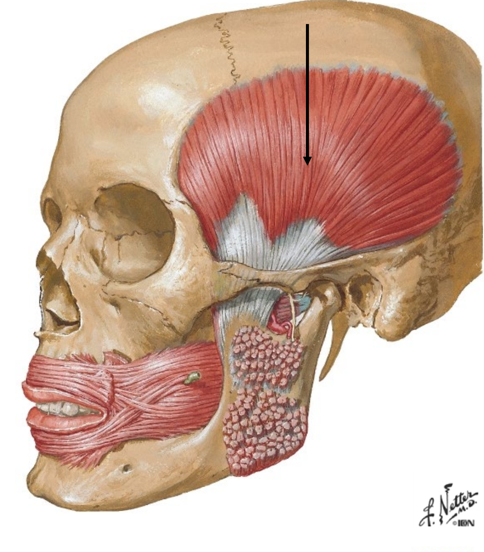
Temporalis m. (Insertion)
Insertion: Coronoid process & anterior border of ramus of mandible

Temporalis m. (Innervation)
Innervation: Deep temporal nn. (from CN V3)
Temporalis m. (Actions)
Actions: Elevate, retract (retrude) mandible
Masseter m. (Origin)
Origin:
Superficial head: Zygomatic arch, anterior 2/3
Deep head: Inner aspect of zygomatic arch, posterior 1/3

Masseter m. (Insertion)
Insertion: Lateral surface of angle & ramus of mandible

Masseter m. (Innervation)
Innervation: Masseteric n. (from CN V3)
Masseter m. (Actions)
- Both heads: Elevate mandible
- Superficial head: Protrude (protract) mandible
- Deep head: Retrude (retract) mandible from a protruded position
Medial Pterygoid m. (Origin)
Origin:
- Deep head: Medial surface of lateral pterygoid plate
- Superficial head: Maxillary tuberosity and posterior wall of maxilla

Medial Pterygoid m. (Insertion)
- Insertion: Medial surface of ramus of mandible

Medial Pterygoid m. (Innervation)
Innervation: n. to medial pterygoid (from CN V3)
Medial Pterygoid m. (Actions)
Bilateral actions: Elevate and protrude mandible
Unilateral actions: Lateral excursion (move mandible laterally) toward the contralateral side
Lateral Pterygoid m. (Origin)
Origin:
Superior head: Infratemporal roof – greater wing of sphenoid
Inferior head: Lateral surface of lateral pterygoid plate

Lateral Pterygoid m. (Insertion)
Insertion:
Superior head: Joint capsule and articular disc of TMJ
Inferior head: Pterygoid fovea of mandible

Lateral Pterygoid m. (Innervation)
Innervation: Nerve to lateral pterygoid (from CN V3)
Lateral Pterygoid m. (Actions)
Bilateral actions: Protrude mandible and pull the articular disc forward → depress mandible
Unilateral actions: Lateral excursion (move mandible laterally) toward the contralateral side
Medial & Lateral Pterygoid mm.: Posterior View

Buccinator m. (Origin)
Origin: Pterygomandibular raphe, buccal alveolar processes of mandible and maxilla

Buccinator m. (Insertion)
Insertion: Orbicularis oris m

Buccinator m. (Innervation)
Innervation: Facial n. (CN VII)**
Buccinator m. (Actions)
Compress cheeks to push food onto the teeth
Maxillary artery (3 Divisons)
Enters the infratemporal fossa.
Passes over the lateral pterygoid muscle.
Enters the pterygopalatine fossa.
.
Divided into 3 parts:
First part: Lateral to lateral pterygoid muscle.
Second part: Anterior to lateral pterygoid muscle.
Third part: Medial to lateral pterygoid muscle (in the pterygopalatine fossa).

Maxillary artery (1st Branch)
Selected branches from the 1st part:
Inferior alveolar artery: Passes through the mandibular foramen and mandibular canal.
Mylohyoid artery: Travels along the mylohyoid groove of the mandible with the mylohyoid nerve (CN V3); supplies the mylohyoid muscle.
Mental artery: Exits the mental foramen with the mental nerve (CN V3) and supplies the chin and lip.

Maxillary artery (2nd Branch)
Selected branches from the 2nd part:
Pterygoid arteries: Supply the pterygoid muscles.
Buccal artery: Supplies the buccinator muscle and cheek.
Deep temporal arteries: Supply the temporalis muscle.
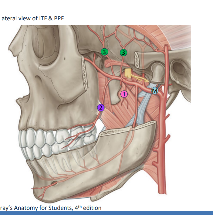
Maxillary v. & Pterygoid Venous Plexus (2)

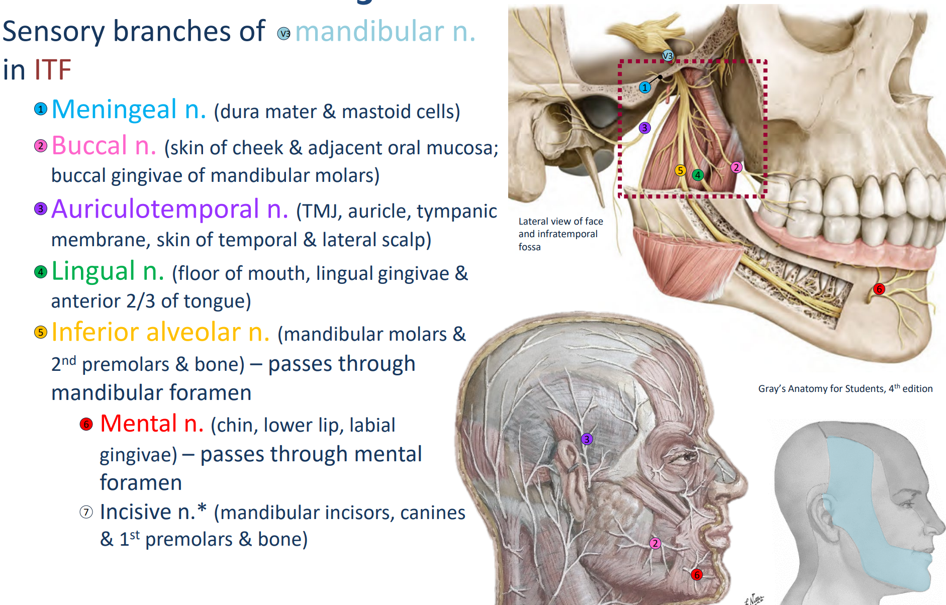
Nerves of the Infratemporal Fossa (3)
1. Branches of CN V3
2. Chorda tympani from CN VII
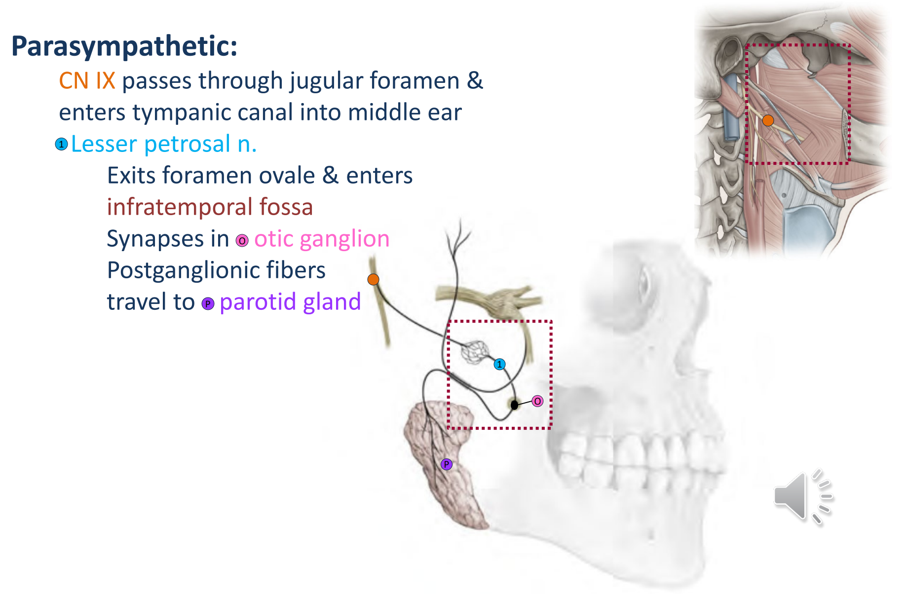
3. Lesser petrosal n. from CN IX
Branches of CN V3
- General sensory
- Branchial motor
Chorda tympani from CN VII
- Special sensory (taste)
- Parasympathetic (submandibular & sublingual glands)
Lesser petrosal n. from CN IX
- Parasympathetic (parotid gland)
CN V3 Mandibular n. (Types of fibers)
Types of fibers:
Branchial motor – supplies masticatory and suprahyoid muscles.
General somatic sensory – supplies the temporal region and lower part of the face.
CN V3 Mandibular n. (Pathway)
Pathway:
Passes through the foramen ovale.
Enters the infratemporal fossa (ITF) and gives off branches

CN V3 Mandibular n.

CN V3 Mandibular n.
CN VII Facial n.

CN IX Glossopharyngeal n.