The cycle of life: Gastrulation and Tubulation ( Birds and Mammals )
 Call Kai
 Learn
 Practice Test
 Spaced Repetition
 Match
 Flashcards
 Knowt Play
1/77

There's no tags or description
Looks like no tags are added yet.
78 Terms
First step of gastrulation of birds
From two layered blastoderm. The EPIBLAST (embryo proper) and HYPOBLAST (extraembryonic membranes)
The mechanisms of blastoderm formation
Cells delaminate from the epiblast and migrate as “islands” into the sub germinal cavity (Primary hypoblast)
Cells near the posterior end migrate as a sheet toward the anterior end (inside the sub germinal cavity) (Secondary hypoblast)
Second step of gastrulation of birds
Formation of germ layers: Chick embryos form a structure called the Primitive streak , where the formation of germ layers begins. Formation of the primitive streak begins after 3 to 4 hours of incubation as a thickening of the epiblast near the posterior end. In 16 hours of incubation, primitive streak is fully visible. As the primitive streak extends, a depression forms at the midline, called the primitive groove • It is through the primitive groove that cells will migrate into the blastocoel. At the anterior end, a thickening called Hensen’s nodeis a funnel- shaped depression where cells begin to migrate by ingression into the blastocoel.

1
Anterior

2
Margin

3
Kollers sickle

4
Posterior

5 & 8
Area opaca

6 & 9
Area pellucida

7
Thickening area of blastoderm

10
Primitive streak taking shape
Primitive streak consists of:
Primitive pit
Primitive ridge
Primitive groove
Henson node
Area pellucida
Fetal arena
Area pellucida adjacent to the primitive streak become thicker
Creation of the primitive streak established the
Anterior-Posterior axis of the embryo beginning at the tail ends and moves toward the head end

1
Primitive pit

2
Primitive groove

3
Primitive ridge

4
Hensen node

5
Area pellucida

6
Area opaca

7
Surface view of a 16 hour chick embryo
Formation of the three germ layers
during gastrulation - epiblast cells move toward the midline of the blastoderm
epiblast cells migrate through the primitive streak toward the yolk.
some cells push the hypoblast to the side to become the endoderm
those remaining cells form the mesoderm
leftover epiblast cells is the ectoderm
Triploblastic
Animals with three germ layers

1
Primitive streak

2
Epiblast

3
Future ectoderm

4
Migrating cells (mesoderm)

5
Blastocoel

6
Endoderm

7
Hypoblast

Cells continue to ingress through the primitive streak, laying down the mesoderm layer as a following steps:
It arises on both sides of the primitive streak and then quickly spreads in the form of two wings
these two mesodermal wings begin to deviate gradually to the front
It extends over the peripheral edges of the pellucida area below intermediate zone that lies in front of the receiving head of fetus, where it is completely devoid of the mesoderm. This area is known as pro-amnion
.At this point, the primitive streak regresses

1
Posterior epiblast cells change shape and thicken, forming the primitive streak

2
Cells migrate, converging at the primitive streak and causing it elongate.

3
The primitive streak narrows and lengthens

4
forming the primitive groove-the chick blastopore. Cells migrate inward through the primitive groove and Hensen's node.

5
Cells generate in Hensens node and passing into the gastrula migrate anteriorly and form head structures and notochord

6
Cells moving over the sides of the primitive groove from mesoderm and endoderm

7
The hypoblast is displaced by spreading endoderm

8
Surface cells move toward the groove and into the gastrula

Stereogram of a gastrulating chick embryo, showing the relationship of the primitive streak, the migrating cells, and the hypoblast and epiblast of the blastderm.

1
Cleavage

2
Blastula

3
Gastrulating stage

4
Gastrula

5
Primitive streak
Tubulation in birds
During regression of the primitive streak, the dorsal axis of the embryo is laid down (Notochord, neural become established, head ectoderm begins to develop and Hensen's node regresses to the posterior end, becomes anus). In this case, the result is actually a flat embryo with 3 germ layers, rather than a spherical embryo : The avian embryo is still atop the yolky mass and Endoderm separates the embryo from the yolk
Notogenesis (formation of notocord) in birds
A narrow strip of blastoderm just in front of primitive knot, consist of presumptive notochordal cell responsible for the formation of notochord.
Tubulation at 17hpf in birds
rapid divisions occur in cells of the Hensen, node these cells are pushed forward in midline to form the. cells of notochord in the embryo of 18 hpf As the notochord is distinguished in the anterior region, hensen ’not retracts back

Notogenesis in birds (18hpf)
Neurulation: neural tube formation in chick embryo
The upper surface of the 18 hpf (hours post fertilization) emberyo, which is the neural ectoderm , in front of the Hensen's node, starts thickening due to the change in cell shape, called neural plate.
At 21 hpf, The neural plate invaginates to form a neural groove flanked by neural folds.
At 22-23 hpf, the neural groove and the two nerve folds become more clearly.
soon the groove is closed into a tube from anterior to posterior end of the embryo.(60 hpf)

Neurulation: neural tube formation in chick embryo
Mammalian gastrulation
By the end of blastula stage, the blastula consist of : Inner cell mass and trophoblast. The inner cell mass that Develops into two layers: Epiblast and Hypoblast

1
Inner cell mass

2
Trophoblast
The trophoblast develops
Through several stages, eventually becoming the chorion, the embryonically derived portion of the placenta. It also induce the mother’s uterine cells to form the maternal portion of the placenta, the decidua. The decidua becomes rich in the blood vessels that will provide oxygen
and nutrients to the embryo.
The inner cell mass
gives rise to the epiblast and the hypoblast (primitive endoderm). The hypoblast will generate yolk sac cells, while the epiblast will generate the embryo, the amnion, and the allantois.
Gastrulation in mammals begins after
the germinal vesicle has settled into the (uterine )amniotic cavity - where it is characterized on inner surface of inner cell mass facing the vesicle lumen, a layer of flat cells that represents the beginning of a appearance of the endoderm layer At first it is confined to the lower surface of inner cell mass, then it spreads to line the entire inner wall of germinal vesicle, its cavity will be called the yolk sac
Amniotic cavity
During the lining process, and with onset of the endodermal layer differentiation, a small cavity appears within inner cell mass separating an upper cell layer consisting of one row of cells and a thicker layer below.

1
Inner cell mass

2
Amniotic cavity

3 & 6
Trophoblast

4 & 8
Extraembryonic endoderm

5
Germinal vesicle

7 & 9
Ectoderm

10
Somatic mesoderm

11
Splanchnic mesoderm

12
Embryonic disc
Inner Cell mass was distinguished into two layers
Outer layer has columnar cells and is called the Ectoderm layer
An inner layer, the cells of which are flat at first, then are cubic and called the endoderm layer, and each layer is like a disk, as they are two adjacent discs and together they are called the embryonic disk
Rest of the endodermal layer lines the inner surface of the trophic ectoderm and is known as: the extraembryonic endoderm
Primitive streak formation in mammals
begins as a thickening of the epiblast (ectoderm cells )near the posterior end of the embryonic disc. it is relatively short and occupies only the posterior half of the length of the embryonic disc.
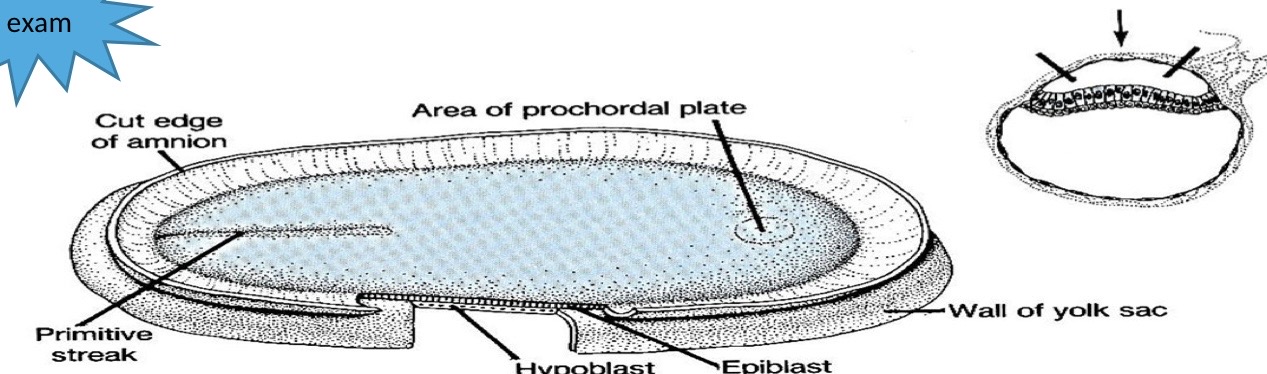
Primitive streak formation
Notogenesis- formation of notochord in mammals
a node forms as a thickened bulb at the anterior end of the primitive streak. Those cells arising from the node give rise to the notochord.

Notogenesis- formation of notochord in mammals
Mesoderm formation
Surface cells migrate in the direction of the primitive streak, where they sheath inwards, so some of these cells contribute to a formation of the endoderm layer, the remainder of it enters the composition of the mesoderm layer
Mesoderm layer
It spreads down between a trophic octoderm( trophoblast) and the Extraembryonic endoderm (endoderm lining a yolk sac), and up between the trophic octoderm ( trophoblast ) and amnion membrane
Mesoderm surrounding the yolk sac splits under the embryonic disc into two layers
External: Somatic mesoderm or Somatopleure
Internal: visceral mesoderm or Splanchnopleure, there is a cavity called the extra Coelom lumen between them.
upward-spreading mesoderm that surrounds the amnion cavity does not split and is known as a Somatopleure

Neural Tube formation in mammals (1)

Neural tube formation in mammals (2)
Differentiation of mesodermic (somatic) pieces
Formation of first somite (mesodermic) on both sides of a neural tube begins at about the twentieth day of formation, at a rate of 3 pairs every day until the end of fifth week, when there are 44 pairs of body pieces