CVS100 Introduction to Imaging and Patient Care
 Call Kai
 Learn
Learn Practice Test
 Spaced Repetition
 Match
Match Flashcards
 Knowt Play
1/63

There's no tags or description
Looks like no tags are added yet.
Name | Mastery | Learn | Test | Matching | Spaced | Call with Kai | Chat |
|---|
No analytics yet
Send a link to your students to track their progress
64 Terms
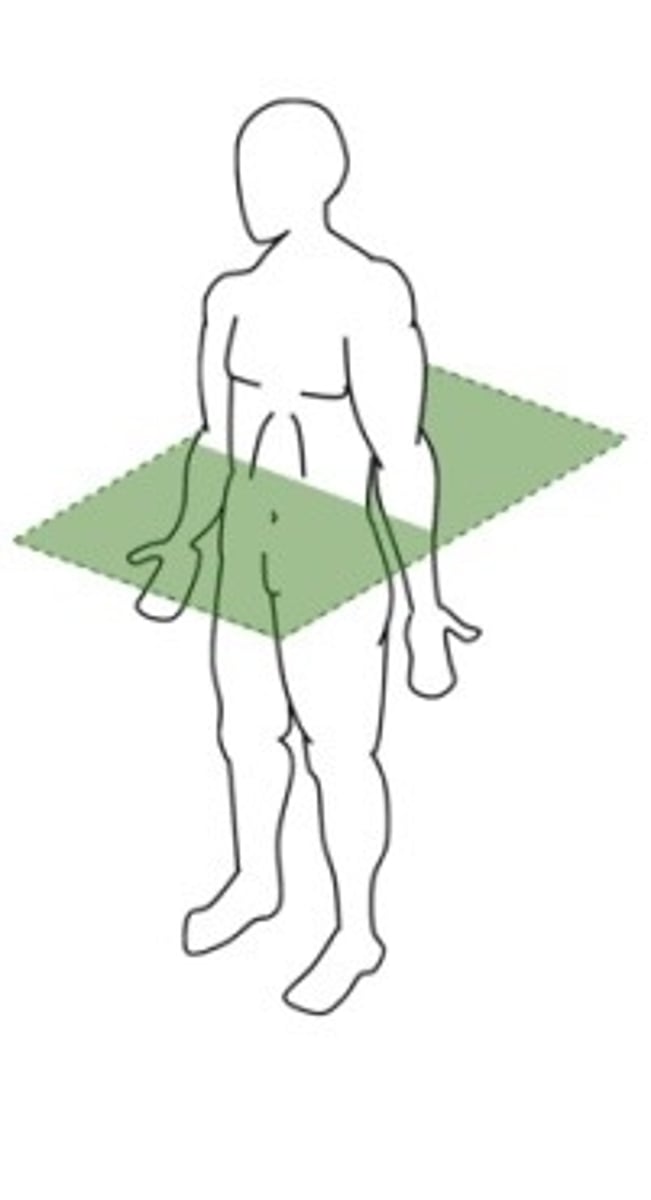
Which body plane divides the body into upper and lower portions?
transverse plane horizontal
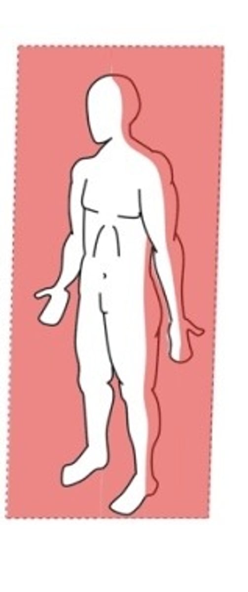
Which body plane divides the body from an anterior and posterior view?
Coronal (Frontal) plane vertical
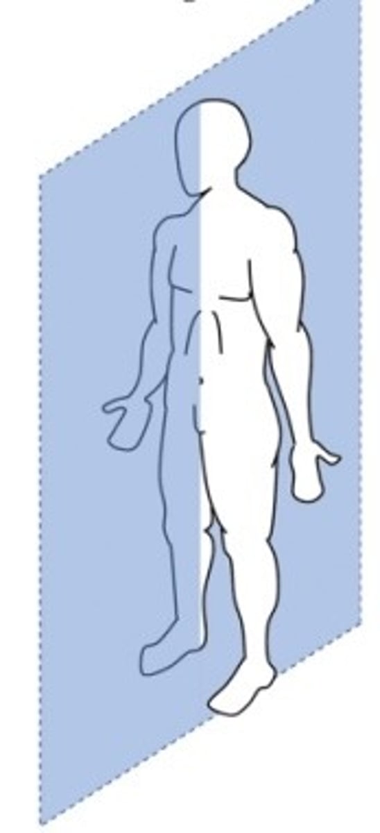
Which body plane divides the body from left to right?
Sagittal plane vertical longitudinal view
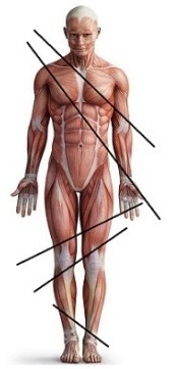
Which body plane cuts through the body through a diagonal angle?
Oblique plane (not vertical or horizontal, not parallel, "odd angles")
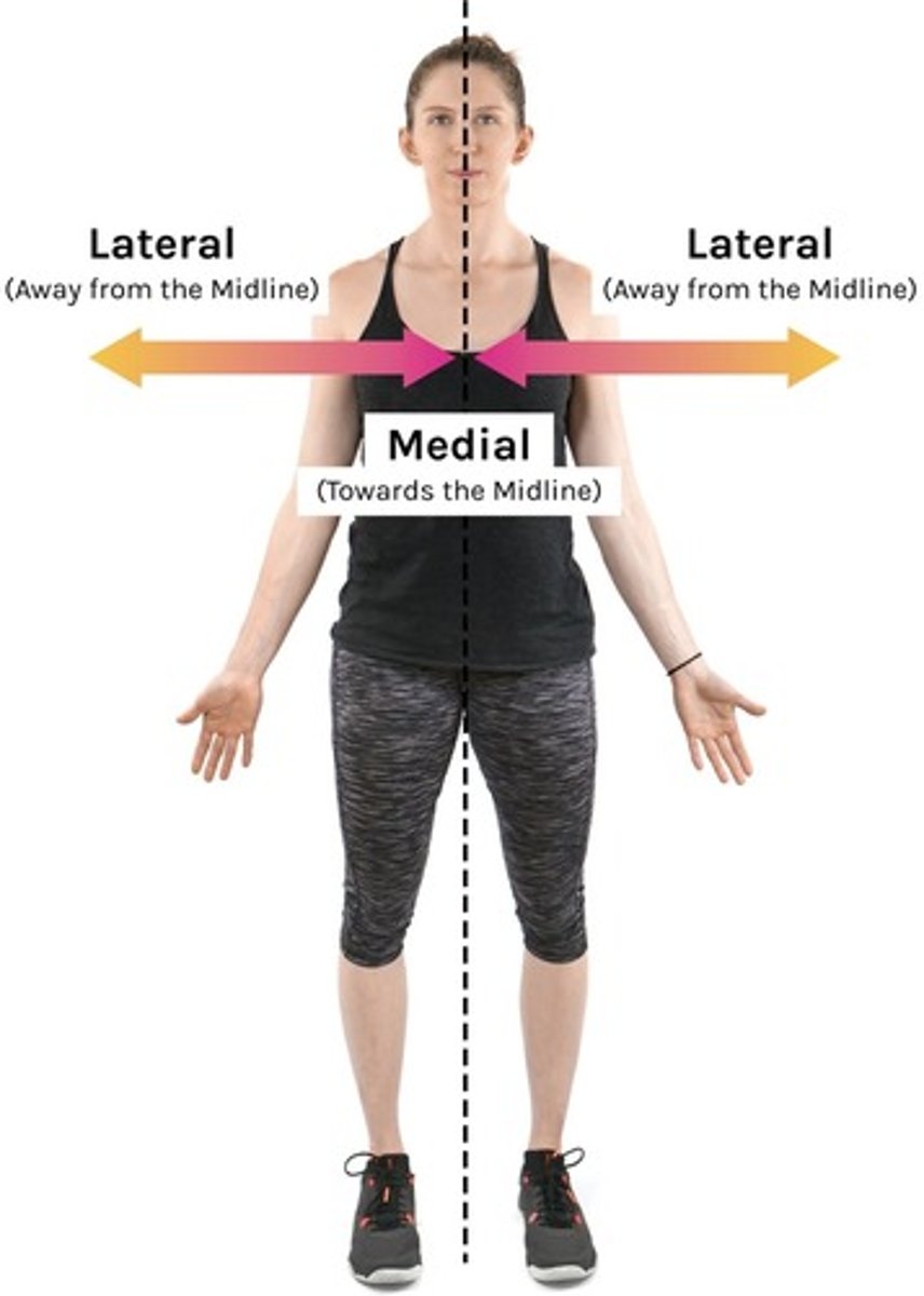
Body Orientation Terms: medial/lateral
Medial- towards the midline of the body
Lateral- away from the midline of the body
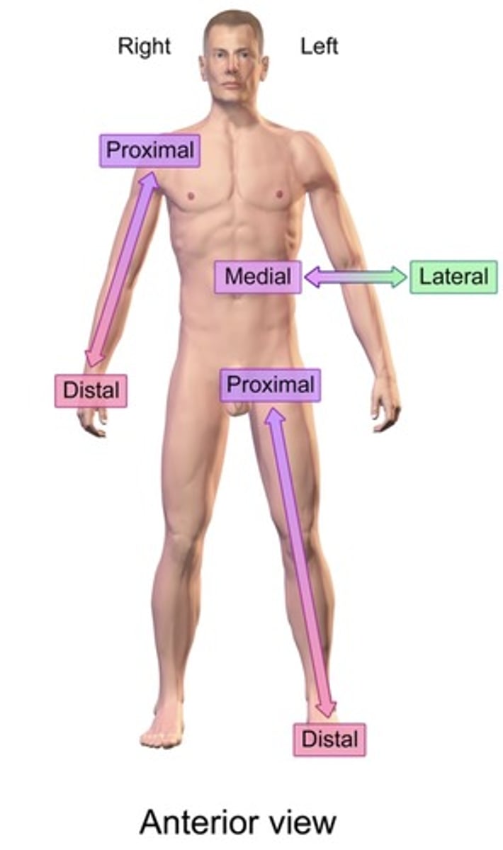
Body Orientation Terms: proximal/distal
Proximal- closer to the origin
Distal- farther away
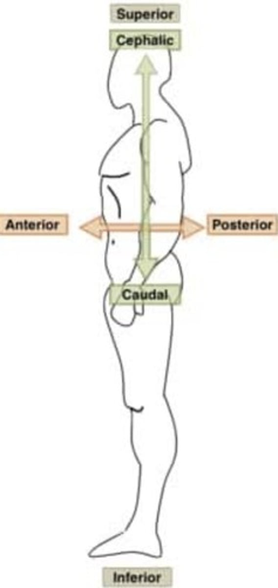
Body Orientation: cephalad/cadual
Cephalad- refers to the direction towards the head or anterior end of body (superior)
Cadual- refers to the direction towards the tail, feet, or inferior end of the body (inferior)
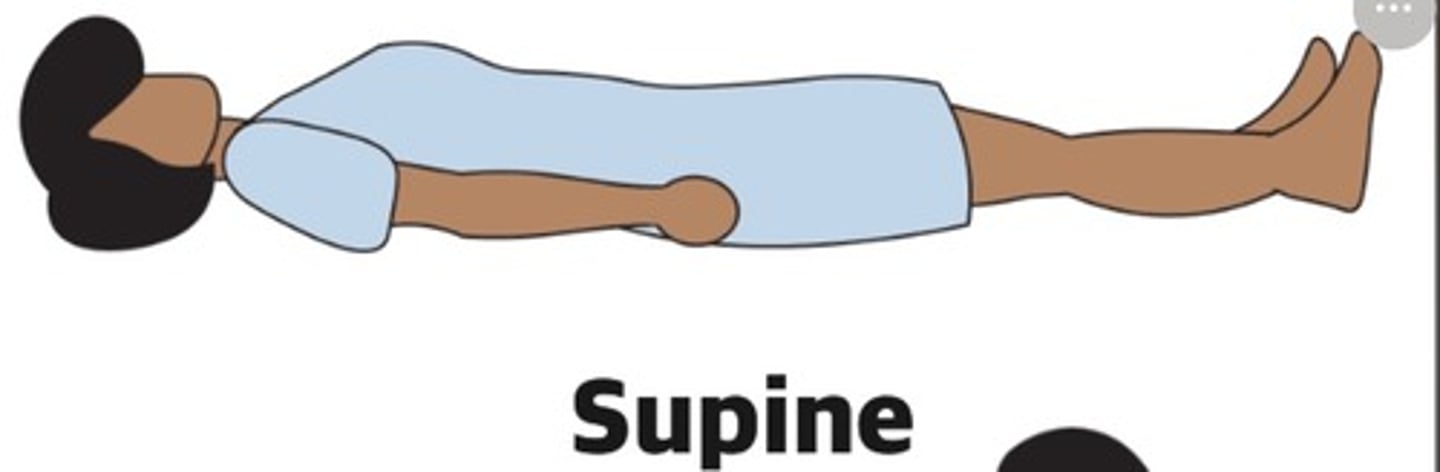
What is the patient positioning called where the patient is laying face up on back?
Supine (vascular)
What is the patient Positioning called where the patient is laying face down on stomach?
Prone
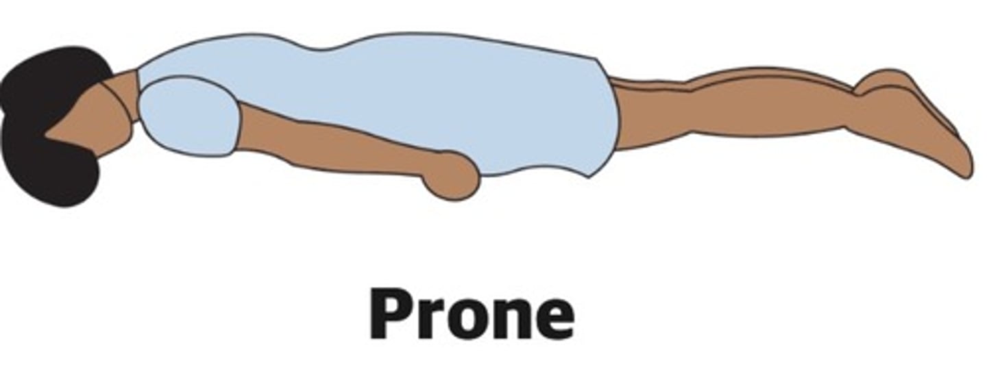
What is the positioning called where the patient is laying on their right side?
Right lateral recumbent decubitus (echo)
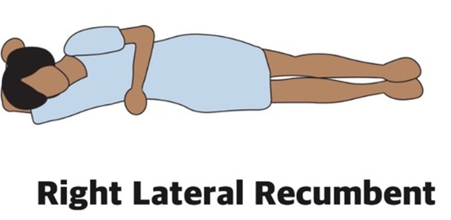
What is the positioning called where the patient is laying on their left side?
Left lateral recumbent decubitus (echo)
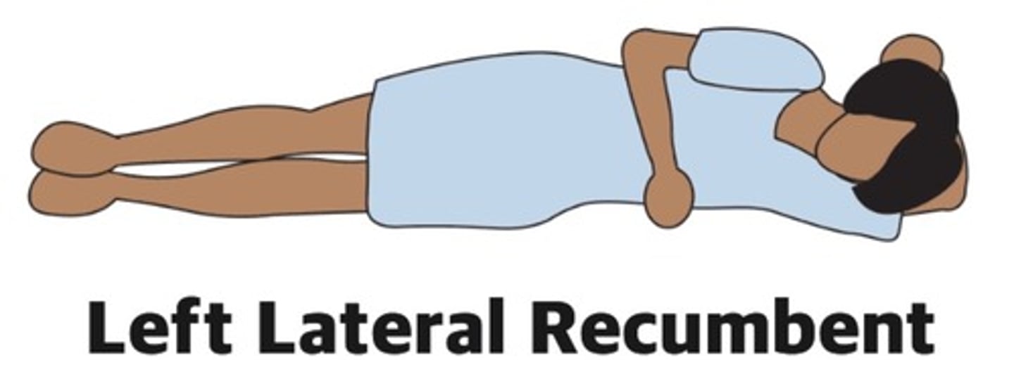
What's the position called where the patient is in a semi-sitting position where the head of the bed is raised between 45 and 60°?
Fowlers
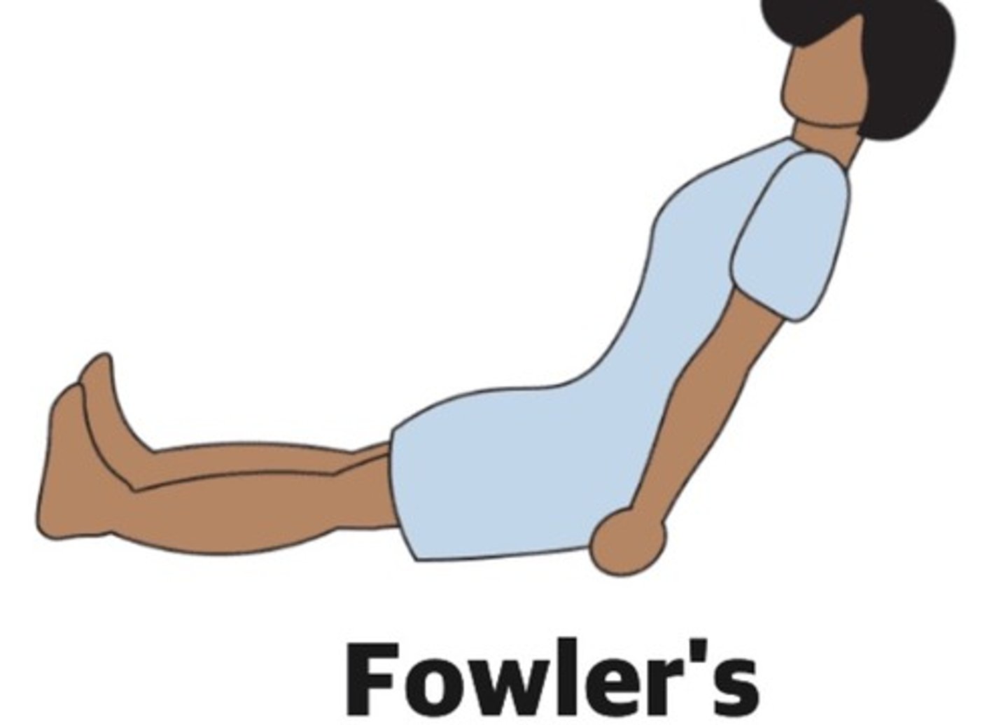
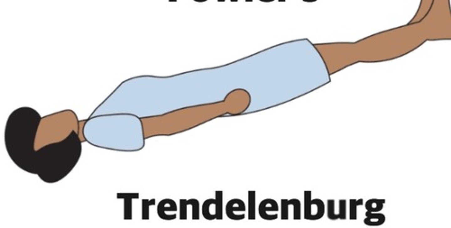
What position is called where the patient is laying supine with the feet elevated? Can also be referred to as shock position
Trendelenberg
Know anatomical locations
What is ultrasound and what is it used for?
Very high frequency sound waves using millions of hertz. They bounce off of structures and blood inside the body to obtain images and flow signals. It's used for diagnostic purposes.
What is a transducer?
Any device that changes energy from one form into one another ultrasound probes turn electrical energy into mechanical vibrations of the crystals, producing ultrasound waves, and which also changes the echo goes back into electrical energy for display on screen
Explain what the beam is
Ultrasound emitting from the transducer, whatever the beam transects through will appear on your image
When do you use a lower frequency transducer/probe?
To scan deeper structures. Loss of some resolution meaning image will become less sharp and detailed. (2.5-4 MHz for echo and abdominal)
When do you use a higher frequency?
To scan more superficial structures. Better resolution images are more clear and detailed. (5-10 MHz Vascular
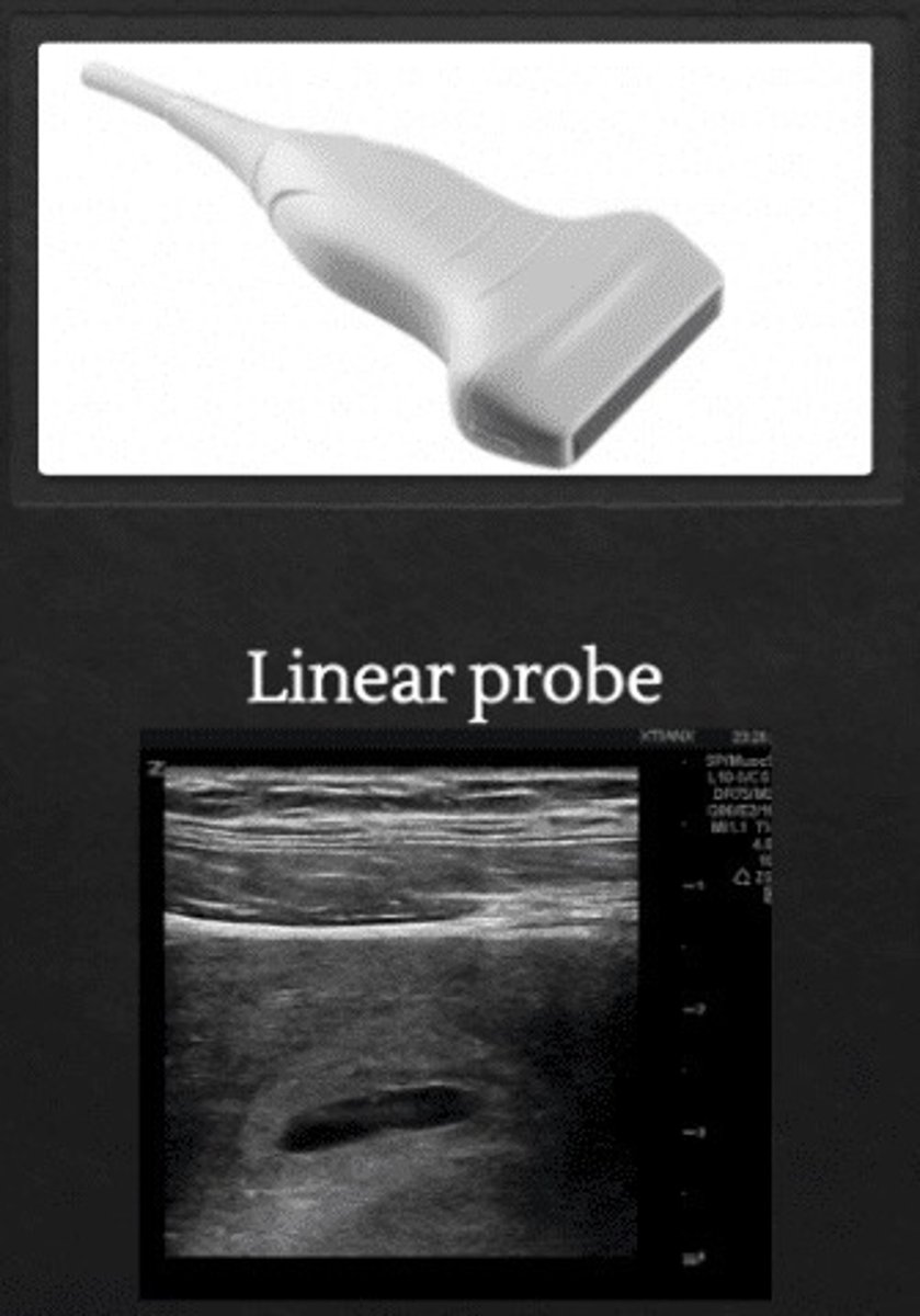
Linear array transducer (superficial-vascular)
transducer with multiple rectangular elements arranged in a linear format; creates a rectangular image field. It's created by a line of many little crystals.
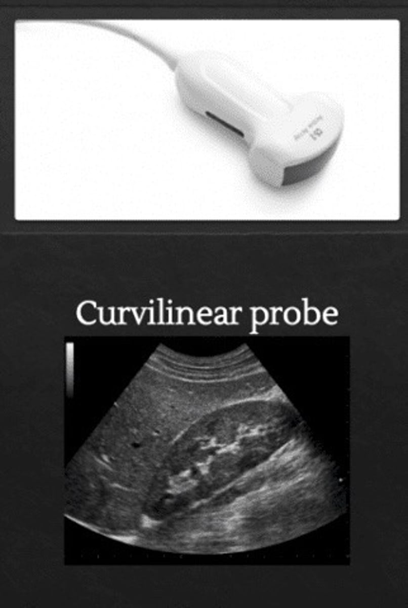
Curved Linear Array (deep- OB/Abd)
Line of crystals, but it is curved to create a sector shaped field of view
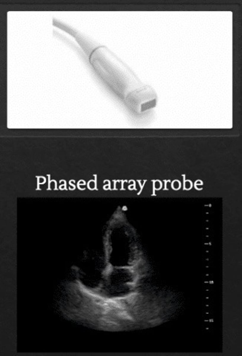
Phased array (echo)
Pie shaped sector, smaller probe face and creates a wider sector than curved
The index always faces ...
The right side of the Pt
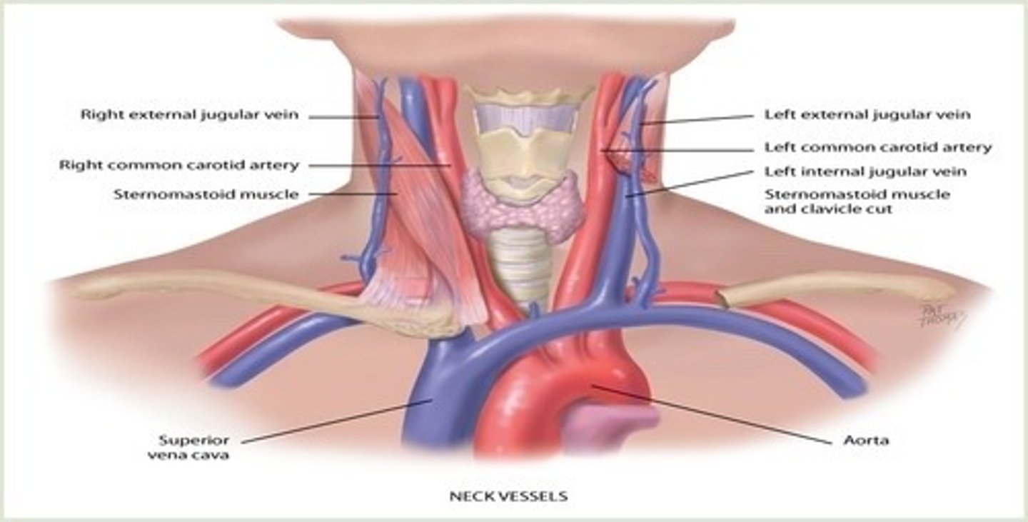
Long view Medial angle shows
IJV and CCA on top of each other
Long view Intima to intima internal diameter
0.5-1.25cm if over that range, then you're most likely on the IJV
The mark on the screen is known as the
screen index orientation marker
The mark on the transducer is known as the
index
Time Gain Compensation TGC controls
Ability to compensate for attenuation of the transmittal beam as the sound wave travels through tissue in the body
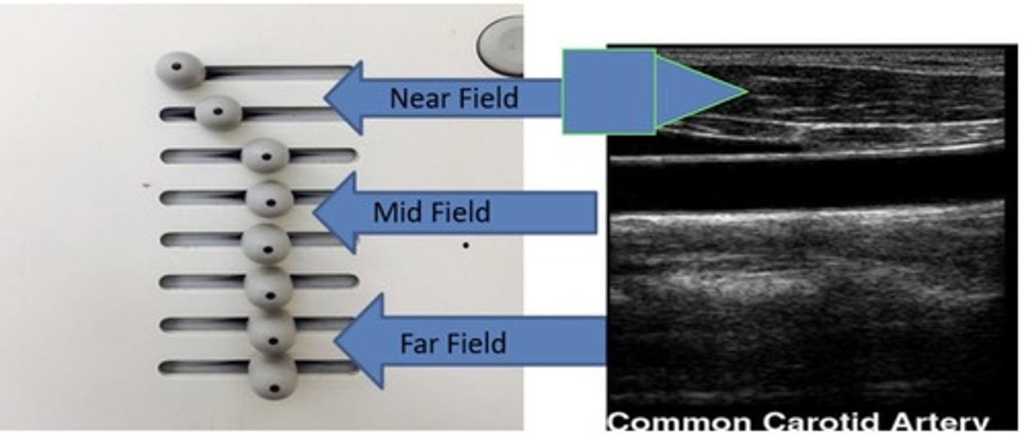
The overall gain/2D gain controls
overall brightness of the image
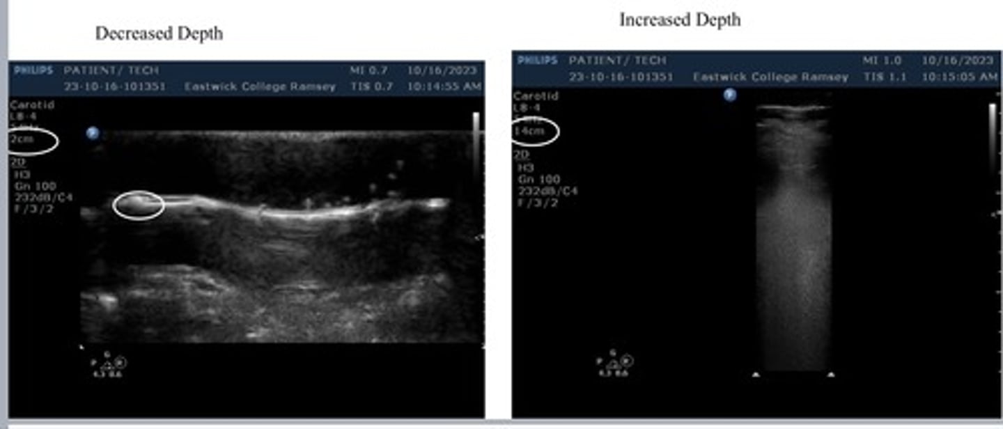
Depth
Describes the maximum distance into the body that an ultrasound system is imaging. It also adjust how far or how close we see the body.
Increase depth- image is farther showing a more zoomed out view
Decreased depth- images closer showing a more zoomed in view
Focal Zone: Focus/ Trans Zone
Always adjust to the area of interest. Used to enhance the focus and resolution for specific anatomical structure.
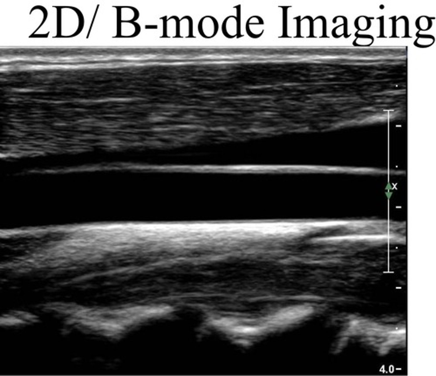
2D B-mode imaging
Brightness-mode or Two-Dimensional mode which provides structural information utilizing different shades of gray in a two-dimensional image.
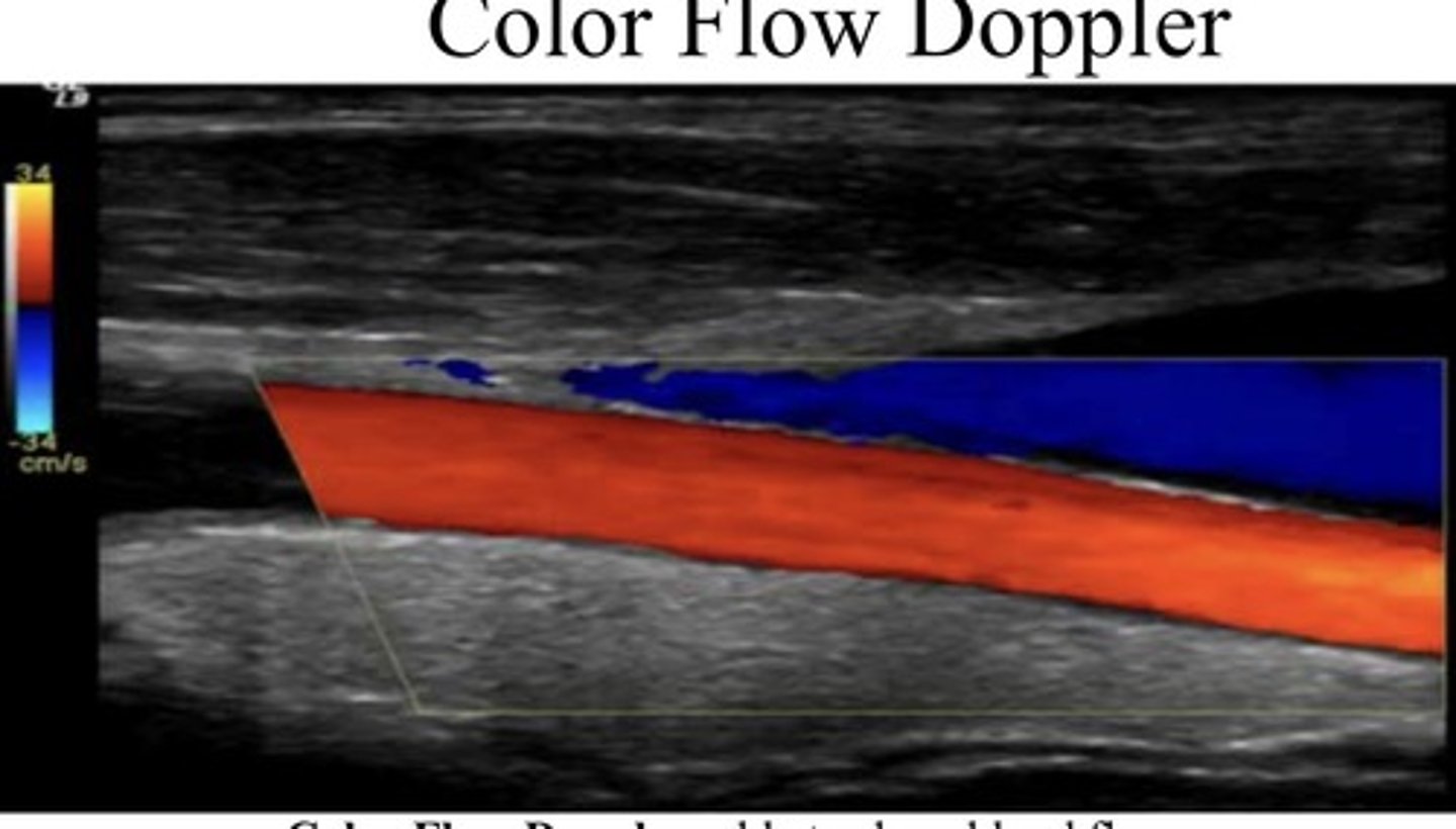
Color Flow Doppler
Able to show blood flow motion in a selected two dimensional area. Direction in velocity blood flow are color-coded and superimposed on the corresponding B mode image
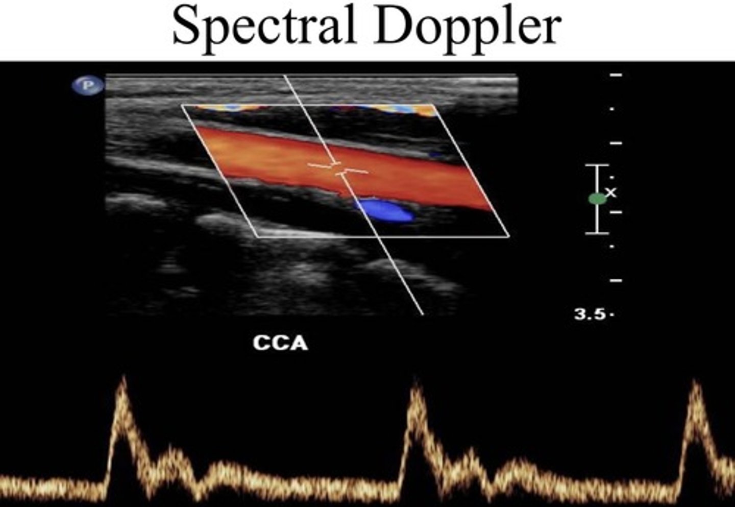
Spectral Doppler
Consist of a continuous and pulsed wave form shows the velocity of moving RBC's. Shown to us as a spectral waveform. Uses a 1D graph
shows us the velocity of the blood flow moving through the vessels
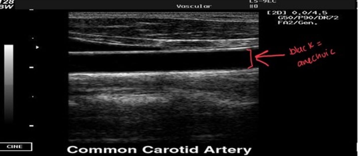
Anechoic
free from echoes and reverberations (black)
- sound waves pass through an echoic structures because there is not enough density to stop the beam. This structure is fluid filled and transmitted sound easily.
Echogenic
Capable of producing echoes
white/grey indicating solid tissues, calcifications, or fat
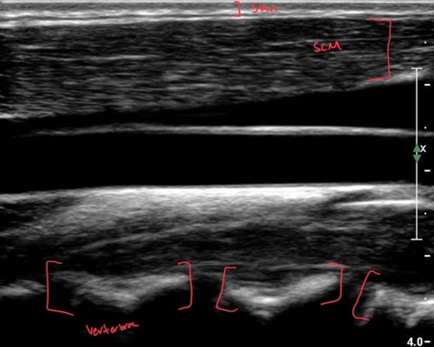
Hyperechoic
Producing echoes of higher amplitude than the surrounding medium
specific type of echogenicity meaning the structure appears brighter (whiter) then surrounding tissues because it reflects more sound
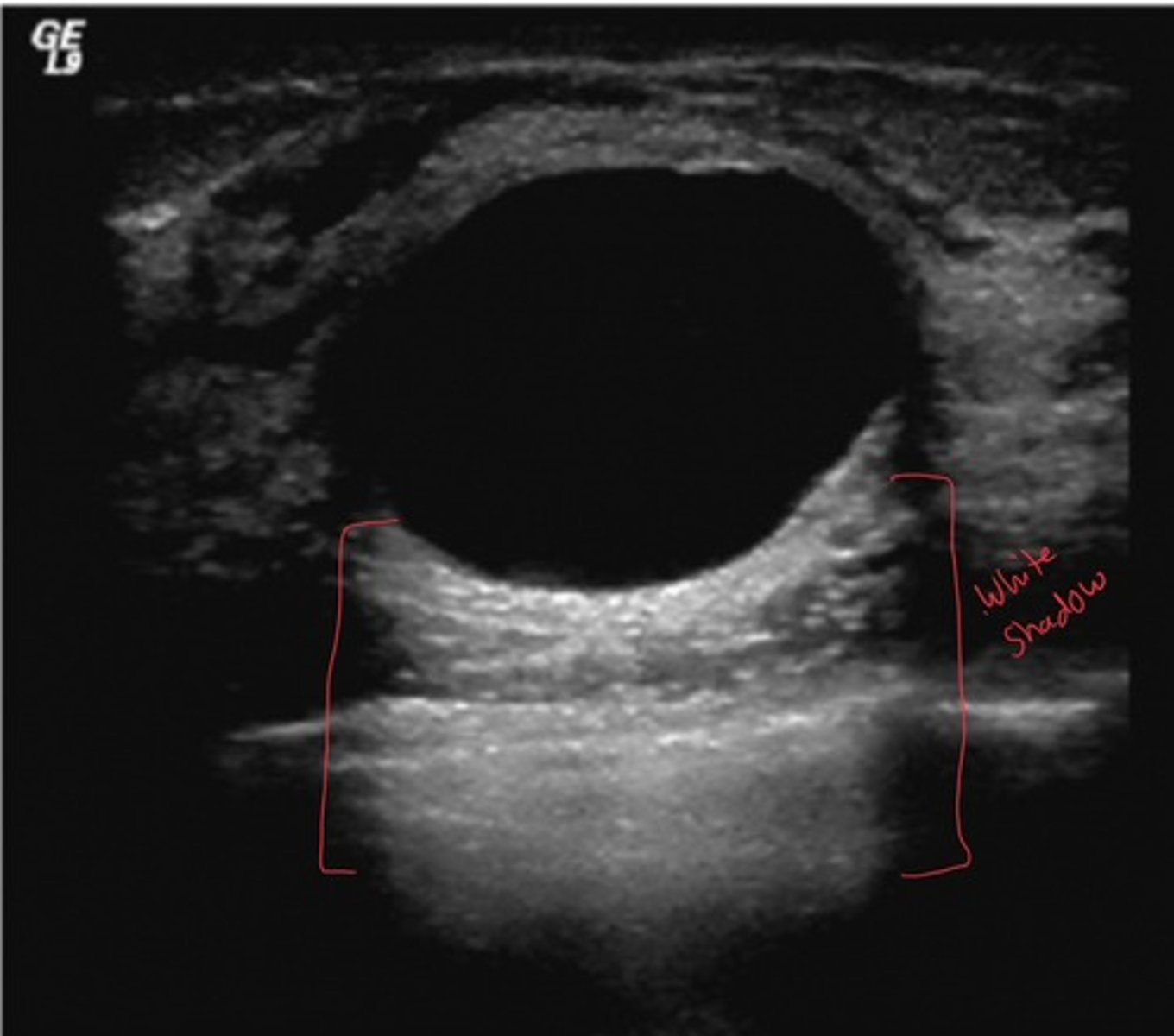
Hypoechoic
Producing echoes of lower amplitude than normal for the surrounding medium
portions of an image that are not as bright as surrounding tissues, or tissues that appear less bright than normal
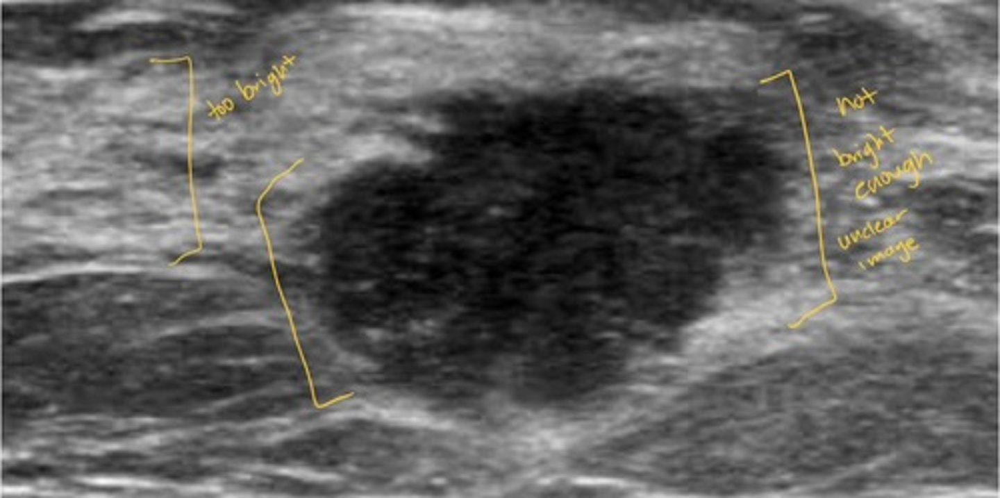
homogeneous mixture
Completely uniform in texture or composition, same grayscale
image comes out uniform, smooth, and consistent creating a smooth looking image usually a good sign no abnormalities
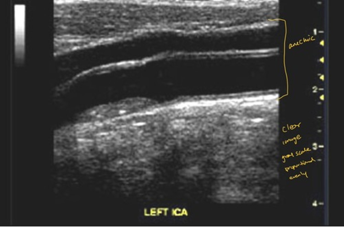
Heterogeneous
Refers to an uneven echo pattern or reflection of varying gray scale, not uniform and texture or composition.
image comes out looking patchy, mixed or irregular often indicates in need for further investigation
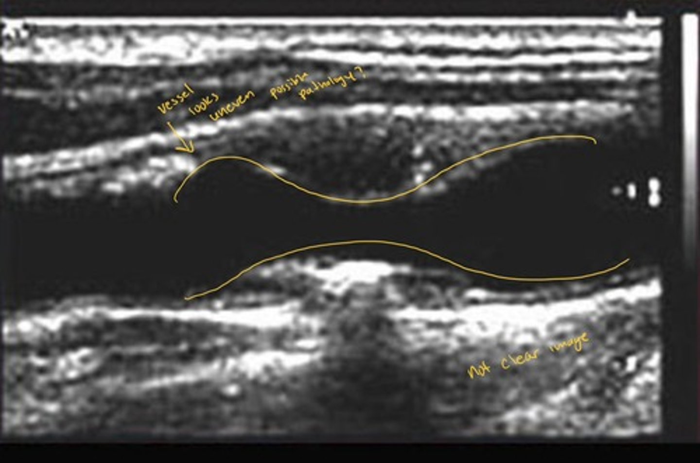
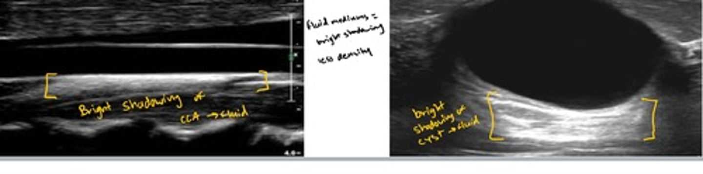
acoustic enhancement
When ultrasound passes through a fluid medium, the intensity of the sound energy is not diminished. Tissue behind the fluid collection are more echogenic (brighter because there is more acoustic power to reflect back to the transducer)
Ex://sound travels easily through liquid, such as cysts or bladder. The area behind the fluid looks brighter because the ultrasound energy didn't get absorbed. A simple cyst in the liver or breast is a classic example, appearing black in the center with a brightly lit area immediately behind it.
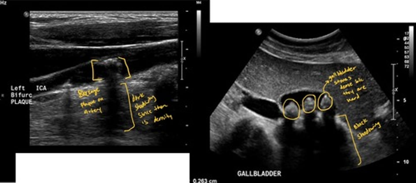
posterior acoustic shadowing
When a very dense structure, such as a gallstone or bone, reflects so many echoes that virtually no sound waves are transmitted, the tissue deeper to this structure is displayed as hypoechoic
Ex:// sound travels poorly through solid/hard items such as gallstones bones, hard tumors. The area behind the item looks darker because the sound was blocked creating a shadow.
Color flow Doppler CFD
variation of Doppler; converts recorded flow frequencies into different colors allowing us to examine the direction and velocity of blood flow through chambers and valves
* CCA is red
* IJV is blue
When scanning CCA's, if you see the IJV, how should you move the probe?
You're most likely to lateral and need to slide more medial
When scanning CCA if you see the thyroid, how should you move the probe?
You're most likely too medial and need to slide more lateral
What are the modes on the ultrasound machine and what are their functions?
2-D - base grayscale, anatomy and cardiac windows
M mode - motion overtime for fast linear measurements
color - maps flow direction interference on the 2-D image
PW Doppler - samples velocity at one specific location
CW Doppler - measures higher velocity along the full beam
TDI/CPA - add tissue velocity or low flow sensitivity when needed
Making a positive patient care experience
Empathy and compassion
Communication
Listen to your patient
Be supportive
Be respectful
Be friendly
Explain the ultrasound
Ask questions
When a patient cannot communicate
•If a patient is unconscious, you can communicate with a nurse and/or family member instead of the patient
•If the patient is unable to speak or difficult to understand, provide the patient with something to write on or only ask yes or no questions
•If a patient is clinically deaf or hard of hearing, use writing instead of speaking
•Use an interpreter or family member to interpret if there is a language barrier
What Not to Communicate to a Patient
There can be legal consequences if the wrong information is given
Student and staff sonographers should avoid telling a patient the results of an ultrasound
Avoid telling the patient anything that may add stress or anxiety
Intracavitary ultrasound
Involves the use of a specially designed ultrasound transducer that is placed inside a body cavity.
invasive procedure
a test or treatment that requires incisions to the skin or the insertion of instruments or other materials into the body
Obstretic sonographic examination
Healthy patient with an uncomplicated pregnancy depend depending on which protocol your facility follows will typically have anywhere from 2 to 4 ultrasounds during the pregnancy.
- 1st examination is done in the early first trimester. A confirms due date and viability
- 2nd examination, (in some cases, the first ultrasound) is non-invasive and determines whether the fetus is at risk
- 3rd examination (sometimes the second ultrasound), feta is evaluated from head to toe for fetal abnormalities, as well as uterine and placental abnormalities
Aortocaval compression syndrome
compression of the aorta and inferior vena cava (IVC) by the gravid uterus resulting in symptoms of nausa, hypotension, light headedness, and syncope
Fetus Cardiac arrhythmia during pregnancy
Bradycardia rhythms of less than 110 bpm
Tachycardia rhythms of more than 160 bpm
Ethical dilemmas
- ultrasound is used to figure out how far along a pregnancy is. That information can be used to plan an abortion.
- chorionic villus sampling (CVS) and amniocentesis test the fetus for genetic conditions or abnormalities.
doctors may face ethical concerns when test helped determine pregnancy timing for abortion, decisions, or revealed fetal conditions I could influence whether a pregnancy continues. Going about the situation is very crucial as a healthcare professional.
3 d and 4 d ultrasound
Used in scanning the heart
HIPPA
The Health Insurance Portability and Accountability Act, a federal law protecting the privacy of patient-specific health care information and providing the patient with control over how this information is used and distributed.
OSHA (Occupational Safety and Health Administration)
a government agency in the Department of Labor to maintain a safe and healthy work environment
Consent/Refusal of Treatment
Consent is mandatory when the patient is having an invasive procedure. Patient has the right to except or decline treatment.
Disinfecting Equipment
Prevents disease spread between patients.
Practice universal precautions, use hand sanitizer before, and after every patient encounter.
* disinfecting probes, beds, stretchers, materials use on a patient, wearing protective gear
Vascular-Specific Patient Care Considerations
The vascular laboratory will usually perform the nonimaging Doppler examinations
Vascular sonographers will need to be able to work with patients with open sores, ulcers, and gangrenous digits
Vascular sonographers will have patients that have amputated limbs or are being worked up for an amputation
Certain studies will require obtaining blood pressures and tracings using blood pressure cuffs that will cause the patient pain
Empathy and good communication are important
Echocardiography specific patient care consideration
- may perform the majority of their studies as portable examinations
- continuous wave Doppler used
- 3-D and 4-D ultrasound used in scanning the heart
Moving patients
Find out how the patients will travel to the department
Ask if the patient needs assistance getting up on and down off the stretcher
Lock wheels on wheelchairs and stretchers
Offer a bedpan or urinal if the patient needs to use the restroom and requires too much assistance
Be careful of IV lines, tubes, drains, and urinary bags
Portable Ultrasound
Communication with nurse is essential
Ergonomic challenge for tech