ex phys exam 3
 Call Kai
 Learn
 Practice Test
 Spaced Repetition
 Match
 Flashcards
 Knowt Play
1/44

There's no tags or description
Looks like no tags are added yet.
45 Terms
how much of the human body is skeletal muscle?
human body contains over 600 skeletal muscles
40-50% of total body weight
what are the functions of skeletal muscle?
force production for locomotion, breathing, and postural support
heat production during cold stress
skeletal muscle gross structure
epimysium (whole muslce), perimysium (bundle of muscle fibers- fascicle), endomysium (between fibers)
tendons connect muscle to bone
microstructure of skeletal muscle
muscle fiber (muscle cell)
myofibrils
sarcomeres
myofilaments (actin and myosin)
actin = thin filament — troposin complex and tropomyosin
myosin = thick filament
arrangement of myosin and actin
resting: myosin and actin overlap with myosin heads pointing outward from M line
actin extends inward from Z discs
myosin is centered around M line
topomyosin blocks the myosin binding sites on actin
during contraction:
actin slides toward M lines
myosin heads move through cross bridge interactions

sarcolemma
muscle fiber’s cell membrane
t-tubules
inward extensions of sarcolemma, carry electrical signal (AP) from the surface deep into muscle fiber
SR
specialized network of membrane bound tubules that surrounds the myofibrils
stores calcium ions and releases them when muscle fiber is stimulated
takes calcium back up when the muscle needs to relax
the sliding filament model
proposes that muscle shortens or lengthens because thick and thin filaments slide past each other without changing length
myosin crossbridge cycling
myosin continually attaches and detaches from actin filaments with energy from ATP hydrolysis
contractions in sliding filament model
Concentric contraction: fiber length shortens during force generation
Isometric contraction: fiber length remains unchanged during force generation
Eccentric contraction: fiber lengthens during force generation
cross bridge formation during muscle contraction
AP triggers Ca2+ release from SR → Ca2+ binds to troponin → tropomyosin shifts to expose myosin binding sites on actin
myosin head (carrying ADP and Pi) binds to actin, forming cross bridge
myosin head pivots and pulls actin toward the M line (releasing Pi and ADP) → generates force (POWER STROKE, causing filaments to slide)
new ATP binds to myosin, causing myosin head to detach from actin
myosin hydrolyzes ATP into ADP and Pi → energy released re energizes the head and returns it to its cocked position, ready to bind to actin again if binding sites remain exposed
**cycle repeats as long as calcium remains elevated and ATP is available

explain the sarcomere length tension curve
shows that the amount of force (tension) a muscle can generate is dependent upon the initial length of the sarcomere before contraction (which is determined by the degree of overlap between actin and myosin)
if a muscle is over stretched or overly shortened before contracting, its ability to generate force is compromised
steps of muscle excitation
sac-like vesicles within axon terminal release Ach → diffuses across synaptic cleft + attaches to specialized Ach receptors on sarcolemma
MAP depolarizes transverse tubules at the sarcomere’s A-I junction
t-tubule system depol causes CA2+ release from SR lateral sacs
steps of muscle contraction
Ca2+ binds to troponin-tropomyosin in actin filaments
actin joins myosin ATPase to split ATP with energy release during muscle action → tension from energy release produces myosin cross bridge movement
muscle shortening occurs after ATP binds to the myosin cross bridge, which breaks the actin myosin bond and allows cross bridge dissociation from actin and sliding of thick and thin filaments
cross bridge activation continues when Ca2+ concentration remains high (From membrane depol) → this prevents troponin-tropomyosin action
when muscle stimulation ceases, Ca2+ moves back into the SR lateral sacs through active transport via ATP hydrolysis
Ca2+ removal restores troponin-tropomyosin inhibitory action → with ATP present, actin and myosin remain in the dissociated relaxed state
how do muscle fiber types differ
skeletal muscle contains two main types of fibers that differ in three characteristics:
primary mechanism to produce ATP
motor neuron innervation type
myosin heavy chain type expressed
how do the proportions of each type of muscle fiber vary?
they vary from muscle to muscle and from person to person:
on average, how many slow twitch fibers to men, women, and children possess in their arm and leg muscles?
45-55%
what is the muscle biopsy technique?
removes cross sectional area of muscle to determine muscle fiber type
how does the muscle biopsy technique work?
Serial cross sections obtained by muscle biopsy- using acidity (pH), different muscle fiber types stain different colors
More basic solution shows more slow twitch; more acidic shows more fast twist
type I fibers (slow twitch)
SO (slow oxidative)
aerobic, highly fatigue resistant (endurance activities)
type IIa (fast twitch)
FOG (fast oxidative glycolytic)
fast contraction speed, utilizes both aerobic and anaerobic energy pathways
type IIx (fast twitch)
fast twitch intermediate fiber type bridging type IIa and type IIb
type IIb (fast twitch)
FG (fast glycolytic)
primarily anaerobic, generates high force rapidly, but fatigues very quickly
contraction time of fibers
type I- slow
type IIa- moderately fast
type IIx- fast
type IIb- very fast
motor neuron size of m fibers
type I- small
type IIa- medium
type IIx- large
type IIb- very large
fatigue resistance of m fiber types
type I- high
type IIa- fairly high
type IIx- intermediate
type IIb- low
activity of m fiber types
type I- aerobic
type IIa- long term anaerobic
type IIx- short term an
type IIb- short term an
useful duration of m fiber types
type I- hours
type IIa- <30 minutes
type IIx- <5 minutes
type IIb- <1 min
force production of m fiber types
type I- low
type IIa- medium
type IIx- high
type IIb- very high
mitochondrial density of m fiber types
type I- high
type IIa- high
type IIx- medium
type IIb- low
capillary density of m fiber types
type I- high
type IIa- intermediate
type IIx- low
type IIb- low
oxidative capacity of m fiber type
type I- high
type IIa- high
type IIx- intermediate
type IIb- low
glycolytic capacity of m fiber types
type I- low
type IIa- high
type IIx- high
type IIb- high
major storage fuel of muscle fiber types
type I- triglycerides
type IIa- PCr glycogen
type IIx- PCr glycogen
type IIb- PCr glycogen
myosin heavy chains of each muscle fiber type
type I- MYH7
type IIa- MYH2
type IIx- MYH1
type IIb- MYH4
primary subtypes of fast twitch fibers (type II)
type IIa- represents fast-oxidative glycolytic fibers (FOG)
type IIx- midway between a and b types in physiologic and metabolic characteristics
type IIb- possesses greatest anaerobic potential and most rapid shortening velocity; represents ‘true’ fast glycolytic fiber
key features of fast twitch fibers
high electrochemical transmission of APs
high myosin ATPase activity
rapid Ca2+ release and uptake by SR
high cross bridge turnover rate
key features of slow twitch fibers
low myosin ATPase activity
slow calcium handling ability and shortening speed
less glycolytic capacity than fast twitch fibers
large and numerous mitochondria
generate energy for ATP resynthesis through aerobic system of energy transfer
fiber type differences among athletic groups
fast twitch fibers distribute equally between type IIa and type IIx subdivisions
although no gender differences exist in fiber distribution, larger inter individual variation occurs
endurance athletes possess predominantly slow twitch fibers predominate for elite sprint athletes
force velocity relationship
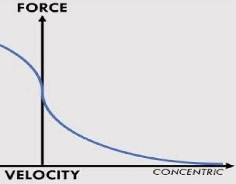

explain this: force power relationship
Peak power happens at something less than maximum force and less than maximum velocity
If force x velocity = power, then if im at max force, then velocity is zero, and power is zero (more force doesn't always mean more power)