Anatomy Lab Final
 Call Kai
 Learn
 Practice Test
 Spaced Repetition
 Match
 Flashcards
 Knowt Play
1/167

Earn XP
Description and Tags
Eye/Ear, Brain/Cranial Spinal Nerves, Heart/Circulatory System
168 Terms
dura mater
Parts: Periosteal layer, Meningeal layer, Falx cerebri & cerebelli, Tentorium cerebelli

periosteal layer
outer layer of dura mater; attach to skull

meningeal layer

falx cerebri
vertical fold of the meningeal layer; seperate left and right cerebrum in upper cranium

falx cerebelli
vertical fold of the meningeal layer; seperate left and right cerebrum in lower cranium

tentorium cerebelli
crescent-shaped fold of the dura mater; covers posterior cranial fossa

arachnoid mater
“spider-like”

pia mater
Follow each gyrus and sulcus

gyrus
fold in cerebral cortex; increases surface area

sulcus
shallow groove between elevations

cerebrum
Organization: cerebral cortex, white matter, basal nuclei

diencephalon
Major brain region

thalamus
Part of diencephalon

hypothalamus
part of diencephalon
pituitary gland, optic chiasm, olfactory bulb; homeostasis and hormones

epithalamus
part of diencephalon, pineal gland

central sulcus
Seperate frontal and parietal

longitudinal fissure (large sulcus)
separates hemispheres

transverse fissure
separates cerebrum and cerebellum

frontal lobe

parietal lobe
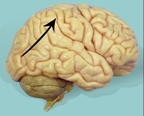
occipital lobe

temporal lobe
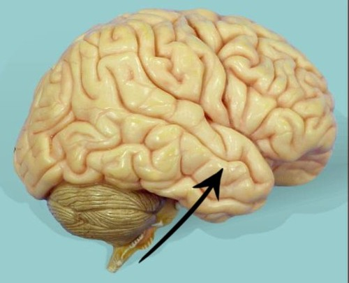
Brocas area

Wernickes area
understanding spoken and written language

Primary motor area
precentral gyrus located here
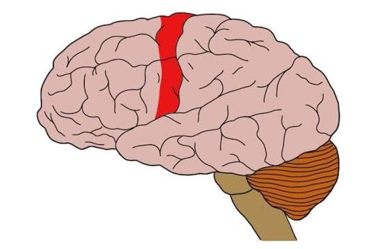
Primary somatosensory area
postcentral gyrus located here

Primary auditory area
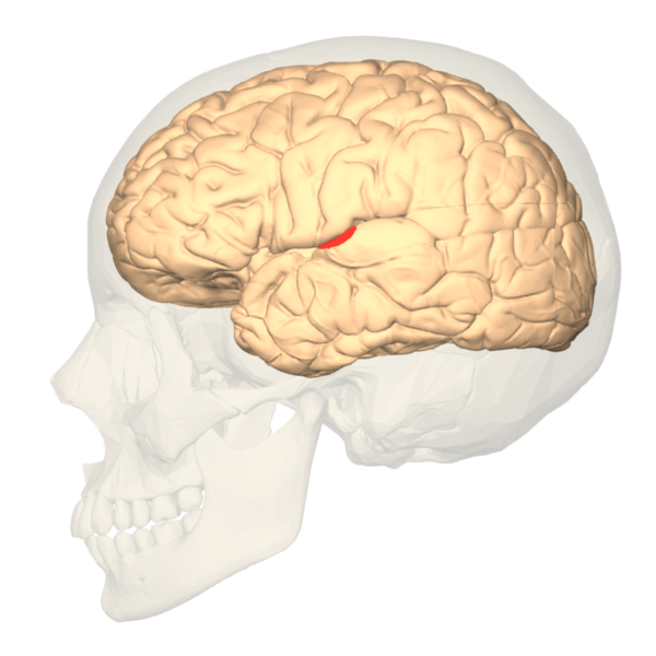
primary olfactory area
pink area

Primary visual area

medulla oblongata
part of brain stem: respiratory and cardiovascular control

pons
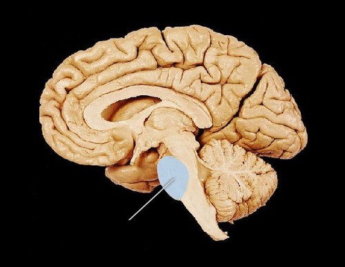
midbrain
part of brain stem: reflex centers for visual and auditory stimuli

corpora quadrigemina
on dorsal roof of midbrain
superior colliculi - visual & spatial tracking
inferior colliculi - auditory processing

cerebellum
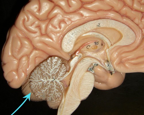
vermis

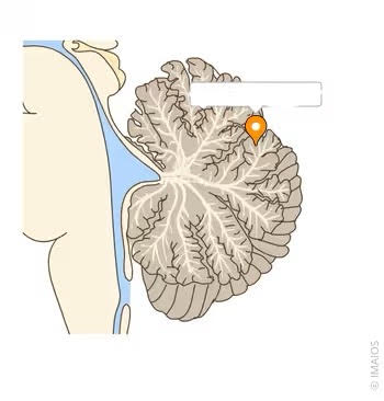
arbor vitae
White matter

Cerebellar cortex
outer layer; tightly folded gray matter
corpus callosum
bridge between hemispheres to share information

lateral ventricles
in each cerebral hemisphere
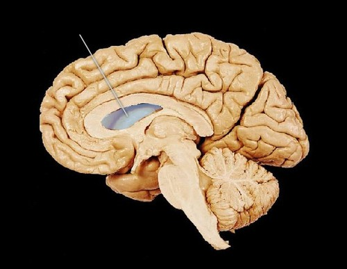
third ventricle
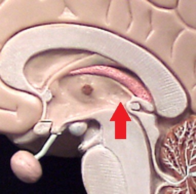
cerebral aqueduct
connect 3rd ventricle to 4th ventricle

fourth ventricle
between pons and cerebellum

Pituitary gland

Optic chiasm
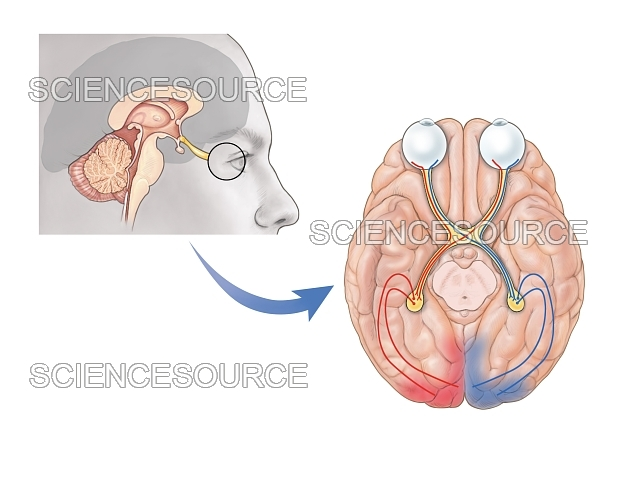
Olfactory bulb

Infundibulum
pituitary stalk

olfactory nerve

optic nerve

oculomotor nerve

trochlear nerve

trigeminal nerve
facial sensation (sensory); facial expressions (motor)
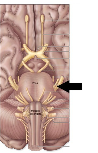
abducens nerve

facial nerve

auditory nerve

glossopharyngeal nerve

vagus nerve

spinal accessory nerve
head and shoulder movement (motor)

hypoglossal nerve

cervical enlargement
C5 to T1; House motor and sensory neurons to control/sense shoulders, arms, hands

lumbar enlargement
L2 to S3; House extra s/m to get input from lower limbs and pelvis

Cauda equina
L2-S5; house peripheral nerves floating in CNS

conus medullaris
spinal cord ends in

spinal cord cross section
Info not from lecture:
lateral portions - sensory and motor
columns - major data ways
commissure - connect left and right halves

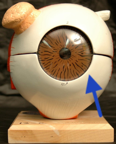
sclera
iris

cornea
outer clear layer

lens
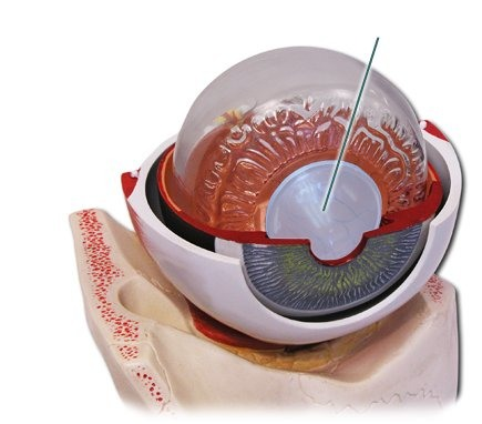
pupil

ciliary body
green portion

conjunctiva

superior oblique of eye

superior rectus of eye

inferior rectus of eye

medial rectus of eye

lateral rectus of eye

inferior oblique of eye

optic nerve

vitreous body
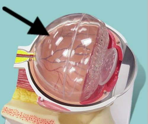
choroid
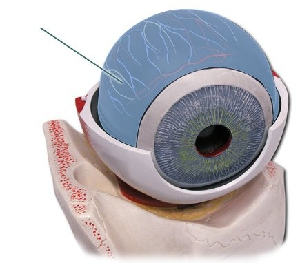
retina

fovea

optic disc/papilla
blind spot

aqueous humor

auricle/pinna
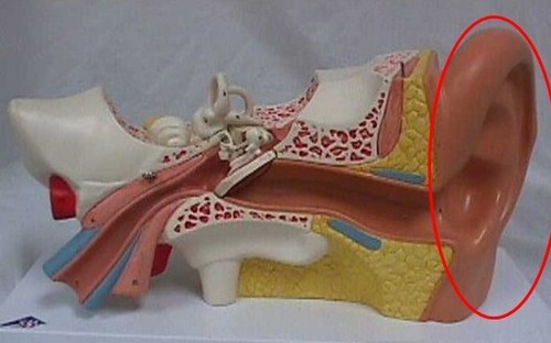
external auditory canal

tympanic membrane
eardrum

malleus
hammer

incus
anvil
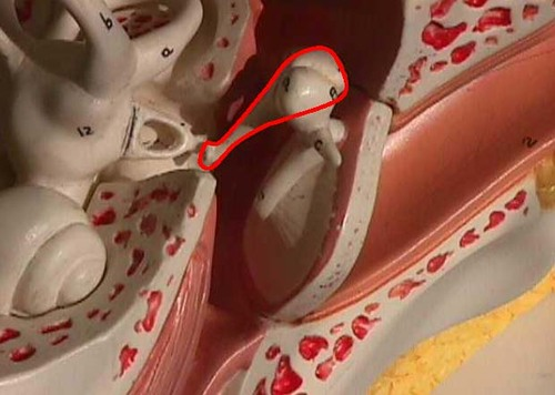
stapes
stirrups

semicircular canal
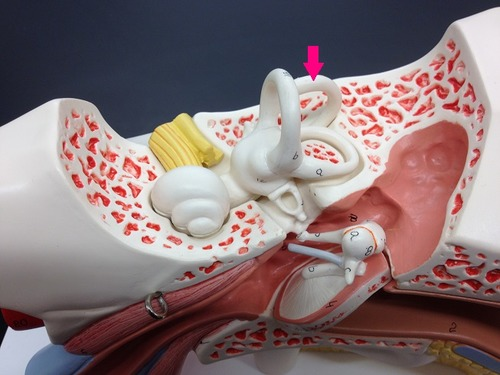
internal auditory canal

cochlea

auditory/eustachian tube

vestibular nerve
square blank

cochlear nerve

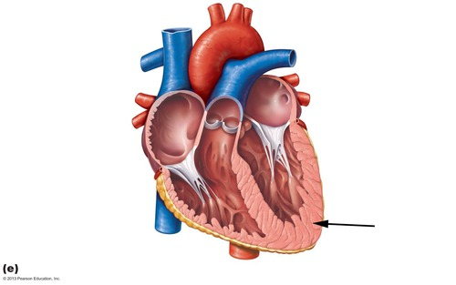
apex of heart

base of heart

mediastinum
space between the lungs
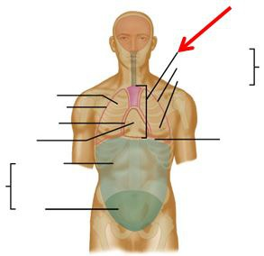
epicardium
visceral layer of pericardium

myocardium
cardiac muscle tissue