Central & Peripheral Nervous Systems 325 L
 Call Kai
 Learn
 Practice Test
 Spaced Repetition
 Match
 Flashcards
 Knowt Play
1/98

There's no tags or description
Looks like no tags are added yet.
99 Terms
CNS organization
consists of the brain (including cerebellum, diencephalon, cerebrum, and cerebral hemispheres) and spinal cord
olfactory bulb

optic chiasm

dura mater
composed of two layers (periosteal and meningeal layers)

pia mater

arachnoid mater
2

falx cerebri

falx cerebelli

tentorium cerebelli

subarachnoid space

cerebrospinal fluid
fills the ventricles within the brain

cerebral hemispheres

diencephalon
consists of the thalamus, hypothalamus, and epithalamus

brain stem
consists of midbrain, pons, and medulla

cerebellum

gyri

sulci

central sulcus

precentral gyrus

postcentral gyrus

primary motor area

primary somatosensory area

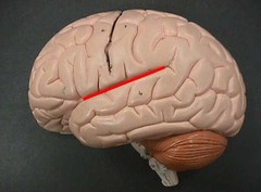
lateral sulcus
longitudinal fissure

frontal lobe

parietal lobe

temporal lobe

occipital lobe

cerebral cortex
made up of gray matter (unmyelinated axons)

corpus callosum

anterior commissure

septum pellucidum

lateral ventricles
orange part

intraventricular foramen

choroid plexus
makes cerebral spinal fluid (found in diencephalon)

fornix

basal nuclei
little "islands" of gray matter stuck within the white matter of nervous tissue

thalamus

internal medullary lamina of thalamus

cerebral white matter
area filled with myelinated axons

hypothalamus

infundibulum

pituitary gland

epithalamus

third ventricle

mammillary body

pineal gland

midbrain

pons

medulla oblongata

cerebral peduncles

cerebral aqueduct
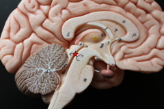
pyramids
corpora quadrigemina

inferior colliculi

superior colliculi
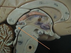
fourth ventricle

olives
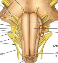
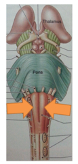
middle cerebellar peduncles
B

vermis

folia
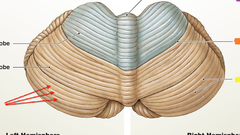
cerebellar nuclei

arbor vitae

anterior lobe of cerebellum
1

posterior lobe of cerebellum
2

olfactory cranial nerve
cranial nerve I
sensory fibers
responsible for smell
optic cranial nerve
cranial nerve II
sensory fibers
responsible for vision
oculomotor cranial nerve
cranial nerve III
mixed fibers (mainly motor)
responsible for eyeball and eyelid movement
trochlear cranial nerve
cranial nerve IV
mixed fibers (mainly motor)
responsible for eyeball movement
trigeminal cranial nerve
cranial nerve V
mixed fibers
responsible for chewing
abducens cranial nerve
cranial nerve VI
mixed fibers (mainly motor)
responsible for eyeball movement
facial cranial nerve
cranial nerve VII
mixed fibers
responsible for facial expression
vestibulocochlear cranial nerve
cranial nerve VIII
sensory fibers
responsible for hearing
glossopharyngeal cranial nerve
cranial nerve IX
mixed fibers
responsible for swallowing
vagus cranial nerve
cranial nerve X
mixed fibers
responsible for efferent output of skeletal muscles of pharynx and larynx and for smooth muscle and lands of thoracic and abdominal organs and for cardiac muscle
accessory cranial nerve
cranial nerve XI
motor fibers
responsible for efferent output of skeletal muscles of pharynx, larynx, neck, and shoulder
hypoglossal cranial nerve
cranial nerve XII
motor fibers
responsible for tongue movement
spinal meninges
consist of pia mater, dura mater, and arachnoid mater

white matter of spinal nerves

spinal nerve

dorsal roots

ventral roots

dorsal root ganglia

central canal

posterior median sulcus

anterior median fissure

gray matter of spinal nerve
consists of the anterior, posterior, and lateral horns

gray commissure

cervical plexus
formed by C1-C4 spinal nerves
innervates the skin and muscles of the neck, shoulder, and arm
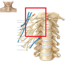
brachial plexus
formed by C5-C8 and T1 spinal nerves
supplies sensory and motor nerve fibers to the chest, shoulder, arm, forearm, and hand
major branches include the musculocutaneous, median, ulnar, axillary, and radial nerves

thoracic nerves
formed by T1-T12 spinal nerves
provide both somatic sensory and motor innervation to their corresponding intercostal spaces and some adjacent structures
nerves run their own course after emerging from the cord, innervating a narrow strip of skin in the anterior and posterior portion of the body in line with their emergence point from the spinal cord

lumbar plexus
formed by L1-L4 spinal nerves
innervates lower anterior abdominal wall, groin, thighs, knees, and calves
major branches include femoral and obturator nerves

sacral plexus
formed by L4-S3 spinal nerves
motor control to and receive sensory information from most of the pelvis and leg, along with upper and lower foot
major branch includes the sciatic nerve and inferior/superior gluteal nerves

somatic nerves
nerves that provide voluntary control to skeletal muscle for movement and receive sensory information such as touch, pain, and temperature from the skin
autonomic nerves
nerves that supply involuntary control to internal organs
PNS organization
consists of nerves (cranial, spinal) and ganglia (roots and branches, peripheral nerves, neuromuscular junctions)

association tract
communication of information from one region to another within the same cerebral hemisphere

commissural tract
communication of information from one cerebral hemisphere to the other

projection tract
communication of information between higher and lower brain areas and spinal cord centers
