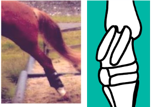
Hock and Distal Limb Muscles
 Call Kai
 Learn
 Practice Test
 Spaced Repetition
 Match
 Flashcards
 Knowt Play
1/27

Earn XP
Description and Tags
M.8R, W.3, L.8
28 Terms
Describe the tarsus/hock (bones its composed of, type of bones, the different rows and what they’re composed of)
composed of tarsal bones
Type
short bones
Proximal row
Talus
Calcaneus
Middle row
Central & 4th TBs
Distal row
I, 2, 3 & 4 TBs
4th TB bridges middle and distal rows

Which part of the tarsus/hock is palpable and what row is it in?
Calcaneus
proximal row

List the species differences of the tarsus/hock in regards to the equine
Distal Row
1 & 2 fused
3 very large

List the 4 joints of the tarsus/hock
Tibio-tarsal joint (TT):
Talus - tibia & fibula
Proximal intertarsal joint (PIT):
Talus & calcaneus - Central & 4 Tbs
Distal intertarsal joint (DIT):
Central – 1, 2 & 3 TBs
Tarso-metatarsal joint (TMT):
1,2,3 & 4 TBs - metatarsal bones

Which joint of the tarsus has the most movement
Tibio-tarsal joint
How is the communication between the compartments of the tarsus/hock?
poor communication between compartments
which joint of the tarsus is palpable and at what anatomical aspects is it palpable
Tibio-tarsal joint
Dorsomedial aspect
Plantarolateral aspect
Plantaromedial aspect
List the structures that provide stability for the tarus/hock
Collateral ligaments:
Fibrocartilagenous reinforcement of joint capsule
Retinaculum
Plantar ligament
Where are the collateral ligaments of the tarus/hock located?
Long: Tibia – metatarsal bone
Short: Bridge bone – bone
How many centers of ossification do the bones of teh tarsus/hock have and list the exception
All single center of ossification
Except calcaneus = 2
List the distal hindlimb muscles
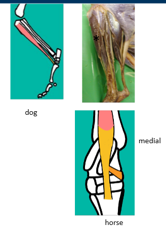
Cranial tibial muscle
Describe the Cranial tibial muscle (origin, insertion, function, nerve supply)
Origin
proximal tibia
Insertion
metatarsal bones (all species)
Insertion
medial aspect hock (horse) = Cunean tendon
Function
Hock flexion
Nerve Supply
Peroneal / Fibular Nerve (branch of Sciatic)
Describe the
Origin
lateral tibia & fibula
Insertion
Plantar aspect tarsus
Function
Hock flexion
Nerve Supply
Peroneal / Fibular Nerve (branch of Sciatic)

Origin
lateral femoral condyle
(with long digital extensor muscle)
Insertion
3rd MT (with cranial tibial muscle)
Insertion
lateral aspect tarsus
Function
Hock flexion
Nerve Supply
Peroneal / Fibular Nerve (branch of Sciatic)

Long Digital Extensor Muscle
Origin
extensor fossa (femur)
Tendon of Origin
incorporated into stifle capsule
provides lateral collateral support to stifle
Insertion
all digits
distal phalanx - extensor process
Function
Hock flexion
Digital extension
Nerve Supply
Peroneal / Fibular Nerve (branch of Sciatic)

list the distal hindlimb muscles (caudal aspect of limb)
Common calcanean tendon
Describe the Common Calcanean tendon
Components:
Biceps femoris
Semitendinosis
Gracilis
Gastrocnemius
Superficial digital flexor
I – calcaneus
Tuber calcis / calcanean tuberosity
Acts as lever for distal limb
Plantar ligament
Function
hock extension
Nerve supply
reflects contributors
mixed nerve supply
mostly sciatic and tibial nerves contributing

Gastrocnemius muscle
Origin
femur
Insertion
calcaneous
Function
hock extensor
stifle flexor
Nerve supply
Tibial Nerve (branch of Sciatic)
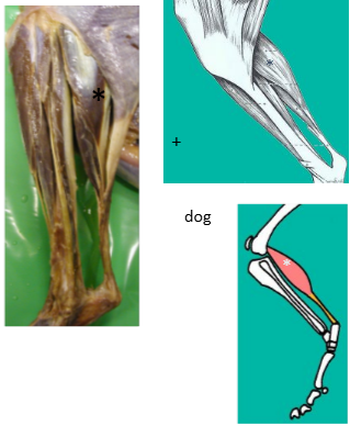
Describe the Superficial Digital Flexor Muscle (Origin, Insertion, Function and Nerve Supply)
Origin
distal femur
Insertion
calcaneus
Insertion
branches to all digits (middle phalanx)
Function
hock extension
digital flexion
Nerve supply
Tibial Nerve (branch of Sciatic)

Describe the Deep Digital Flexor Muscle (origin, insertion, function, and nerve supply)
Origin
Tibia
Insertion
branches to all digits (middle phalanx)
Function
hock extensor
digital flexor
Nerve Supply
Tibial Nerve (branch of Sciatic)

What is the natural weightbearing position of the hindlimb stay apparatus?
Extension
What do the MTP, PIP and DIP joints prevent?
hyperextension
List the muscles that create a combined force for hindlimb extension
Gluteal muscles
hip extensors / limb retractors
stifle extensors
hock extensors
Where are the forces of the muscles during hindlimb extension directed?
caudally in axial plane