A&P Lab 1 Practical #2: Nervous System - Chapters 15, 17, & 19
 Call Kai
 Learn
Learn Practice Test
 Spaced Repetition
 Match
Match Flashcards
 Knowt Play
1/132

Earn XP
Description and Tags
This flashcard set covers all of the content we'll be tested on for our A&P Lab 1 Practical Exam #2. It covers Chapters 15, 17, and 19 which cover the Histology of Nervous Tissue (Ch. 15), Gross Anatomy of the Brain and Cranial Nerves (Ch. 17), and The Spinal cord and Spinal Nerves (Ch. 19) respectively. Practical Exam #2 is 92% labeling.
Name | Mastery | Learn | Test | Matching | Spaced | Call with Kai | Chat |
|---|
No analytics yet
Send a link to your students to track their progress
133 Terms
To-Do List for flashcard deck:
Anatomical language flashcards
Diagram / labeling w/ location, physical description (function, etc as needed)
Cranial Nerves 1-12
Add any additional flashcards I need
Edit and organize and re-order and clean up flashcards AND finish description
IGNORE THIS
REVIEW: Anatomical terminology
Toward the front
Toward the back
Toward the middle
Toward the side
Toward the head
Toward the feet
Closer to the point where a limb attaches
Farther from the point where a limb attaches
Near the surface of the body
Away from the surface of the body
Toward the front → Anterior / Ventral
Toward the back → Posterior / Dorsal
Toward the middle → Medial
Toward the side → Lateral
Toward the head → Superior / Cranial
Toward the feet → Inferior / Caudal
Closer to the point where a limb attaches → Proximal
Farther from the point where a limb attaches → Distal
Near the surface of the body → Superficial
Away from the surface of the body → Deep
What is the Nervous System?
Master integrating and coordinating system of the body
Continuously monitors and processes sensory information from the external and internal environment
Every thought and action of human body is a reflection of the nervous system’s processes
How does the Nervous System work?
Processes and integrates new information (“inputs”) with information previously fed into it to produce a response.
Has two major divisions: CNS and PNS
What are the major divisions of the Nervous System?
Two major divisions:
Central Nervous System (CNS)
Peripheral Nervous System (PNS)
What organs/structures are found in the Central Nervous System (CNS)? Function?
Brain and spinal cord
Interprets sensory information based on that information and past experiences
What organs/structures are found in the Peripheral Nervous System (PNS)? Function?
Everything in the nervous system except for the brain and spinal cord including: Spinal nerves, ganglia, and cranial nerves
Connects the CNS to the rest of the body
Sensory vs Motor vs Mixed Division of Nervous System
Sensory (Afferent) Division: UP
Sensory = Afferent division
Carries nerve impulses to CNS from sensory receptors
Motor (Efferent) Division: DOWN
Motor = Efferent division
Transmits impulses away from the CNS to effector organs (muscles and glands)
Splits into SNS and ANS
Mixed Nerves (Both Sensory and Motor)
Nerve bundles containing both afferent (sensory) and efferent (motor) fibers traveling in parallel pathways.
Ex: All 31 pairs of spinal nerves and mixed cranial nerves (CN V, VII, IX, and X).
What is a neuron? Function?
Neuron = Nerve cell
Transmits electrical and chemical messages throughout the body to control movement, senses, and thinking

What are the important structures of an neuron?
8 Structures:
Cell Body (Soma)
Dendrites
Axon
Myelin Sheath (via Schwann cells/oligodendrocytes)
Nodes of Ranvier
Axon Terminals
Myelin Sheath
Axon Terminals / Terminal Buttons
Additionally, the cytoskeleton is important for structural support and substance transport.


What is an axon?
Do this for the other parts of neuron later
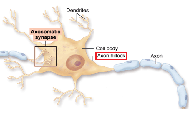
Long, thin, thread-like fiber projection / extension of nerve cell (neuron)
Sends electrical signals away from the cell body to other cells


Label: Axon hillock
*Do this for the other parts of the neuron as well
Location: At the junction where the cell body (soma) of a neuron transitions into the axon
What are the major divisions of the human nervous system?
REPEAT QUESTION
Two major divisions:
Peripheral Nervous System
Everything but spinal cord and brain
Central Nervous System
Spinal cord and brain
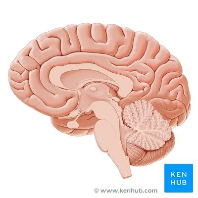
Label: Cerebrum
Location: The “big brain” inside the skull at the top and front of the skull
The largest, topmost portion of the human brain
Function: Sensory processing, voluntary muscle movement
*Don’t confuse with the cerebellum (“the little brain”)


Label: Cerebellum
Location: lower back of the brain, directly beneath the larger cerebrum and behind the brainstem
“Small brain” (think -ellum = small)
The cerebellum is located at thelower back of the head, beneath the large cerebral cortex and behind the brainstem
Function: Maintain balance and posture, coordinate voluntary muscle movements


What is a nerve plexus?
A nerve plexus is a complex network of intersecting nerves that route sensory and motor signals between the spinal cord and specific regions of the body

What are the major nerve plexuses in the body?
5 Major Plexus:
Cervical Plexus
Brachial Plexus
Lumbar Plexus
Sacral Plexus
Coccygeal


Label: Cervical plexus
First plexus of human body / at top (?)
Superior to brachial plexus


Label: Cervical enlargement
The region of the c-spine that gets wider


Label: Brachial plexus
Think: Located at brachial artery / shoulder
*Not on the inside where the musculocutaneous nerve is
Inferior to cervical plexus

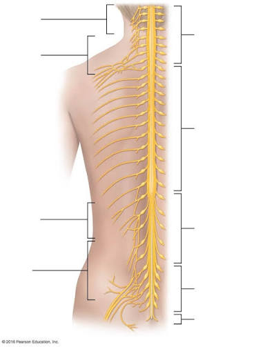
Label: Intercostal nerves
Intercostal = Between the ribs


Label: Lumbar plexus
Location: Lower back (lumbar region), lying in the retroperitoneal space (behind the abdominal cavity lining).


Label: Lumbar enlargement
AKA lumbosacral enlargement
A widened, thickened region of the spinal cord that provides the neural connections for the lower limbs


Label: Sacral plexus
Inferior to lumbar plexus
Think: Above coccygeal / above area that goes into / behind pelvic bones
A web of intersecting nerves located on the back wall of the pelvis that supplies motor and sensory function to the pelvis, buttocks, and lower limbs


Label: Conus medullaris
Think: Looks like little cones
The tapered, cone-shaped lower end of the spinal cord


Label: Filum terminale
Fibers at the very end of the spinal cord // Delicate strand of fibrous tissue that anchors the lower end of the spinal cord to the coccyx


Label: Cauda equina
Think: “Horse hair-like projections” AFTER spine ends
A bundle of nerve roots that continues below where the spinal cord ends.


Label: Intervertebral discs
Inter = Between
Vertebral = Vertebrae
Discs between vertebrae
A fibrocartilaginous cushion that sits between adjacent vertebrae in the spinal colum
Function: Shock absorber, allows spinal flexibility

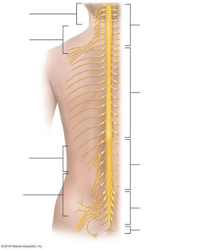
Label: Intervertebral foramen
Foramen = Opening
Think: Small openings in spine near vertebrae
A small, oval-shaped opening between adjacent vertebrae where spinal nerves and blood vessels pass from the spinal cord to the rest of the bod
Spinal nerves exit spinal column through intervetebral foreman


Label: Spinal nerve
Location: Along the length of the spinal cord
Humans have 31 pairs of spinal nerves (and 12 CN) extending from the brain to the tailbone
Spinal nerves exit spinal column through small openings called intervertebral foramina (think: “small holes'“) located between adjacent vertebrae
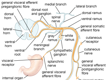
What is the breakdown for the spinal nerves?
31 Spinal Nerves in human body split into 5 sections:
Cervical: C1-C8
Neck region
Thoracic: T1-T12
Upper and mid-back
Lumbar: L1-L5
Lower back
Sacral: S1-S5
Pelvic region (sacrum)
Coccygeal (Co1)
Started as 3 bones fused throughout childhood
Located a the very bottom of the spine - the coccyx
CAN THE LADY SAVE CATS to remember the names + order of SN

Label: Cervical plexus
A network of nerve fibers formed by the anterior rami of the first four cervical spinal nerves (C1–C4) located deep in the neck.
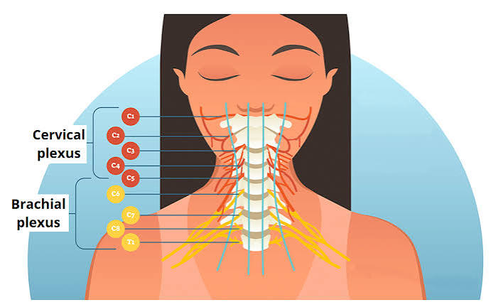

Label: Brachial plexus
Inferior to cervical plexus


Label: Lumbar plexus
Superior to sacral plexus


Label: Sacral plexus
Inferior to cervical plexus and superior to coccygeal
Collection of nerves in sacral spine


Label: Spinal curve
The natural or abnormal alignment, direction, and degree of curvature of the backbone
Label: Dorsal root ganglion
Dorsal = Back
Looks like a bulge (Think: ganglion = bulge)
Positioned in the intervertebral foramina (the small bony openings between adjacent vertebrae) AND found at very spinal level between spinal column
A cluster of sensory neuron cell bodies located on the dorsal root of a spinal nerve just outside the spinal cord

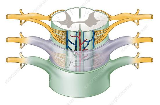
Label: Dorsal root
Dorsal = Back
Connected to dorsal root ganglion and closer to the grey matter than the dorsal root ganglion is
Emerges from the posterior lateral sulcus of the spinal cord
A sensory nerve bundle that transmits information from the body's peripheral receptors to the spinal cord


Label: Ventral root
Ventral = Front
Location: Located at the front (anterior side) of the spinal cord where small groups of nerve fibers emerge to carry motor signals out to the body
CLOSER TO VENTRAL MEDIAN FISSURE (big opening / slit in spinal cord cross section)
Starts as tiny thread-like rootlets coming out of a groove from the front of the spinal cord
Runs outward and meets the dorsal root (which handles sensory signals) just before they pass through a tiny opening in the backbone to form a regular spinal nerve
Function: Motor control


Label: Epidural space
Cylindrical, fat-filled compartment located outside the dural sac inside the bony vertebral canal
Has a distinct yellowish hue and a highly vascularized, soft, compartmentalized texture
Outside the protective dural sac of the spinal cord
Location: Inside the spinal canal between the bones of the vertebrae and the outermost layer of the spinal meninges (the dura mater)
is the outermost part of the spinal canal, located between the tough protective membrane covering the spinal cord (called the dura mater) and the inside walls of the bony vertebrae
Runs the entire length of the spine


Label: Dura mater
Blue layer
Dura mater = “Tough mother”
Outermost, thick protective meningeal layer
Thick, tough, leather-like or rubberized membrane
DAP from superior to inferior

Label: Dura mater
BLUE LAYER


Label: Falx cerebri
Top of skull inside
Dural fold separating cerebral hemispheres

Label: Tentorium cerebelli
Think: The “floor” of the skull / floor of “tent” if placed in skull
Dural fold seperating cerebrum from cerebellum
Location: Sits horizontally inside the cranium like a tent roof

Label: Falx cerebelli
Small fold separating cerebellar hemispheres
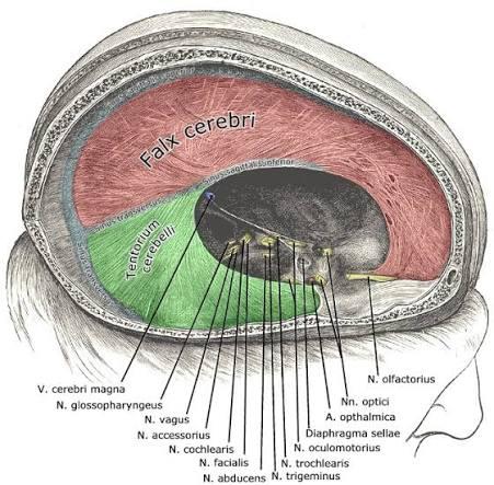
Label: Subdural space
BLUE LAYER

Label: Arachnoid mater
SPIDER WEB-Y LOOKING
VERY HALLOWEEN-Y

Label: Subarachnoid space
LIGHT BLUE
CSF inside of it
Label: Pia mater
PINK (PIA = PINK)
innermost, highly vascularized protective layer that forms a tight, form-fitting seal over the nervous tissue
DELETE
DELETE
DELETE

Label: Gray matter
Gray Matter = Cell bodies and terminals
Location
The gray matter of the spinal cord is a butterfly- or H-shaped region in the center of the cord that contains nerve cell bodies, unmyelinated fibers, and glial cells. It processes spinal reflexes and integrates incoming sensory and outgoing motor signals. You can learn more about these internal structures through the The Internal Anatomy of the Spinal Cord guide
Three main regions with gray matter in the spinal cord

What regions of the spinal cord are made up of gray matter?
3 Main Regions:
Posterior (Dorsal) Horn
Anterior (Ventral) Horn
Lateral Horn

Label: Posterior Horn
AKA: Dorsal horn
Location: Posterior (back) section of the spinal cord’’s butterfly / “H-shaped” gray matter
Position: Found on the back or rear side of the spinal cord inside the central gray matter region
Orientation: Forms posterior projection (the upper or rear “wings”) of the butterfly-shaped gray matter
*Extends along the length of the spinal cord on the left and right sides

What is the function of gray matter?
Signal Integration
Controlling reflexes
Sending motor commands


Label: Gray commissure
Location: At the center of the spinal cord, surrounding the small central canal (which contains CSF) and bridges the H-shaped columns of gray matter (between the anterior/ventral horn and posterior/dorsal horn)
Description: Thin, horizontal strip of gray matter that connects the left and right halves of the spinal cord
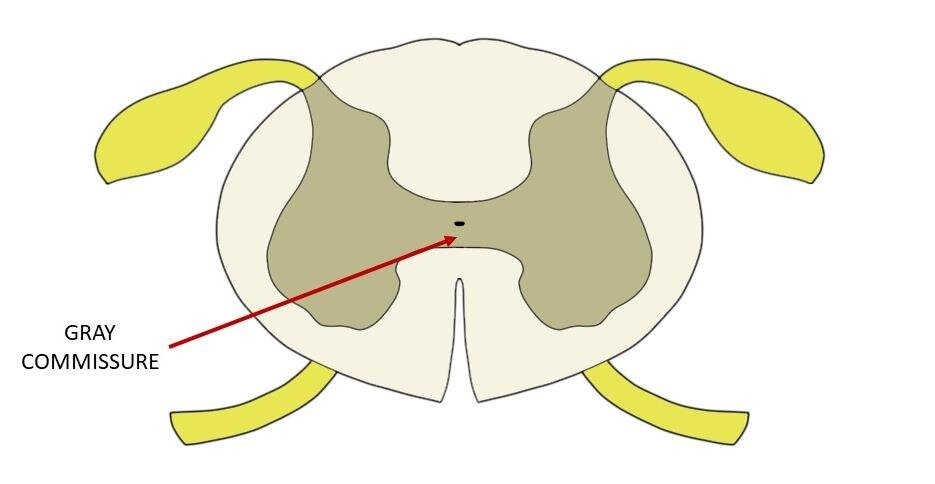

What are the subdivisions for the gray commissure?
Two subdivisions:
Anterior gray commissure (in front of the central canal)
Posterior gray commissure (behind the central canal)

Label: White matter
White matter = Axon therefore it transmits signals
Axons give it the white appearance
Location: On the outside (periphery) of the spinal cord surrounding the butterfly-shaped gray matter in the center forming the superficial boundary
Divided into 3 main regions in spinal cord
white matter of the spinal cord is the outer tissue layer that surrounds the central, butterfly-shaped gray matter and acts as a signal highway connecting the body to the brain. [1, 2, 3]
Structure and Composition
Appearance: It looks white because it is rich in myelin, a fatty insulating cover around nerve fibers. [1, 2]
Contents: It consists mostly of myelinated axons (nerve fibers) rather than cell bodies. [1, 2]
Organization: It is divided into three paired columns or funiculi:
Dorsal (posterior) column: Located in the back.
Lateral column: Located on the sides.
Ventral (anterior) column: Located in the front. [1, 2, 3, 4]
Function and Pathways
The white matter contains bundles of nerve fibers called tracts that transmit signals
![<ul><li><p>White matter = Axon therefore it transmits signals </p></li><li><p>Axons give it the white appearance </p></li><li><p>Location: On the outside (periphery) of the spinal cord surrounding the butterfly-shaped gray matter in the center forming the superficial boundary</p></li><li><p>Divided into 3 main regions in spinal cord</p></li></ul><p></p><p><span>white matter of the spinal cord is </span><strong><mark>the outer tissue layer that surrounds the central, butterfly-shaped gray matter and acts as a signal highway connecting the body to the brain</mark></strong>. [1, 2, 3]</p><p><strong>Structure and Composition</strong></p><ul><li><p><span><strong>Appearance:</strong> It looks white because it is rich in myelin, a fatty insulating cover around nerve fibers.</span> [1, 2]</p></li><li><p><span><strong>Contents:</strong> It consists mostly of myelinated axons (nerve fibers) rather than cell bodies.</span> [1, 2]</p></li><li><p><span><strong>Organization:</strong> It is divided into three paired columns or funiculi:</span></p><ul><li><p><span><strong>Dorsal (posterior) column:</strong> Located in the back.</span></p></li><li><p><span><strong>Lateral column:</strong> Located on the sides.</span></p></li><li><p><span><strong>Ventral (anterior) column:</strong> Located in the front. [1, 2, 3, 4]</span></p></li></ul></li></ul><p><span><strong>Function and Pathways</strong></span></p><p><span>The white matter contains bundles of nerve fibers called tracts that transmit signals</span></p>](https://assets.knowt.com/user-attachments/9d1c0449-5d74-4e2b-9f2d-87b1e75c28c5.png)
What regions of the spinal cord does white matter make up?
3 Funiculi:
Funiculi = Column
Dorsal (posterior) column
Lateral column
Ventral (anterior) column


Label: Dorsal/posterior funiculi
Funiculus = Column
Dorsal = back
TWO COLUMNS


Label: Ventral/anterior funiculus
Funiculus = Column


Label: Lateral funiculus
Funiculus = Column

Label: Posterior median sulcus
Posterior = Behind
sulcus = shallow groove

Label: Anterior median fissure
Fissure = Deep section

Label: Central canal
Hole in the center running through the entire spinal cord

Label: Spinal nerve on spinal cord cross section of thoracic region
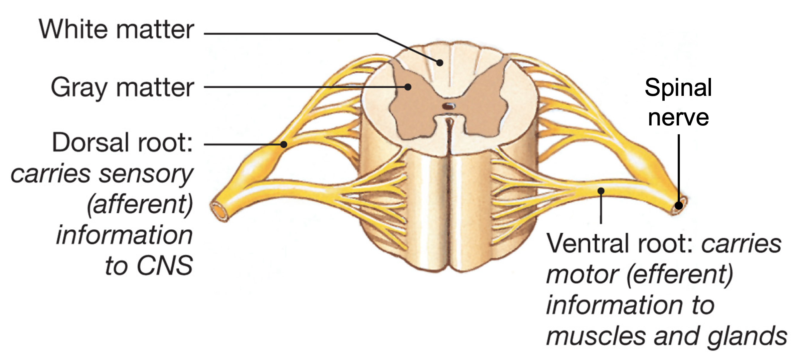
Label: Dorsal root ganglion
Dorsal= back
Ganglion = bulge

Label: Dorsal root
Label: Ventral root
Ventral = front
Label: Dorsal horn

Label: Ventral horn
Dorsal = back

Label: Ventral horn

Label: Lateral horn
Label: Posterior median sulcus
Label: Anterior median fissure
Label: Gray matter
Label: Gray commissure
Label: White matter
Label: Dorsal funiculus
Funiculus = Column
Label: Ventral funiculus
Funiculus = Column
Label: Lateral funiculus
Funiculus = Column
Label: Central canal
Label: Cervical plexus
Label: Brachial plexus
Label: Lumbar plexus
Label: Sacral plexus
Label: Conus medullaris
Description: Tapered, cone-shaped lower end of the spinal cord
What are the peripheral nerves?
9 Main Peripheral Nerves:
Radial nerve
Median nerve
Axillary nerve
Musculocutaneous nerve
Ulnar nerve
Femoral nerve
Sciatic nerve
Fibular nerve
Tibial nerve
Label: Radial nerve
Think: Thumb / “where I take a radial pulse”

Label: Median nerve
Median = Middle (of arm in this case)

Label: Axillary nerve
Nerve in shoulder
*not musculocutaneous
Label: Musculocutaneous nerve

Label: Ulnar nerve
Think: Pinky
Location: Refers to structures in the forearm and hand located on the same side as the pinky finger


Label: Femoral nerve
Think: Femur
Location: Located in lower back, pelvis,and travels down the front of each thigh
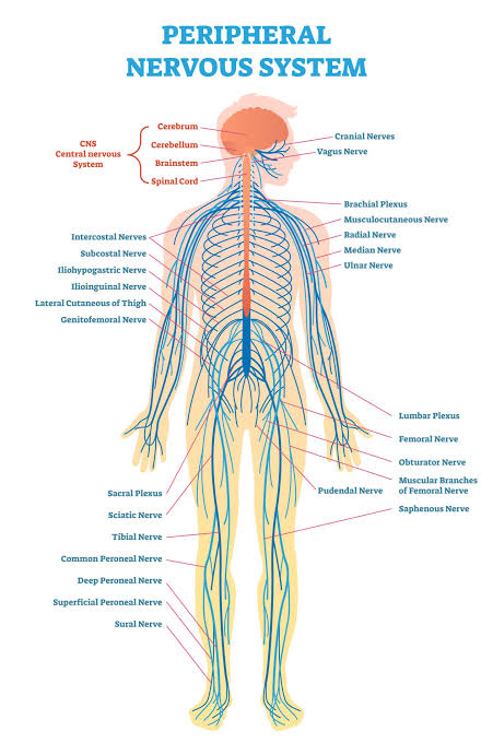

Label: Sciatic nerve
THIGH
Think: Runs from butt to leg
Location: In the lower spine and runs down the back of each leg

Label: Fibular nerve
BUTT

Label: Tibial nerve
OUTSIDE


Label: Cerebrum
Location: Located at the lower back of the brain, directly beneath the rear of the cerebrum and behind the brainstem
Split into left and right hemispheres
Has a bundle of nerves called corpus callosum that joins the two halves to send signals back and forth
Description: Large, upper, wrinkled part of brain

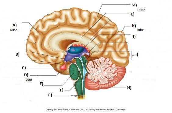
Label: Cerebellum
Location: Lower back of the skull and sits at the back of head, below the cerebrum and behind brainstem
Has two hemispheres and a middle section called the vermis
An inner branched white matter pattern → the arbor vitae or tree of life (Think: Looks like tree branches)
Description: Small, tightly folded structure located underneath the back of the cerebrum
