H&N exam 1
 Call Kai
 Learn
Learn Practice Test
 Spaced Repetition
 Match
Match Flashcards
 Knowt Play
1/106

There's no tags or description
Looks like no tags are added yet.
Name | Mastery | Learn | Test | Matching | Spaced | Call with Kai | Chat |
|---|
No analytics yet
Send a link to your students to track their progress
107 Terms
Viscerocranium
Face
Neurocranium
Cranial base and calvaria
Coronal suture
Joins frontal and parietal bones
Sagittal suture
Joins the two parietal bones along the midline.
Lamdboid suture
Joins the occipital bone with the parietal bones at the posterior of the skull.
Paired bones
Pariteal and temporal
Unpaired bones
Fronta, occipital, sphenoid, ethmoid
Fontanelles
Gaps between incompletely joined bones in infants
Pterion
Region in temporal fossa where many skull bones meet
Middle meningeal artery travels deep to the pterion

A fracture in this region can cause a epidural hematoma (blood btwn skull and dura mater)
Pterion
because it damages the middle meningeal artery
Hypophyseal fossa (pit in the sella turcica)
Houses the pituitary gland

Dorsum sellae
Posterior boundary of the sella turcica.

Petrous ridge
Part of the temporal bone separating the middle and posterior cranial fossae.

Foramen magnum
Large opening in the occipital bone
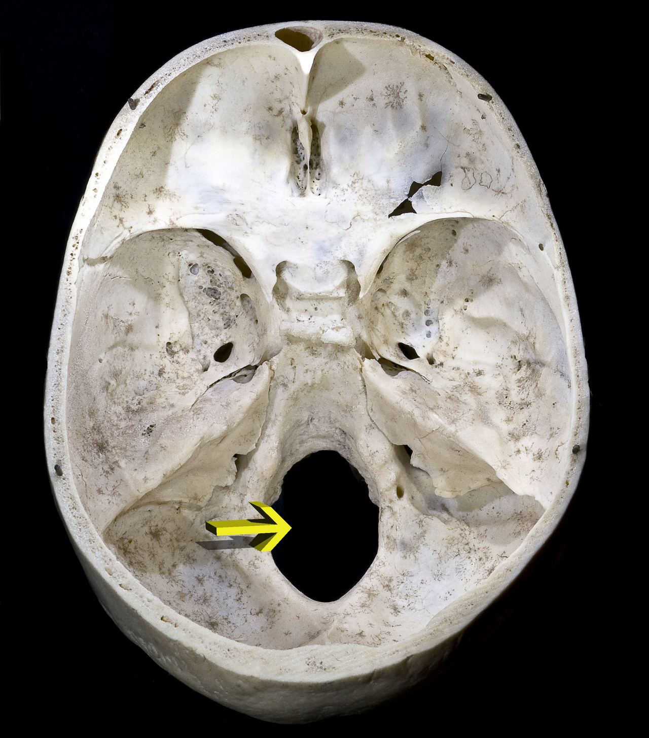
Occipital condyles
Articulate with the atlas (C1)

CN VII nerve route
enters the internal acoustic meatus
passes through the facial canal in the temporal bone
its motor branches exit via the stylomastoid foramen.
CN XI
has spinal roots that enter the skull through the foramen magnum before exiting through the jugular foramen
Layers of scalp superficial to deep
· S — Skin
· C — Connective tissue (dense)
· A — Aponeurosis
· L — Loose connective tissue
· P — Periosteum
Dense connective tissue
Contains major nerves and blood vessels supplying the scalp
Aponeurotic layer
Aponeurotic layer
Loose connective tissue
Allows movement of the superficial scalp
Periosteum
Covers the skull bones
Outside to inside of the skull
Scalp
Calvarial bone
Meningeal vessels
Meninges
Subarachnoid space
Cerebral arteries
Structure of the Cranial Vault
Outer compact table: Hard, dense bone
Diploë: Spongy bone containing diploic veins
Inner compact table: Hard, dense bone
Emissary veins:
Veins that pass through openings in the skull and connect veins outside the skull with intracranial venous sinuses.

3 meningeal layers
Dura mater
Arachnoid mater
Pia mater
2 Layers of the Dura mater
1. Periosteal layer: Outer layer attached to the skull.
2. Meningeal layer: Inner layer that forms the dural folds.
When the two layers separate, they create dural venous sinuses.
Epidural space
Between skull and dura
potential space in the cranial cavity
Subdural space
Between dura and arachnoid
Potential space
Subarachnoid space
Between arachnoid and pia
contains CSF and major cerebral blood vessels
Dural fold
Dural folds are formed by the meningeal layer of dura mater.
They divide the cranial cavity into smaller compartments and help support the brain.
Dural fold: Falx cerebri
Separates the right and left cerebral hemispheres

Tentorium cerebelli
Separates the cerebrum from the cerebellum

The superior sagittal sinus travels along..
the superior attachment of the falx cerebri.
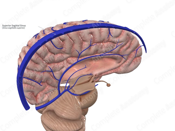
Dural venous sinus
Dural venous sinuses are endothelium-lined spaces formed between the periosteal and meningeal layers of dura mater.
They receive:
Venous blood from the brain, scalp and other connected regions
CSF through arachnoid granulations (villi)
Superior sagittal sinus
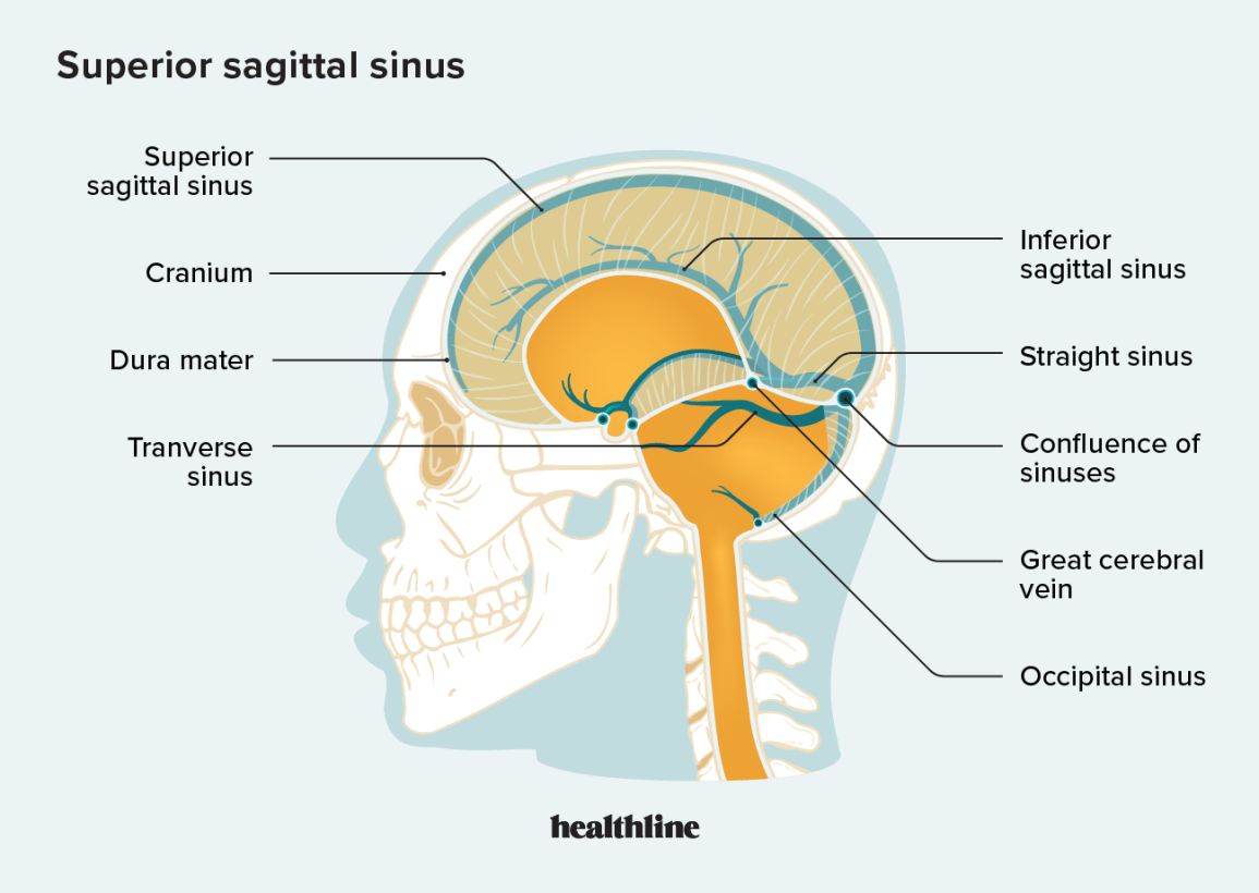
Confluence of sinuses
Transverse sinus

Sigmoid sinus

Arachnoid granulations
allow CSF to return to venous circulation.
CSF passes from the subarachnoid space into the dural venous sinuses.

The cavernous sinus is located
on either side of the sella turcica.
why is CN VI (Abducens nerve) vulnerable to damage in the cavernous sinus
because it travels directly beside the internal carotid artery
structures in the cavernous sinus
Structure | Location |
CN III — Oculomotor | Lateral wall |
CN IV — Trochlear | Lateral wall |
CN V1 — Ophthalmic | Lateral wall |
CN V2 — Maxillary | Lateral wall |
CN VI — Abducens | Within sinus |
Internal carotid artery | Within sinus |
2 arterial systems that supply blood to the brain
Anterior: internal carotid
Posterior: vetebral arteries
Pathway 1: Internal carotid arteries (anterior)
Common carotid artery
Internal carotid artery
Enters skull through carotid canal
ACA
Anterior cerebral artery
MCA
Middle cerebral artery
The internal carotid artery also gives off the posterior communicating artery and ophthalmic artery.
Pathway 2: Vertebral arteries (posterior)
Right and left subclavian arteries
Right and left vertebral arteries
Travel through transverse foramina C6–C1 and enter the foramen magnum
One basilar artery
Formed when the two vertebral arteries join; travels along the pons
Right and left posterior cerebral arteries (PCA)
Anterior cerebral arteries (ACA)
Right and left branches of the internal carotid system.
Anterior communicating artery (ACom)
Connects the right and left anterior cerebral arteries.
Internal carotid arteries
Contribute to the anterior circulation.
Posterior communicating arteries (PCom):
Connect the internal carotid arteries to the posterior cerebral arteries.
Posterior cerebral arteries (PCA):
Arise from the basilar artery and contribute to the posterior portion of the circle.
Anastomosis:
A connection between blood vessels.
Collateral circulation:
Alternate circulation that allows blood to travel around an obstructed vessel.
structures that pass through Optic canal
CN II and ophthalmic artery
structures that pass through Superior orbital fissure
CN III, IV, V1, VI and superior ophthalmic vein
structures that pass through Inferior orbital fissure
Maxillary nerve (V2) and inferior ophthalmic vein
structures that pass through Anterior/posterior ethmoidal foramina
Ethmoidal neurovascular structures
structures that pass through Nasolacrimal canal
Nasolacrimal duct
Branches of V1 associated with the superior orbital fissure
Frontal nerve
Lacrimal nerve
Nasociliary nerve
Orbital adipose tissue
· Protection of orbital structures
· Support of eye movement
Orbital septum
· Preseptal space: Contains eyelid, skin, and fascia.
· Postseptal space: Located behind the orbital septum.

The postseptal space is further divided into
· Intraconal space: Within the muscle cone.
· Extraconal space: Outside the muscle cone.
Medial palpebral commissure
Medial junction of upper and lower eyelids

Lateral palpebral commissure
Lateral junction of upper and lower eyelids

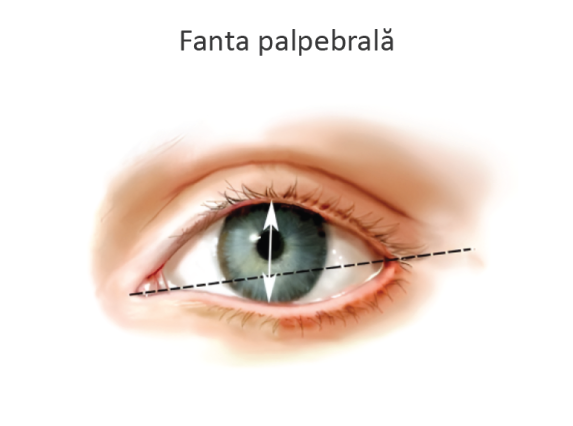
Palpebral fissure
Opening between the eyelids
Sclera
White outer covering of the eye

Cornea
Transparent anterior covering

Iris
Colored portion of the eye
Pupil
Opening in the center of the iris
Conjunctiva
Mucous membrane covering portions of the eye and eyelids
Palpebral conjunctiva: Lines the inner surface of the eyelids.
Bulbar conjunctiva: Covers the anterior sclera.
Levator palpebrae superioris
raises upper eyelid
Inn: CN III
Superior tarsal muscle
assists upper eyelid elevation
Inn: sympathetic
Orbicularis oculi
Closing eyes or blinking
CN VII
lacrimal apparatus
produces and drains tears
Lacrimal gland
Located in the superolateral portion of the orbit.
Secretes lacrimal fluid (tears).
Receives parasympathetic innervation associated with CN VII (facial nerve).

Tear drainage pathway
Lacrimal gland
Produces tears
Conjunctival sac / surface of eye
Tears move medially across the eye
Lacrimal puncta
Lacrimal canaliculi
Lacrimal sac
Nasolacrimal duct
Inferior meatus of nasal cavity
Outer fibrous layer
Sclera and cornea
Middle vascular layer
Choroid, ciliary body and iris
Inner neural layer
Retina
Choroid
Pigmented, vascular layer.
Supports the retina.

Ciliary body
Anterior continuation of the choroid
Connected to the lens through zonular fibers.
Contains the ciliary muscle.

Retina
Neural layer of the eye.
Contains rods and cones (photoreceptors).
Associated with the optic nerve (CN II).
Anterior chamber
Between cornea and iris
Posterior chamber
Between iris and lens
Aqueous humor
anterior and posterior chambers
Vitreous humor
large cavity behind the lens.
Superior rectus
Elevation; also adduction and intorsion
CN III
Inferior rectus
Depression; also adduction and extorsion
CN III
Medial rectus
Adduction (eye moves medially)
CN III
Lateral rectus
Abduction (eye moves laterally)
CN VI
Superior oblique
Intorsion; also depression and abduction
CN IV
Adducted eye looks down
Inferior oblique
Extorsion; also elevation and abduction
CN III
Adducted eye looks up
Intorsion (Adduction)
Top of the eye rotates toward the nose.
Extorsion (ab-duction)
Top of the eye rotates away from the nose.
H test: Rectus muscles
test vertical movement when eye is abducted.
H test: Oblique muscles
test vertical movement when eye is adducted
CN III somatic motor
moves and lifts eyeball
Innervates:
· Superior rectus
· Inferior rectus
· Medial rectus
· Inferior oblique
· Levator palpebrae superioris
CN III parasympathetic
Controls pupil size and helps the eye focus on nearby objects.
parasympathetic functions:
Ciliary muscle | Changes lens shape to focus on nearby objects (accommodation) |
Sphincter pupillae | Constricts the pupil (makes it smaller) |