Exercise 23 & 24 special senses: visual anatomy and visual experiments
 Call Kai
 Learn
 Practice Test
 Spaced Repetition
 Match
 Flashcards
 Knowt Play
1/68

Earn XP
Description and Tags
69 Terms

identify this structure and its function
structure: Lacrimal glands
function: produces tears
what enzyme does the Lacrimal glands contain?
contains the antimicrobial enzyme

identify this structure and its function
structure; excretory ducts
function: Several small ducts that continually liberate tears from the lacrimal gland

identify this structure
structure: Lacrimal canaliculi

identify this structure
lacrimal punctum (open at corner of eye)

identify this structure
lacrimal sac


identify this structure
nasolacrimal duct

identify this structure
palpebrae (DO NOT CALL IT EYELID)

identify this structure and function
Lateral commissure
where the eyelid meet laterally

identify this structure and function
medial commissure
where the eyelids meet medially

identify this structure and function
caruncle
fleshy raised are at the medial commissure, produces a whitish, oily secretion
identify this structure
eyelashes
describe what a ciliary glands is
Modified sweat/sebaceous gland found in between the eyelash follicles
Secretes an oily substance that helps lubricate the surface of the eye
what’s a stye
Acute inflammation of a ciliary gland

describe what a tarsal gland is
Modified sebaceous gland posterior to the eyelashes
Secretes an oily substance that helps lubricate the surface of the eye
describe what conjunctiva is
It is a mucous membrane that secretes mucus that lubricates the eye
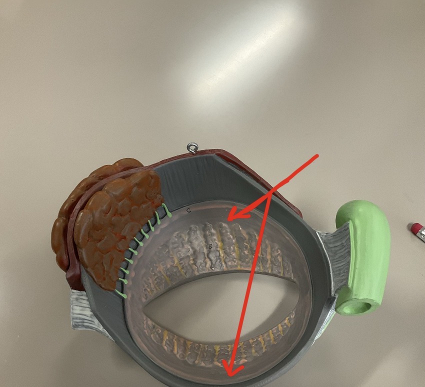
identify this structure
palpebral conjunctiva
lines the internal surface of the eyelid
identify this structure
bulbar conjunctiva
Lines anterior surface of the eyeball to its junction with the corneal epithelium
what is conjunctivitis
Inflammation of the conjunctiva

identify this structure, action, and cranial nerve #
Lateral rectus
moves eye laterally / VI (abducens)

identify this structure, action, and cranial nerve #
Medial rectus
moves eye medially / III (oculomotor)

identify this structure, action, and cranial nerve #
Superior rectus
elevates eye and turns it medially / III (oculomotor)

identify this structure, action, and cranial nerve #
Inferior rectus
depresses eye and turns it medially / III (oculomotor)

identify this structure, action, and cranial nerve #
inferior oblique
extorsion; roll eye laterally / III (oculomotor)

identify this structure, action, and cranial nerve #
Superior oblique
Intorsion; roll eye medially IV (trochlear)

Identify this structure
Superior oblique tendon

identify this structure
sclera (posterior)
white of the eye

Identify this structure
Pupil

identify this structure
Cornea (anterior)
transparent, anterior most portion

what structure is this and what’s its function
structure: iris
function:regulates the amount of light entering the eye
what does the circular smooth muscle do
constricts pupil (close vision, bright light)
what does the radial smooth muscle do
dilate pupil (distant visions, dim light)

identify this structure and its function, which is part of the BLANK
ciliary muscle, ciliary body, vascular tunic
control the shape of the lens

identify this structure and its function
suspensory ligaments
connect the lens to the ciliary body

identify this structure and its function
ciliary processes
capillaries that secretes aqueous humor

identify this structure and its function
choroid
contains dark pigment that prevents light scatter


identify this structure
optic nerve
identify this structure, location, and its function
pigmented layer
pigmented cells absorb light to prevent light scatter
Located between the choroid and neural layer

identify this structure and its function
optic disc
blind spot, where the optic nerve emerges (no rods/cones)

identify this structure and its function
fovea centralis
pit within the macula, has the highest visual acuity


identify this cell structure and its function
Pigmented part of retina

identify this cell structure and its function
rods
photoreceptors cells for dim light perceives gray tones

identify this cell structure and its function
cones
photoreceptors cells for color, need high amount of light

identify this cell structure
bipolar neurons

identify this cell structure and function
ganglion cell
axons converge and leave the retina as the optic nerve

identify this structure and its function
lens
crystallin structure held in place by suspensory ligaments attached to the ciliary body
identify this structure and its function
cataracts
lens becomes hardened and opaque

identify this structure and its function
anterior segment / anterior cavity
filled with aqueous humor

identify this structure and its function
posterior segment / posterior cavity
filled with vitreous

Identify this structure
Fovea centralis

Identify this structure
Macula lutea

what is aqueous humor
clear, watery fluid continually formed by the ciliary process
provides nutrients for lens and cornea / maintains intraocular pressure

identify this structure and its function
scleral venus sinus (canal of schlemm)
absorbs the aqueous humor
what is a glaucoma
inability to drain aqueous humor resulting in dangerously high intraocular pressure
what is vitreous humor
gel-like and formed before birth, helps to keep the retina in place

what is this structure of the cow eye
adipose (fatty) cushion

what is this structure of the cow eye
cornea

what is this structure of the cow eye
sclera

what is this structure of the cow eye
optic nerve

what is this structure of the cow eye
extrinsic muscle attachments

what is this structure of the cow eye
ciliary body

what is this structure of the cow eye
lens

what is this structure of the cow eye
optic disc

what is this structure of the cow eye
sclera

what is this structure of the cow eye
retina
what does the vascular tunic contain
iris
ciliary body
choroid
what does the neural tunic (retina) contain
neural part
pigmented part
what does the fibrous tunic contain
cornea
sclera

identify this structure
ora serrata