Anatomy Practical 2 Exercise 18
 Call Kai
 Learn
 Practice Test
 Spaced Repetition
 Match
 Flashcards
 Knowt Play
1/116

There's no tags or description
Looks like no tags are added yet.
117 Terms

What is A?
dural venous sinus (superior sagittal sinus)

What is B?
Skull

What is C?
Periosteal dura

What is D?
Meningeal dura

What is E?
Subdural space

What is F?
Arachnoid mater

What is G?
Subarachnoid space

What is H?
Vessels in subarachnoid space

What is I?
Pia mater (directly attached to brain surface and not removable)

What is J?
Cerebrum
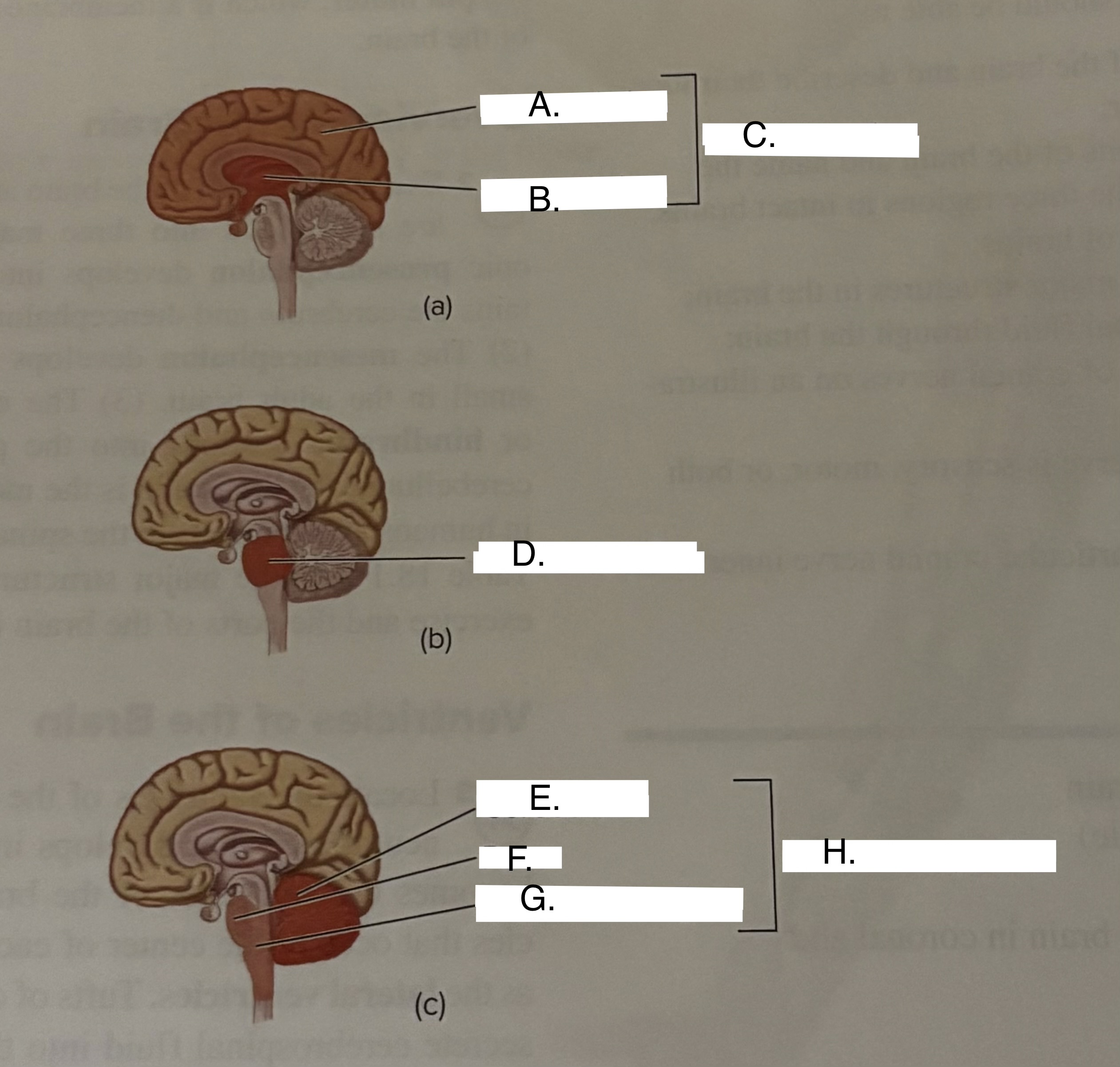
What is A?
Telencephalon

What is B?
Diencephalon

What is C?
Prosencephalon

What is D?
Mesencephalon

What is E?
Cerebellum

What is F?
Pons

What is G?
Medulla oblongata

What is H?
Rhombencephalon
What is A?
Anterior horn of lateral ventricle
What is B?
Interventricular foramen
What is C?
Third ventricle
What is D?
Inferior horn of lateral ventricle
What is E?
Posterior horn of lateral ventricle
What is F?
Cerebral aqueduct
What is G?
Fourth ventricle
What is H?
Central canal of spinal cord
the dura matter is divided into two things…
periosteal dura and the meningeal dura
three main regions of the brain and what do they develop into
Prosencephalon(forebrain), mesencephalon(midbrain), and rhombencephalon(hindbrain).
what are lateral ventricle
two ventricles that occupy the center of each cerebral hemisphere
what are tufts of capillaries that secrete CSF
choroid plexus
Fluid flows from lateral ventricles into x thru y
interventricular foramina and into third ventricle
what wall separates the of lateral ventricles and third ventricle
setum pellucidum
the third ventricle drains into x by y
fourth ventricle through the cerebral aqueduct
what the cerebral aqueduct becomes back up what happened
CSF accumulates in lateral and third ventricles increasing size and pressure- called internal hydrocephalus
CSF returns to the rest of cardiovascular system by
internal jugular veins
in the cerebrum, fold and ridges are
convolutions
ridges of convolutions are called
gyri
depressions in between are called x or y
sulci or fissures
what separates the frontal lobe and has functions of higher purposes
central sulcus
formation of speech area
Broca’s area
another work for Broca’s area
motor speech area
area in front of central gyrus is called
precentral gryus or primary motor cortex
region behind central gryus is
post-central gryus or primaru somatosensory cortex
association areas is
regions of the cerebral cortex that integrate information from different sensory modalities, playing a critical role in higher cognitive functions.
what does Wernicke area do and where is it located
formation of speech and in both parietal and temporal lobes
occipital lobe is considered as
visual area
what is responsible for separating the occipital lobe and cerebellum
transverse fissure
what is the temporal love separate from frontal and parietal lobe by
lateral fissure
the temporal lobe contains an area important called
primary auditory cortex
what senses are important in temporal lobe
smell and taste
what mass is deep to the temporal bone
insula
what separates the left and right hemispheres
longitudinal fissure
the left hemisphere is responsible for
language and analytical tasks/reasoning
the right hemisphere is responsible for
integrates space and pattern perceptions, artistic awareness, imagination and music comprehension
the inferior forebrain includes
pituitary gland, optic chiasm, and mammillary bodies
the hindbrain includes
pons, medulla oblongata, and cerebellum
what connects the two hemispheres
corpus callosum
anterior corpus callosum
genu
posterior corpus callosum
splenium
dicephalon consists of
thalamus and hypothalamus
what is involved with hypothalamus
pituitary gland
what does the pineal gland do
secrete melatonin which regulates body rhythm
what are the small area posterior to the dicephalon called
cerebral peduncles
the peduncles are anterior to
tetum
posterior to the tectum are 4 hemispheric process known as
corpora quadrigemina
the corpora quadrigemina consists of
superior and inferior colliculi
the superior colliculi is important for
visual and auditory reflexes
the inferior colliculi is important for
auditory pathways
the midbrain contains a center called
substantia nigra
when the substantia nigra is not functioning well, what disease can you get
parkinson's disease.
the hindbrain consists of a anterior budge called
pons
pons are importnat for
respiratory centers and involved in controlling the breathing rate
the medulla oblogata has centers for
respiratory rate control, heart rate, and other vital centers
where motor tracts from left brain cross over to right in medulla is called
decussion of pyramids
the cerebellum consists of an outer area called
cerebellar cortex
an inner branched patter of white material in cerebellum is called
arbor vitae
what is the branches called in cerebellum
folia
what is the triangular space anterior to the cerebellum called
fourth ventricle
what is mylienated
white matter
what is unmyelinated
gray matter
white matter consists of
tracts that take information from one region to another for processing
deeps islands of gray matter are called
basal nuclei or basal ganglia.
Nerve I
Olfactory nerve
Nerve II
optic nerve
Nerve III
oculomotor nerve
Nerve IV
Trochlear nerve
Nerve V
Trigeminal Nerve
Nerve VI
Abducens nerve
Nerve VII
Facial nerve
Nerve VIII
Vestibulocochlear Nerve
Nerve IX
Glossopharyngeal nerve
Nerve X
Vagus Nerve
Nerve XI
Accessory Nerve
Nerve XII
Hypoglossal nerve
Function of Olfactory (I)
receives sensory info from the nose
Function of Optic (II)
receives visual sensory information from the retina
Function of Oculomotor (III)
transmits motor info to move the eye muscles particularly the medial, superior and inferior rectus muscles and to the inferior oblique muscle
Function of Trochlear (IV)
transmits motor info to move the superior oblique muscle of the eye.
Function of Trigeminal (V)
three branched nerve, transmits both sensory and motor info to head
Function of Abducens (VI)
transmits motor info to move the lateral rectus muscle of the eye.