Lymphoid System Pathology
 Call Kai
 Learn
 Practice Test
 Spaced Repetition
 Match
 Flashcards
 Knowt Play
1/96

There's no tags or description
Looks like no tags are added yet.
97 Terms
Where are foetal lymphocytes produced?
yolk sac (birds)
liver (mammals)
Where are adult lymphocytes produced?
Bursa of Fabricus (birds)
bone marrow (mammals)

What does this image show?
journey of a lymphocyte
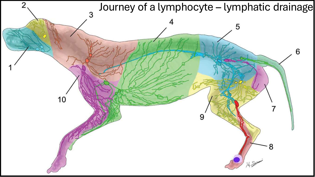
1
submandibular

2
parotid
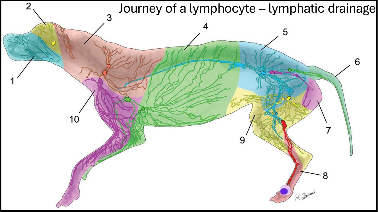
3
dorsal superficial cervical (*prescapular*)

4
axillary

5
medial iliac

6
lateral sacral

7
hypogastric

8
popliteal

9
superficial inguinal
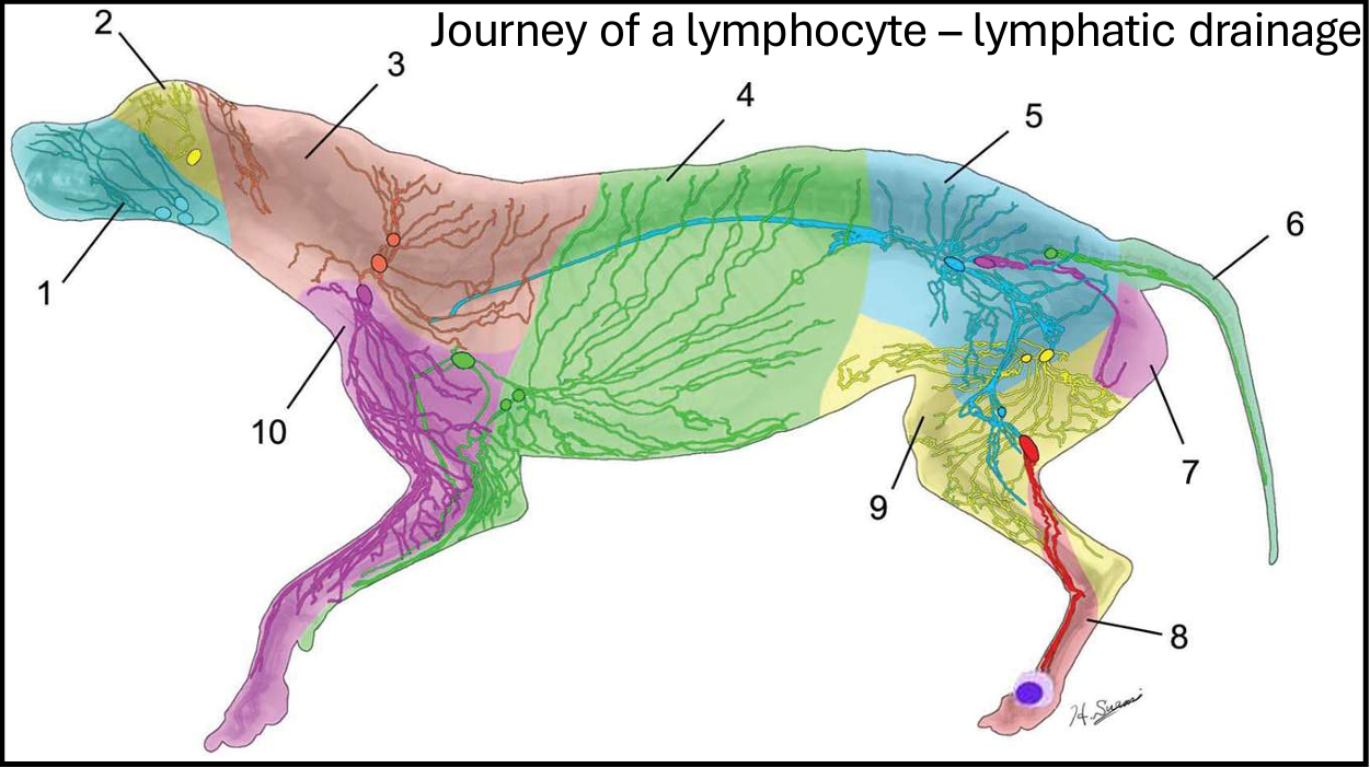
10
ventral superficial cervical (*prescapular*)

What does this image show?
lymph node size - bath tub analog

What does this image show?
hydrops foetalis
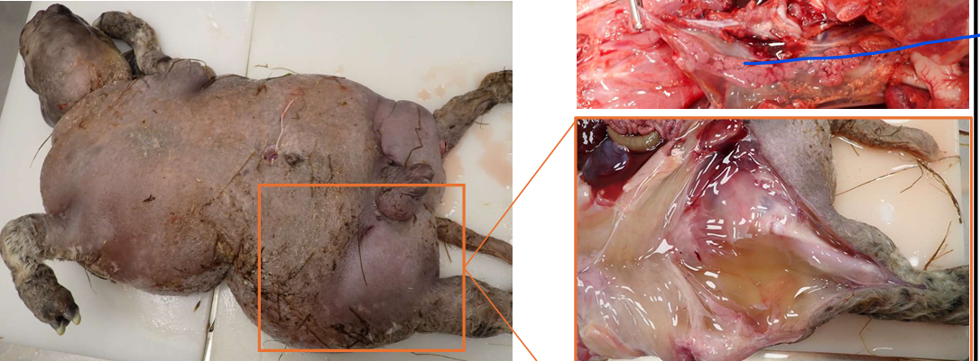
What is hydrops foetalis?
lack of development of lymph nodes lymphatic drainage
What is the role of the thymus?
primary lymphoid organ
development and maturation of T lymphocytes
adaptive immunity
What does injury to the thymus result in?
reduced adaptive immune response - increased susceptibility to secondary infections
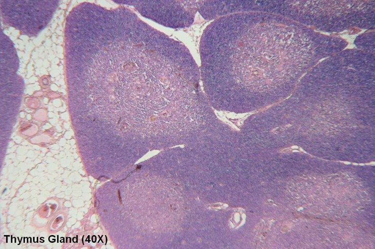
What do the purple and pink colours indicate?
H&E stain
H - stains DNA and bacteria purple - where nucleus & cells are
E - proteins pink
What happens to T cells in the thymus gland?
immature T cell in thymus
CD4, CD8, TCR present???
no - apoptosis
yes - reactive to MHC1 (self)???
no - mature T-cell enters circulation
yes - apoptosis


Describe the colouring of the parts of the thymus (cortex and medulla) on histology.
cortex = outer region of each lobule, darker
medulla = outer region of each lobule, lighter
What types of pathology do you see in the thymus?
atrophy
neoplasia
hypoplasia
haemorrhage/haematomas
inflammation
hyperplasia
What is atrophy of the thymus hard to distinguish between?
physiological involution
What is physical involution (thymus)?
thymus undergoes natural involution after sexual maturity
When is a small thymus considered abnormal?
if in neonates
What is thymic atrophy?
alteration of its cellular density and/or cellular composition and subsequent lobular shrinkage
What are common causes of thymic atrophy?
stress - immunosuppression
environmental toxins
malnutrition
infectious agents - predominantly viral
immunotoxicants - chemotherapy/radiation

What does this image show?
gelatinous texture of thymic atrophy
Why do viruses cause thymic atrophy?
viral tropism for immune cells
rapidly dividing cells in thymus - virus uses host machinery
inducing apoptosis - bystander effect
What are examples of viruses that cause thymic atrophy in cats?
FIV
FeLV
What are examples of viruses that cause thymic atrophy in dogs?
canine distemper virus (CDV)
parvovirus
What are examples of viruses that cause thymic atrophy in cattle?
BVDV
What are examples of viruses that cause thymic atrophy in horses?
equine herpes virus 1 (EHV1)
What are examples of viruses that cause thymic atrophy in pigs?
classical swine fever
porcine reproductive and respiratory syndrome (PRRS)
porcine circovirus 2
What are the names of thymic neoplasia?
thymoma
thymic lymphoma
thymic carcinoma (rare)
What are the characteristics of thymomas?
benign (-oma) epithelial cell tumour
goats, cats and dogs (older animals)
slower growing
can lead to autoimmune diseases (e.g. myasthenia gravis)
In what species do you see thymic lymphomas?
cats
occasionally beef cattle
What clinical signs do you see with thymic neoplasias (both thymomas and thymic lymphomas)
compression of cranial mediastinal structures (trachea, oesophagus & blood vessels)
dyspnoea
oedema
coughing
regurgitation
What’s the primary function of the spleen?
filter blood through series of sinuses containing large numbers of macrophages which remove old rbcs
act as reservoir for RBC and platelets
What’s the secondary function of the spleen?
secondary lymphoid tissue
contains lymphoid follicles that act as B&T cell maturation and multiplication sites

(spleen anatomy) What is the grey arrow?
capsule

(Spleen anatomy) What is the green arrow?
trabeculae

(Spleen anatomy) What is the white arrow?
white pulp

What is the red arrow?
red pulp
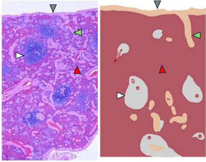
What are the main parts of the spleen anatomy?
capsule (grey arrow)
trabeculae (green arrow)
white pulp (white arrow)
red pulp (red arrow)

What’s the role of the capsule and trabeculae of the spleen?
structural support
contraction for blood expulsion

What’s the role of the red pulp of the spleen?
erythrocyte storage (highest capacity in dog, cat, horse)
filtering old/defective erythrocytes by macrophages
filtering debris & pathogens by macrophages
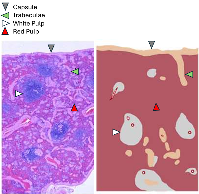
What is the role of the white pulp of the spleen?
macrophages (antigen presenting cells)
B & T lymphocytes
What can cause spleen atrophy?
chronic anaemia
cachexia (starvation)
neoplastic disease
senile atrophy (dogs & horses)

What does this image show?
top - atrophy +
middle - normal
bottom - atrophy ++

Describe the appearance of a spleen with atrophy.
irregular (rough), pale surface
What are commonly mistaken for pathology, fungal infection, neoplasia of the spleen?
siderotic plaques
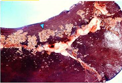
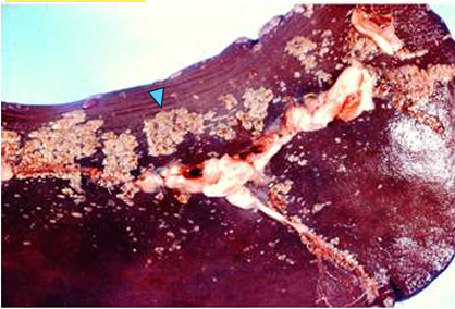
What NORMAL feature of the spleen does this image show?
siderotic plaques - accumulations of iron and calcium
What vascular changes can occur in the spleen?
active hyperaemia
passive congestion
infarction
torsion/volvulus
haematoma
inflammation (splenitis)
splenic hyperplasia
neoplasia
rupture
What is active hyperaemia in the spleen?
acute systemic infections - dilation of blood vessels
What is passive congestion of the spleen?
disorders of systemic circulation
barbiturates
cause splenic relaxation
haemolytic diseases
copper toxicity
babesiosis
What causes infarction of the spleen?
thrombi/thromboembolism
hypercoagulative states
septic emboli

What do these images show?
spleen infarction
What animals are more susceptible to torsion/volvulus of the spleen?
large breed dogs
pigs
Why are large breed dogs and pigs more susceptible to splenic torsion/volvulus?
longer gastrosplenic ligament

What is splenic torsion/volvulus?
twisting of gastrosplenic ligament causes occlusion of the splenic artery and vein

What can severe gastric torsion/volvulus end up causing?
severe congestion & haemorrhagic infarction
What is the appearance of haematoma of the spleen?
usually single and large
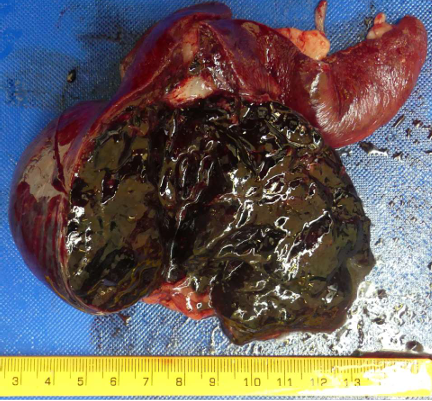

What does this image show?
haematoma of the spleen

What are the causes of haematoma in the spleen?
trauma
older dogs
lymphoid hyperplastic nodules
rupture of splenic vascular tumours
haemangioma
haemangiosarcoma
What’s inflammation of the spleen (splenitis) caused by?
infectious agents in the blood (bacteraemia, viraemia, parasitaemia (rare))

What are examples of bacteria that can cause splenitis?
anthrax (bacillus anthracis)
mycobacterium spp. (nodular)
What are examples of viruses that can cause splenitis?
african and classical swine fever
What’s an example of a parasite that can cause splenitis?
Leishmania
What are the general characteristics of splenic hyperplasia?
diffuse or nodular
result of the response to blood-borne antigens (chronic antigenic stimulation)
What are the characteristics of nodular hyperplasia?
single/multiple nodules
predominantly lymphocytes
can predispose to haematoma
common incidental finding in older dogs

What does this image show?
splenic hyperplasia
What are the types of neoplasia of the spleen?
haemangiosarcoma
lymphoma
haemangioma
sarcoma
metastatic carcinoma/sarcoma
What can cause primary rupture of the spleen?
severe trauma (e.g. RTA)
non-accidental injury (NAI)
What can cause secondary rupture of the spleen?
splenomegaly
neoplasia
haematoma
Sequelle (splenic rupture)
bleed into abdomen - death
haematoma formation
dissemination of splenic fragment (peritoneal splenosis)

How do you tell the difference between different spleen pathologies?
send for histology
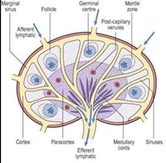
What is the order of flow in lymph nodes?
afferent lymphatic vessels
subscapular sinus
sinuses (through cortex)
medullar sinuses
efferent lymphatic vessels
lymph exits through the efferent lymph vessels

What is this?
lymph node
covered by connective tissue capsule that forms trabeculae penetrating parenchyma (arrows)
outer cortex formed by lymphatic follicles, that are absent in medulla
paracortex between cortex and medulla lacks follicles
medulla contains medullary sinuses & blood & lymph vessels
What pathologies can be seen in the lymph nodes?
hyperplasia
lymphadenitis (e.g. equine strangles, caseous lymphadenitis, mycobacterium bovis)
neoplasia (lymphoma)
What are the features of hyperplasia of the lymph nodes?
lymphadenomegaly (enlargement)
local or generalised
peripheral or internal
reactive process to antigenic stimulation
benign and transient
Why is knowing the drainage of the lymph nodes relevant to lymph node hyperplasia?
knowing drainage of lymph nodes can aid in localising the cause of inflammatory response
What are examples of pathogens commonly associated with inflammation of lymph nodes (lymphadenitis)?
Corynebacterium pseudotuberculosis - caseous lymphadenitis (CLA)
streptococcus equi - strangles
mycobacterium bovis - TB
What’s the causative bacteria of equine strangles?
streptococcus equi
How is equine strangles caused?
bacteria inhaled/ingested, attach & penetrate into mucosa
drain to regional nodes
neutrophil failure to phagocytose
rapid abscessation
compression of local structure → respiratory stridor & dysphagia
rupture & discharge to skin surface & guttural pouches
systemic spread via blood & lymph to other organs

What disease do these images show?
equine strangles
What bacteria causes caseous lymphadenitis?
Corynebacterium pseudotuberculosis
How is caseous lymphadenitis caused?
bacterial entry into wound → drain to local lymph ode
suppurative inflammation & capsulated abscessation
capsule compromised & bacteria escape
suppurative inflammation and reforming of capsule (looks like an onion)
rupture and drain through skin
common to have systemic spread through lymphatics and blood to other organs
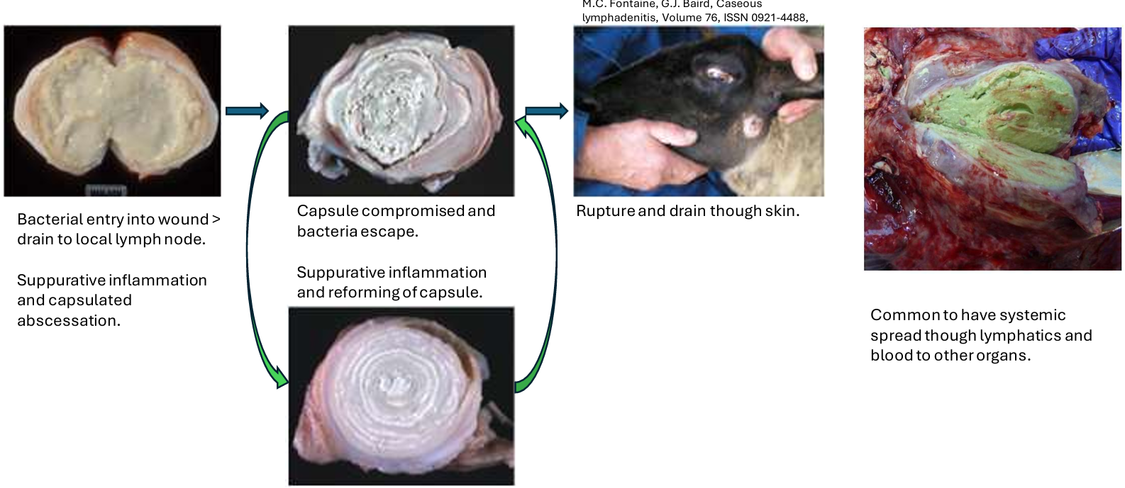
What lymph node pathology does this image show?
caseous lymphadenitis (corynebacterium pseudotuberculosis)
What causes mycobacterium bovis lymph node disease?
inhalation/ingestion of M.bovis bacilli
macrophages phagocytose bacteria
M.bovis resistant to digestion in phagosome (prevents lysosome attachment)
granuloma formation
macrophage recruits more immune cells & fibroblasts
caseous necrosis ± mineralisation
spread to lymph nodes

What lymph node pathology does this image show?
granuloma formation from mycobacterium bovis (bTB)
What pathologies can be seen in lymphatic vessels?
inflammation
obstruction of flow
lymphatic rupture
What can cause obstruction of flow in lymphatic vessels?
lymphangiectasia
lymphoedema
What’s an examples of lymphatic rupture in lymphatic vessels?
rupture of thoracic duct → chylothorax
What are the different types of neoplasia found in lymph nodes?
primary
lymphoma - uncontrolled B/T cell replication
secondary
afferent lymphatic spread (metastasis) e.g. carcinomas
lymphoma
malignant clonal proliferation of lymphoid cells, arising in lymphoid tissue (nodes, spleen, GI tract or extranodal sites)
Describe the mechanism for lymphomas?
single lymphocyte acquired oncogenic changes, escapes normal growth and apoptosis controls & expands clonally
results in accumulation of neoplastic cells in nodes & organs