lecture: developmental disease of the musculoskeletal system (NMSK4)
 Call Kai
 Learn
 Practice Test
 Spaced Repetition
 Match
 Flashcards
 Knowt Play
1/58

There's no tags or description
Looks like no tags are added yet.
59 Terms
Explain the endochondral ossification process
Define what the term osteochondrosis means and recognise that it is a developmental disease
Outline how osteochondrosis may occur
Explain how the clinical signs of osteochondrosis relate to the underlying pathology
List the (four) most common sites of OCD in the dog and horse
what is physis?
the junction between the epiphysis and metaphysis in growing animals
also known as growth plate
what is endochondral ossification prior to birth?
when temporary cartilage ‘bones’ are replaced by bone
endochondral ossification: responsible for the majority of foetal development
whats the primary centre for ossification
the diaphysis
whats the secondary centre for ossification
the epiphysis
what other type of ossification is there?
intramembranous ossification - formation of bone directly from connective tissue (osteoid) with NO cartilaginous precursor
location of intramembranous ossification in foetuses
primarily in craniofacial skeleton (flat bones of face)
what is the role of endochondral ossification after birth
the growth cartilage’s chondrocytes divide at the surface of the existing bone and more bone forms at these locations.
this results in the bone elongating at the epiphysis and metaphysis
centres of ossification expand by articular-epiphyseal cartilage complex
what are the 3 main sites of endochondral ossification post-natally?
1) physis (growth plates)
growth of longitudinal bone
2) articular- epiphysial cartilage complex
radial expansion of the epiphysis
3) cuboidal bone
majority of bones assify from a single centre
can also occur during healing of a fracture
physis anatomy
epiphyseal end = reserve/ resting zone - chondrocytes divide and form the ECM causes increased pressure on this end
proliferative zone → increased pressure and so pushes some chondrocytes downwards towards the metaphysis
real description: Chondrocytes assume columnar arrangement (less obvious in epiphysis), undergo a series of functional and morphological changes, and undergo hypertrophy
hypertrophic zone → have calcified cartilage - the chondrocytes start to form columns parallel to the edge of the bone
cartilage scaffold → blood vessels have access to this layer and osteoblasts can lay on the cartilage surface to create new bone (osteoids → woven bone → lamellar bone)
the vessels act as a conduit for osteoclasts, bone marrow cells and osteoblasts
bone formation at the level of the cartilage scaffold

the role of the ECM
the ECM characteristics change from zone to zone - colminates in synthesis of type X collagen + calcification in hypertrophic zone
the calcified matrix is invaded by blood vessels
where do the osteoblasts come from?
some from hypertrophic chondrocytes
some from surrounding tissues
they deposit osteoid on the spicules of calcified cartilage matrix
what is primary spongiosa?
mix of cartilage and primary bone
it is gradually replaced by lamellar bone as it enters the metaphysis
when does this process end?
it is continuous until puberty ends
all cartilage is replaced by bone - growth cartilage + AECC are completely ossified - no future growth potential
how is endochondral ossification controlled?
highly coordinated process
relies of temporal and spatial organisation of macromolecules
requires - cell to cell signalling + cell differentiation + matrix modifications
what is endochondral ossification controlled by?
systemic factors - primarily growth hormone mediated via insuline-like growth factor 1
if IGF1 is lacking then pituitary dwarfism can occur in dogs + other species (very rare)
paracrine factors
ECM-mediated regulation of cell behaviour + metabolism
transcription factors
*mechanical force
*genetic factors
*nutritional factors
*the factors that we can influence → osteochondrosis: selective breeding can be used to decrease the conditions abundance
types of cartilage involved
articular cartilage
avascular
relies on diffusion of nutrients from synovial fluid
growth cartilage
requires blood vessels in cartilage canals to be viable
cartilage canals
channels within cartilage
contain an arteriole (end artery) + several venules + capillaries
what are factors regarding most of the vessels in the cartilage canals?
perichondrium
run parallel to articular surface
they eventually anastomose (join) with vessels in advancing ossification front
the risk of cartilage canals
relies on only one artery which means if it is damaged there is very limited blood and nutrient supply to the growth cartilage


anastomoses meaning
the joining of blood vessels in the cartilage with those in subchondral bone
forming vessels between these areas
what is osteochondrosis (OCD)
a developmental skeletal disorder primarily in horses, dogs and pigs
there is a similar condition in dogs and humans
can we predict where OCD will occur?
characterised by focal failures of endochondral ossification that occurs in well-defined, species-specific predilection sites in young animals
yes: we can determine where its likely to be based on the most common location for that species
where are the lesions caused by OCD?
initial = often ischaemic chondronecrosis associated with failure of cartilage canal vessels at chondro-osseous junction
the lesions are often BILATERAL
the primary problem in pathogenesis of OCD
focal failure of vessels in cartilage canals as the are incorporated into advancing ossification front
→ can lead to: ischaemic chondronecrosis (cell death due to lack of blood supply)
necrotic cartilage cannot undergo endochondral ossification
what causes the vascular failure in OCD?
it could be micro-forces experienced by arteries as they cross the bone-cartilage junction
or it could be sepsis within the vasculature
what are the steps of OCD?
failure of cartilage canal blood supply →
ischaemia of growth cartilage →
ischaemia chondronecrosis →
failure of endochondral ossification →
osteochondrosis lesion
what area of growth cartilage is OCD more common?
the AECC (under the hyaline cartilage)
the hyaline cartilage stays in tact


what happens after osteochondrosis (OC) lesions + factors?
they could:
heal
progress to cause clinical signs
OCD
subchondral bone cyst (necrotic cartilage travels deeper into the bone)
factors:
the extent of the vascular injury
the diffusion distance to collateral blood supply (to get nutrients when the vessel is damaged)
biomechanical forces - it’s a developmental condition - control the animal’s workload
what radiographic signs are there for OCD?
radiographically evident as deficit in subchondral bone contour
may heal without intervention
intra-articular cartilage fragments
may ossify after separation
subchondral bone cysts
may follow on from ischaemic chondronecrosis lesion
can occur secondary to trauma


OCD terminology
osteochondrosis = failure of endochondral ossification
osteochondritis = extension of lesion to articular surface - cause inflammation of joint
osteochondritis dissecans = presence of dissecting osteochondral flap/ loose fragment in the joint

what are the factors that modify susceptibility in horses for OCD?
genetics
maternal nutrition
nutrition of the individual (growth rate + body size)
exercise regime
what are the clinical signs of OCD?
OCD:
joint effusion
lameness - more likely if:
subchondral bone is involved
synovitis
(the onset of clinical signs is often associated with exercise/ trauma - may convert silently from OC → OCD)
subchondral bone cyst:
lameness (bone involvement)
less likely if deep within the bone
joint effusion

what are the more common sites of OC in horses?
tibiotarsal joint
distal intermediate ridge of tibia (DIRT lesion)
lateral trochlear ridge of talus
femoropatellar joint
lateral trochlear ridge of femur
patella
fetlock joint
(may not need to know the location within the joint)
what are the less common sites of OC in horses?
shoulder
elbow
hip
OC predilection sites in dogs
shoulder
caudal aspect of humeral head
stifle
lateral + medial femoral condyle
elbow
coronoid process of ulna
medial condyle of humerus
what are some other common developmental orthopaedic diseases in horses?
physitis/ physeal dysplasia
angular limb deformities (deviation in long axis of limb in frontal plane)
incomplete ossification of cuboidal bones
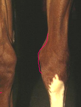
physeal displasia (physitis)
an acquired condition characterised by gross enlargement of the physis and/ or metaphysis
probably caused by mismatch between metaphyseal bone strength + applied loads
rare cases: may lead to early closure and subsequent ALD
(something to do with woven bone being used on hard ground → not strong enough)
physeal closure recap
all physes ossify but their closure timings differ depending on which physis it is - useful to know for treatment of angular limbs
completed when there is no more growth potential
what may cause premature closure?
trauma, infection, severe ALD (angular limb deformity)


Dog: distal radial physis closed (can still see physeal ‘scar’, which will disappear in time)

Dog: distal radial physis open
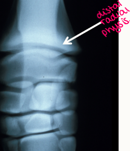
Horse: distal radial physis open
physeal growth recap
the influence of force on growth:
Inverted ‘U-shaped’ relationship
Reduced compression => reduced growth rate
Increased compression (within physiological limits) => increased growth rate
Excessive compression (non-physiological) => reduced growth rate
the rate of growth vs compression graph
can be useful to know whether the limb will self heal.
the rate of growth is highest where compression is highest and so there is more growth at the bent angle of the joint.
this can allow the joint to self correct the issue
what are the categories of angular limb deformity
congenital vs acquired
valgus vs varus
valgus meaning
limb deviates laterally - distal to site of deformity

varus meaning
limb deviates medially - distal to site of deformity

congenital meaning
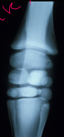
incomplete ossification of cuboidal carpal bones in premature/ dysmature foals
dysmature = born at term but not fully mature
incomplete tarsal bone ossification - leads to decreased hock angle
may also occur 2^y to lax periarticular ligaments/ epiphyseal dysplasia
acquired meaning
Pathogenesis includes premature closure of one side of physis
Trauma
Infection
Physeal dysplasia (equine)
Premature closure of distal ulnar physis (dogs)
Carpal valgus develops due to normal growth of radius

incomplete ossification

normal joint
incomplete ossification