cytology of fluids and less cellular lesions
 Call Kai
 Learn
 Practice Test
 Spaced Repetition
 Match
 Flashcards
 Knowt Play
1/28

There's no tags or description
Looks like no tags are added yet.
29 Terms
fluids
» Fluids may be within cavities
» Fluid filled lesions may also be seen eg cysts
» Fluid cytology may refer to flush/wash samples eg BAL
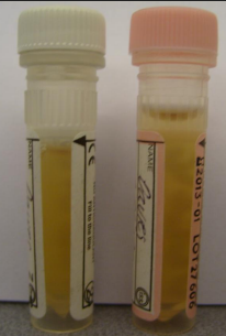
collection of fluids
» Plain tubes (no gel) are required for culture
» For cytology EDTA may preserve cellularity better
• But there will always be some cell swelling in vitro
» Fresh smears made at time of sampling provide the best cytologic evaluation

classification of cavity fluids
» Two systems in use
» (1) Exudate, modified transudate, transudate
» (2) Exudate, protein-poor transudate; protein-rich transudate
» May also have haemorrhagic effusion- not generally included in the above classifications
» Lymphorrhagic effusions may also be considered separately (often associated with chylous effusions)
» Multiple processes can be involved in development of effusions
Exudate
» Effusion produced by increased vascular permeability to plasma proteins because of inflammation
» High cellularity (> 5 x10⁹/L or > 7 x10⁹/L )
» High protein (>20g/L or >30g/L)

transudate
» Effusion produced by passage of fluid or solute through a membrane due to changes in hydraulic or oncotic pressure
» ‘Pure’ transudate- low cellularity (<1.5x 10⁹/L), low protein
» Modified tranusduate- ‘pure’ transudate modified by the addition of protein or cells
» Protein-poor transudates
• Marked hypoalbuminaemia
• Most commonly seen with hepatic cirrhosis or PLN
» Protein rich transudates
• Increased plasma hydraulic pressure in the liver or lungs – often due to venous congestion
• Most commonly seen with congestive heart failure or portal venous hypertension

evaluation of body fluids - gross appearance
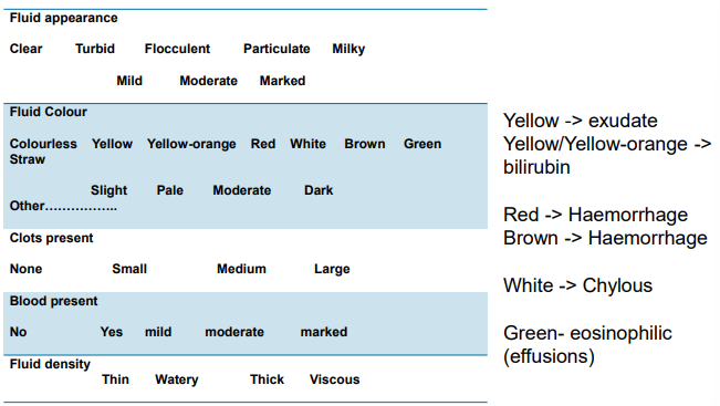
Evaluation of body fluids
» Cell count
» Protein value
» Where relevant
» Cholesterol and Triglycerides (compared to serum)
» Creatinine +/- urea
» Bilirubin

Fluid Preparations
» May use direct smear preparations
» May also use concentrated preparations
• Centrifugation of sample and sediment preparation
• Cytospin preparation
» Microscopic evaluation of the cells
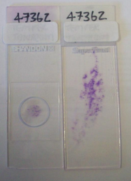
synovial fluid
» Normal cellularity is very low
» Few mononuclear cells
» Rare neutrophils

Chronic Inflammation
» Macrophages
» With trauma also seen increased macrophages and a few neutrophils
» Blood also present

Synovial Fluid Neutrophilic inflammation
» Low numbers of nucleated cells
• Trauma
• Local inflammation
» High cellularity (> 3x10⁹/L)
• Canine – immune mediated polyarthritis (most common- expect multiple joints affected)
• Equine -Probable sepsis (usually only one joint affected)
• Systemic immune mediated disease uncommon in the horse

septic neutrophilic inflammation
» Bacteria may be hard to identify on the smears in synovial fluid
» Growth of bacteria obtained from synovial fluid may be inhibited in the laboratory- the mechanism unclear

blood contamination
» May also increase neutrophil count which may mask inflammation
» If CBC is normal the number of neutrophils added by mild blood contamination are low and shouldn’t significantly alter the cellularity of the fluid
» If in doubt check blood smear/CBC

peritoneal fluid
• Low cellularity with approximately equal numbers of neutrophils and mononuclear cells/macrophages
• However neutrophils may be predominant population in healthy horses

peritoneal and pleural fluid
» Mesothelial cells line these cavity spaces
» Mesothelial cells can exhibit marked atypia when active/reactive – such as in response to fluid presence, or the cause of the fluid presence.
» Unreactive sheets of cells have a fishscale appearance

Septic Peritonitis vs contamination or enterocentesis
• With ongoing sepsis neutrophils are degenerate and bacteria are intracellular
• With contamination bacteria are extracellular and neutrophils may not appear degenerate
• Enterocentesis appears similar to contamination, but may have other organisms such as protozoa present
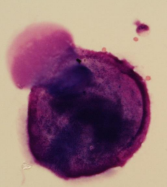

bacterial contamination
septic peritonitis
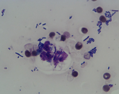

ruptured gut
Haemoperitoneum
• Erythrophagia or haem pigment noted in macrophages
• Often >5% PCV
— Trauma
— Neoplasia
— Idiopathic

Uroperitoneum
• Observe calcium carbonate and oxalate crystals in abdominal fluid in horses
• Other species may have different crystals- or none at all
• May have a neutrophilic inflammatory response

neoplasia
• Not all neoplasms will exfoliate into the fluid
• Careful that you have not aspirated spleen if thinking lymphoma

respiratory wash
» Normally cellularity is low
» Small amount of mucus with a few macrophages and respiratory epithelial cells
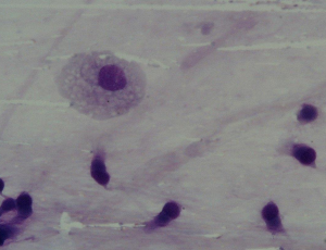

septic inflammation
inflammation
» Curshmann’s spirals
» Inspissated mucus from bronchioles
» Seen with chronic inflammation or irritation

Haemorrhage
» Macrophages containing hemosiderin (hemosiderophages)
» Can be seen with EIPH – Equine Exercise Induced Pulmonary Haemorrhage


Special Stains-Prussian blue
Parasites
» ‘Lungworm’ may be seen in BALs
» Angiostrongylus vasorum in the dog
» Aelurostrongylus abstrusus in the cat

Eosinophilic Inflammation - lung
» Feline allergic airway disease (FAAD) _ (feline asthma)
» Eosinophilic bronchopneumopathy in the dog
» Precise aetiology not elucidated
» Appears allergic in nature
» Marked eosinophils in lung washes

CSF (cerebrospinal fluid)
» Low cellularity (<5 cells/L in the dog, <8 cells/L cat)
» Low Protein ( <0.25g mg/L cisternal; <0.45g/L for lumbar)
» May identify inflammatory processes
» Occasionally may observe neoplastic cells

Seroma, Hygroma, Synovial Cyst, Apocrine Cyst
» There are a number of cutaneous lesions that may be fluid filled and poorly cellular.
» Hygromas will generally occur at pressure points/joints
» Synovial cysts will often have a background similar to synovial fluid
» Apocrine cysts are cutaneous, superficial
» ANY tumour can have a cystic area and a solid area- so if you can palpate a fluid filled space and a thickened tissue area ensure you have sampled both
Keratin containing lesions
» There are a number of benign cutaneous masses that are keratin containing
» These will often present with keratin material or cholesterol crystals only when aspirated.

summary
» Precise interpretation of fluids may depend upon the site of collection
» Inflammation/Inflammatory responses may be clearly identified on fluid samples and in some sites or disease processes may provide clear indication of the underlying cause of the effusion
» Neoplasia may also be identified
» For cytology EDTA tubes AND fresh smears are preferable
» For culture plain tubes are required.