CNS Claude MCQs
 Call Kai
 Learn
 Practice Test
 Spaced Repetition
 Match
 Flashcards
 Knowt Play
1/7

There's no tags or description
Looks like no tags are added yet.
8 Terms
A 16-year-old rugby player is concussed in a match and is allowed to return to play 4 days later against medical advice. He takes a second knock to the head, collapses within minutes, and dies. Which autopsy finding is most likely?
A. A swollen brain with flattened gyri and herniation, possibly with no other intracranial abnormality
B. Biconvex extradural haematoma over the temporal lobe
C. Atrophy of the medial temporal lobes, thalamus and mamillary bodies with ventricular dilatation
D. Acute subdural haematoma from torn bridging veins as the only finding
E. Extensive haemorrhagic lesions in the corpus callosum on gross examination
A. Second impact syndrome. Cerebral oedema may be the only intracranial abnormality. CTE (C) needs repeated concussions over time.
A 52-year-old man falls from a ladder onto his forehead. Over 24 hours he develops bilateral periorbital bruising and a clear watery nasal discharge. Which fracture site and complication are of most concern?
A. Squamous temporal bone; extradural haematoma
B. Occipital bone at the foramen magnum; vertebral artery tear
C. Vertex of the skull; non-accidental injury
D. Petrous temporal bone; bleeding from the ear
E. Anterior cranial fossa (orbital plate/ethmoid) with a dural tear; meningitis

E. Raccoon eyes and CSF rhinorrhoea indicate an anterior fossa (orbital plate/ethmoid) fracture. The scalp and face veins communicate freely with the meninges, so meningitis is the major concern.

A 63-year-old man with poorly controlled hypertension (BP 220/120) develops a sudden severe headache and vomiting, then left hemiparesis. GCS falls from 14 to 8 over a few hours. CT shows a large right putaminal haematoma. Which underlying vascular lesion is most likely?
A. Rupture of a berry aneurysm at the middle cerebral bifurcation with intracerebral extension
B. Rupture of Charcot-Bouchard microaneurysms on deep penetrating arteries
C. Haemorrhagic transformation of an embolic MCA infarct
D. Cerebral amyloid angiopathy
E. Cerebral vasculitis
B. Hypertensive ICH tends to occur in the putamen/external capsule, thalamus, pons and cerebellum, from Charcot-Bouchard microaneurysms.

A 58-year-old hypertensive man dies of myocardial infarction. At autopsy there are multiple small cavitary lesions (3-10 mm) in the putamen and caudate nucleus, with no haemorrhage. What is the most likely pathogenesis?
A. Embolism from an internal carotid thrombus
B. Rupture of small penetrating vessels with later resorption of the haematoma
C. Global hypoperfusion at arterial border zones
D. Arteriolar sclerosis of deep penetrating arteries causing occlusion
E. Malignant hypertension with fibrinoid necrosis causing diffuse oedema
D. Lacunar infarcts: arteriolar sclerosis of deep penetrating vessels causing occlusion and small cavitary infarcts in the basal ganglia and white matter. Slit haemorrhages (B) are haemorrhagic, not infarcts.

A 76-year-old man with AF has an ischaemic MCA stroke. The initial CT shows no haemorrhage and he receives thrombolysis. On day 2 he deteriorates, and repeat CT shows haemorrhage within the infarct. What is the best explanation?
A. Ischaemia-reperfusion injury to vessels within the infarcted territory (haemorrhagic transformation)
B. Rupture of Charcot-Bouchard microaneurysms
C. Rupture of an incidental berry aneurysm
D. Undiagnosed cerebral amyloid angiopathy
E. The original CT was misread, so this was always a primary haemorrhage
A. About 20% of infarcts are haemorrhagic. Secondary haemorrhage follows ischaemia-reperfusion injury. This is why CT before thrombolysis matters, since thrombolytics cannot be given for haemorrhagic stroke.

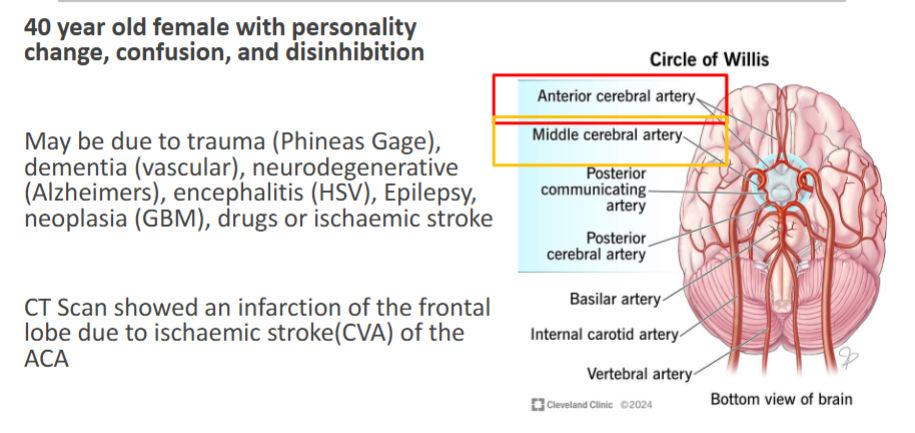
A 66-year-old man has a sudden onset of apathy and disinhibition, contralateral leg weakness greater than arm weakness, and urinary incontinence. Which vessel is most likely occluded?
A. Middle cerebral artery
B. Posterior cerebral artery
C. Anterior cerebral artery
D. Basilar artery
E. Anterior inferior cerebellar artery
C. An ACA infarct gives frontal lobe signs (abulia, disinhibition) and contralateral leg weakness greater than arm. The lecture example of personality change and disinhibition was an ACA infarct
A 34-year-old man has headache, morning vomiting and gait ataxia. MRI shows a vascular cerebellar mass with a cyst. He also has retinal angiomas and a renal mass. Which diagnosis and which lethal complication if untreated?
A. Pilocytic astrocytoma; subfalcine herniation
B. Haemangioblastoma (von Hippel-Lindau); tonsillar herniation
C. Medulloblastoma; transtentorial herniation
D. Meningioma (NF2); subfalcine herniation
E. Ependymoma; uncal herniation
B. Haemangioblastoma (cerebellum, highly vascular), associated with von Hippel-Lindau (retinal angiomas, renal cell carcinoma). A posterior fossa mass leads to tonsillar herniation (“coning”).

A 59-year-old man with a right temporal glioblastoma deteriorates with a fixed dilated right pupil, then loses consciousness and dies. Autopsy shows flame-shaped haemorrhages in the midbrain and upper pons. What is the most likely mechanism of these haemorrhages?
A. Distortion and tearing of penetrating arteries and veins from downward brainstem displacement
B. Rupture of Charcot-Bouchard microaneurysms from hypertension
C. Primary haemorrhage into tumour metastases
D. Diffuse axonal injury
E. Haemorrhagic transformation of a basilar artery infarct
A. Duret haemorrhages accompany progressive transtentorial herniation, from distortion or tearing of penetrating arteries and veins supplying the upper brainstem.
