MMSC200 Exam 2 Chapter 10

0.0(0)
Studied by 0 people
call kaiCall Kai
learnLearn
examPractice Test
spaced repetitionSpaced Repetition
heart puzzleMatch
flashcardsFlashcards
GameKnowt Play
Card Sorting

1/108

encourage image

There's no tags or description

Looks like no tags are added yet.

109 Terms

1
New cards

nervous system

  • memory and movement

  • voluntary and involuntary activities

  • complex: 100 billion nerve cells called neurons (a type of parenchymal cells)

  • active interactions between neurons (pass on information from one to another)

  • nerves carry electrochemical messages

  • external and internal receptors


2
New cards

central and peripheral nervous system

  • central nervous system

    • brain

    • spinal cord

  • peripheral nervous system

    • cranial nerves

    • spinal nerves

    • plexuses

    • peripheral nerves throughout the body


<ul><li><p>central nervous system</p><ul><li><p>brain</p></li><li><p>spinal cord</p></li></ul></li><li><p>peripheral nervous system</p><ul><li><p>cranial nerves</p></li><li><p>spinal nerves</p></li><li><p>plexuses</p></li><li><p>peripheral nerves throughout the body</p></li></ul></li></ul><p></p>
3
New cards

nervous system


<p></p><p></p>
4
New cards

afferent and efferent nerve fibers

  • cranial and spinal nerves

    • sensory nerves (afferent nerve fibers) carry messages toward the brain

    • motor nerves (efferent nerve fibers) carry messages from the brain

  • sensory receptors in organs and glands

    • sight

    • hearing and balance

    • smell

  • mixed nerves may carry both sensory and motor fibers.


<ul><li><p>cranial and spinal nerves</p><ul><li><p>sensory nerves (afferent nerve fibers) carry messages toward the brain</p></li><li><p>motor nerves (efferent nerve fibers) carry messages from the brain</p></li></ul></li><li><p>sensory receptors in organs and glands</p><ul><li><p>sight </p></li><li><p>hearing and balance</p></li><li><p>smell</p></li></ul></li><li><p>mixed nerves may carry both sensory and motor fibers. </p></li></ul><p></p>
5
New cards
<p>linkage between central and peripheral nervous system</p>

linkage between central and peripheral nervous system

knowt flashcard image
6
New cards

simple reflex arc

knowt flashcard image
7
New cards

autonomic nervous system

  • involuntary nervous system

  • carries impulses from the central nervous system to organs; motor nerve

  • sympathetic nerves stimulate body under stress

  • parasympathetic nerves balance sympathetic system

    • slow heart rate

    • lower blood pressure


<ul><li><p>involuntary nervous system</p></li><li><p>carries impulses from the central nervous system to organs; motor nerve</p></li><li><p>sympathetic nerves stimulate body under stress</p></li><li><p>parasympathetic nerves balance sympathetic system</p><ul><li><p>slow heart rate</p></li><li><p>lower blood pressure</p></li></ul></li></ul><p></p>
8
New cards

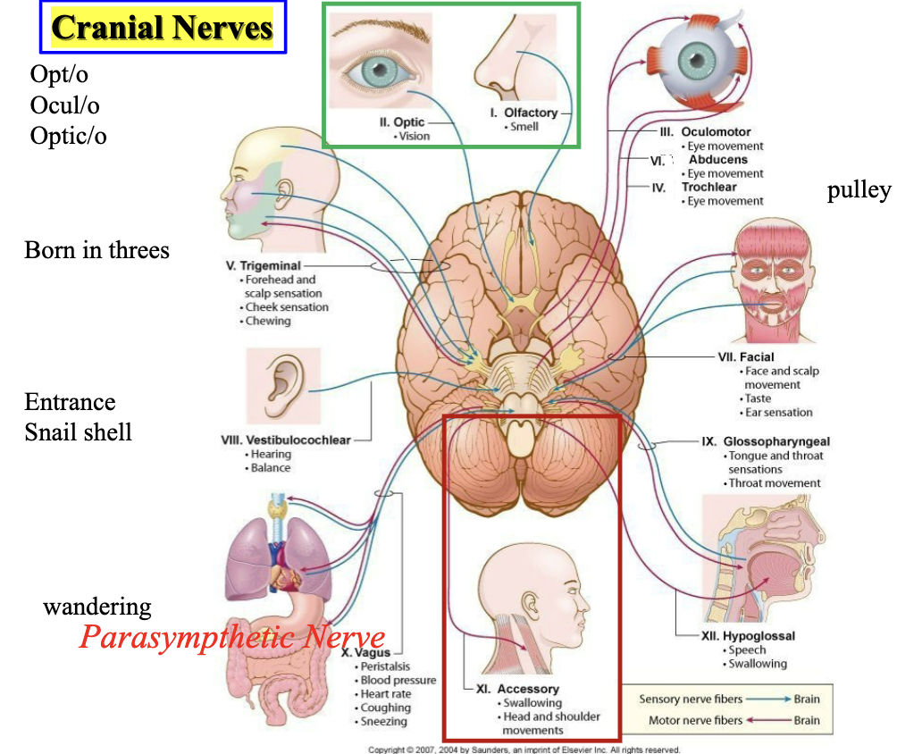
cranial nerves

knowt flashcard image
9
New cards

structure of nervous system

Main Idea:
The nervous system is divided into the Central Nervous System (CNS) and the Peripheral Nervous System (PNS). The CNS processes information, while the PNS carries information to and from the CNS.


1. Central Nervous System (CNS)

Includes:

  • Brain

  • Spinal Cord

Function:

  • Interprets sensory information

  • Makes decisions

  • Sends commands to the body


2. Peripheral Nervous System (PNS)

Includes:

  • Cranial nerves

  • Spinal nerves

Function:

  • Connects the CNS to the rest of the body


Sensory (Afferent) Division

Direction: Body → CNS

Function:

  • Carries information from sense organs (eyes, ears, skin, etc.) to the brain and spinal cord.

  • Allows the CNS to receive information about the environment.

Remember:
Afferent = Arrives at the CNS


Motor (Efferent) Division

Direction: CNS → Body

Function:

  • Carries commands from the brain and spinal cord to muscles and glands.

Remember:
Efferent = Exits the CNS


Motor Division Branches 1. Somatic Nervous System (Voluntary)

Controls:

  • Skeletal muscles

Function:

  • Responsible for conscious movements such as walking, writing, and lifting objects.

Example:

  • Choosing to raise your hand in class.


2. Autonomic Nervous System (Involuntary)

Controls:

  • Cardiac muscle (heart)

  • Smooth muscle (organs)

  • Glands

Function:

  • Regulates automatic body functions without conscious effort.

Sympathetic Division

"Fight or Flight"

  • Increases heart rate

  • Dilates pupils

  • Increases breathing rate

  • Prepares body for stress or emergencies

Parasympathetic Division

"Rest and Digest"

  • Slows heart rate

  • Stimulates digestion

  • Conserves energy

  • Returns body to a calm state

Vagus Nerve:

  • Major nerve of the parasympathetic system

  • Helps regulate heart rate, digestion, and other internal organ functions


Quick Memory Trick

CNS = Control Center (brain & spinal cord)

PNS = Communication Network

Afferent = Arrives at CNS

Efferent = Exits CNS

Sympathetic = Stress ("Fight or Flight")

Parasympathetic = Peace ("Rest and Digest")

<p><strong>Main Idea:</strong><br>The nervous system is divided into the <strong>Central Nervous System (CNS)</strong> and the <strong>Peripheral Nervous System (PNS)</strong>. The CNS processes information, while the PNS carries information to and from the CNS.</p><div data-type="horizontalRule"><hr></div><p> 1. Central Nervous System (CNS)</p><p><strong>Includes:</strong></p><ul><li><p>Brain</p></li><li><p>Spinal Cord</p></li></ul><p><strong>Function:</strong></p><ul><li><p>Interprets sensory information</p></li><li><p>Makes decisions</p></li><li><p>Sends commands to the body</p></li></ul><div data-type="horizontalRule"><hr></div><p> 2. Peripheral Nervous System (PNS)</p><p><strong>Includes:</strong></p><ul><li><p>Cranial nerves</p></li><li><p>Spinal nerves</p></li></ul><p><strong>Function:</strong></p><ul><li><p>Connects the CNS to the rest of the body</p></li></ul><div data-type="horizontalRule"><hr></div><p> Sensory (Afferent) Division</p><p><strong>Direction:</strong> Body → CNS</p><p><strong>Function:</strong></p><ul><li><p>Carries information from sense organs (eyes, ears, skin, etc.) to the brain and spinal cord.</p></li><li><p>Allows the CNS to receive information about the environment.</p></li></ul><p><strong>Remember:</strong><br><strong>Afferent = Arrives at the CNS</strong></p><div data-type="horizontalRule"><hr></div><p> Motor (Efferent) Division</p><p><strong>Direction:</strong> CNS → Body</p><p><strong>Function:</strong></p><ul><li><p>Carries commands from the brain and spinal cord to muscles and glands.</p></li></ul><p><strong>Remember:</strong><br><strong>Efferent = Exits the CNS</strong></p><div data-type="horizontalRule"><hr></div><p> Motor Division Branches 1. Somatic Nervous System (Voluntary)</p><p><strong>Controls:</strong></p><ul><li><p>Skeletal muscles</p></li></ul><p><strong>Function:</strong></p><ul><li><p>Responsible for conscious movements such as walking, writing, and lifting objects.</p></li></ul><p><strong>Example:</strong></p><ul><li><p>Choosing to raise your hand in class.</p></li></ul><div data-type="horizontalRule"><hr></div><p> 2. Autonomic Nervous System (Involuntary)</p><p><strong>Controls:</strong></p><ul><li><p>Cardiac muscle (heart)</p></li><li><p>Smooth muscle (organs)</p></li><li><p>Glands</p></li></ul><p><strong>Function:</strong></p><ul><li><p>Regulates automatic body functions without conscious effort.</p></li></ul><p> Sympathetic Division</p><p><strong>"Fight or Flight"</strong></p><ul><li><p>Increases heart rate</p></li><li><p>Dilates pupils</p></li><li><p>Increases breathing rate</p></li><li><p>Prepares body for stress or emergencies</p></li></ul><p> Parasympathetic Division</p><p><strong>"Rest and Digest"</strong></p><ul><li><p>Slows heart rate</p></li><li><p>Stimulates digestion</p></li><li><p>Conserves energy</p></li><li><p>Returns body to a calm state</p></li></ul><p><strong>Vagus Nerve:</strong></p><ul><li><p>Major nerve of the parasympathetic system</p></li><li><p>Helps regulate heart rate, digestion, and other internal organ functions</p></li></ul><div data-type="horizontalRule"><hr></div><p> Quick Memory Trick</p><p><strong>CNS = Control Center</strong> (brain &amp; spinal cord)</p><p><strong>PNS = Communication Network</strong></p><p><strong>Afferent = Arrives at CNS</strong></p><p><strong>Efferent = Exits CNS</strong></p><p><strong>Sympathetic = Stress ("Fight or Flight")</strong></p><p><strong>Parasympathetic = Peace ("Rest and Digest")</strong></p>
10
New cards

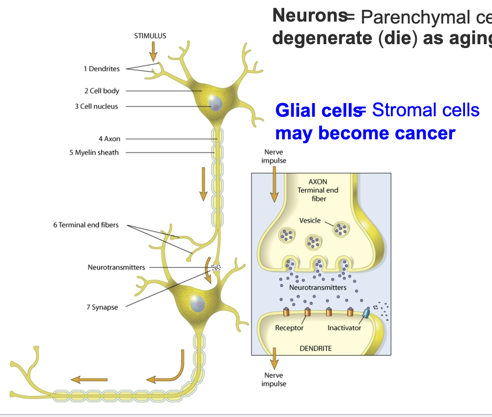
nerve cells (neurons)

  • microscopic

  • collected into macroscopic nerves

  • carry electrical messages all over the body


<ul><li><p>microscopic</p></li><li><p>collected into macroscopic nerves</p></li><li><p>carry electrical messages all over the body</p></li></ul><p></p>
11
New cards

movement of nervous pulse

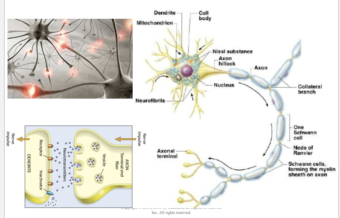
Main Idea:
A neuron is a specialized nerve cell that receives, processes, and sends electrical and chemical signals throughout the body.

Key Parts and Functions:

  • Dendrites: Receive incoming signals from other neurons.

  • Cell Body (Soma): Contains the nucleus and processes information received by dendrites.

  • Nucleus: Controls the activities of the neuron.

  • Axon Hillock: Area where the nerve impulse begins.

  • Axon: Carries the nerve impulse away from the cell body.

  • Myelin Sheath (Schwann Cells): Insulates the axon and speeds up signal transmission.

  • Nodes of Ranvier: Gaps in the myelin sheath that allow the impulse to jump from node to node, increasing speed.

  • Axon Terminals: End branches of the axon that communicate with other cells.

How a Signal Travels:

  1. Dendrites receive a message from another neuron.

  2. The cell body processes the information.

  3. The nerve impulse begins at the axon hillock.

  4. The impulse travels down the axon.

  5. Myelin and nodes of Ranvier help the signal move quickly.

  6. At the axon terminal, neurotransmitters are released into the synapse.

Synapse (Bottom Left Diagram):

  • The axon terminal releases neurotransmitters stored in vesicles.

  • Neurotransmitters cross the tiny gap called the synapse.

  • They bind to receptors on the dendrite of the next neuron.

  • This starts a new nerve impulse in the receiving neuron.

Important Concept: Neural communication occurs through a combination of:

  • Electrical signals within a neuron.

  • Chemical signals (neurotransmitters) between neurons.

Remember:
Dendrites → Cell Body → Axon Hillock → Axon → Axon Terminal → Neurotransmitters → Next Neuron. This is the direction that information flows through a neuron.
<p><strong>Main Idea:</strong><br>A neuron is a specialized nerve cell that receives, processes, and sends electrical and chemical signals throughout the body.</p><p><strong>Key Parts and Functions:</strong></p><ul><li><p><strong>Dendrites</strong>: Receive incoming signals from other neurons.</p></li><li><p><strong>Cell Body (Soma)</strong>: Contains the nucleus and processes information received by dendrites.</p></li><li><p><strong>Nucleus</strong>: Controls the activities of the neuron.</p></li><li><p><strong>Axon Hillock</strong>: Area where the nerve impulse begins.</p></li><li><p><strong>Axon</strong>: Carries the nerve impulse away from the cell body.</p></li><li><p><strong>Myelin Sheath (Schwann Cells)</strong>: Insulates the axon and speeds up signal transmission.</p></li><li><p><strong>Nodes of Ranvier</strong>: Gaps in the myelin sheath that allow the impulse to jump from node to node, increasing speed.</p></li><li><p><strong>Axon Terminals</strong>: End branches of the axon that communicate with other cells.</p></li></ul><p><strong>How a Signal Travels:</strong></p><ol><li><p>Dendrites receive a message from another neuron.</p></li><li><p>The cell body processes the information.</p></li><li><p>The nerve impulse begins at the axon hillock.</p></li><li><p>The impulse travels down the axon.</p></li><li><p>Myelin and nodes of Ranvier help the signal move quickly.</p></li><li><p>At the axon terminal, neurotransmitters are released into the synapse.</p></li></ol><p><strong>Synapse (Bottom Left Diagram):</strong></p><ul><li><p>The <strong>axon terminal</strong> releases <strong>neurotransmitters</strong> stored in vesicles.</p></li><li><p>Neurotransmitters cross the tiny gap called the <strong>synapse</strong>.</p></li><li><p>They bind to <strong>receptors</strong> on the dendrite of the next neuron.</p></li><li><p>This starts a new nerve impulse in the receiving neuron.</p></li></ul><p><strong>Important Concept:</strong> Neural communication occurs through a combination of:</p><ul><li><p><strong>Electrical signals</strong> within a neuron.</p></li><li><p><strong>Chemical signals (neurotransmitters)</strong> between neurons.</p></li></ul><p><strong>Remember:</strong><br><strong>Dendrites → Cell Body → Axon Hillock → Axon → Axon Terminal → Neurotransmitters → Next Neuron</strong>. This is the direction that information flows through a neuron.</p>
12
New cards

multiple sclerosis

a chronic autoimmune condition where the immune system mistakenly attacks the protective myelin sheath surrounding nerve fibers in the brain and spinal cord

most common autoimmune disorder affecting the central nervous system

demyelinating disease in which the insulating covers of nerve cells in the brain and spinal cord are damaged

this damage disrupts the ability of parts of the nervous system to communicate, resulting in a wide range of sign sand symptoms, including physical, mental, and sometimes psychiatric problems

<p>a chronic autoimmune condition where the immune system mistakenly attacks the protective myelin sheath surrounding nerve fibers in the brain and spinal cord</p><p>most common autoimmune disorder affecting the central nervous system</p><p>demyelinating disease in which the insulating covers of nerve cells in the brain and spinal cord are damaged</p><p>this damage disrupts the ability of parts of the nervous system to communicate, resulting in a wide range of sign sand symptoms, including physical, mental, and sometimes psychiatric problems</p>
13
New cards

neurotransmitters

  • acetycholine: neuromuscular synapse

  • dopamine: perception of pleasurable effects

    • opioids, nicotine, amphetamine, ethanol and cocaine increase dopamine release

    • dysfunction involved in Parkinson disease, schizophrenia

  • epinephrine and norepineprhine: ‘fight or flight’, increase heart rate

  • serotonin: regulation of mood, happiness, sleep/wake cycles


<ul><li><p>acetycholine: neuromuscular synapse</p></li><li><p>dopamine: perception of pleasurable effects</p><ul><li><p>opioids, nicotine, amphetamine, ethanol and cocaine increase dopamine release</p></li><li><p>dysfunction involved in Parkinson disease, schizophrenia</p></li></ul></li><li><p>epinephrine and norepineprhine: ‘fight or flight’, increase heart rate</p></li><li><p>serotonin: regulation of mood, happiness, sleep/wake cycles</p></li></ul><p></p>
14
New cards

myasthenia gravis

  • weaknesses, serious

  • chronic neuromuscular disease that leads to fluctuating muscle weakness and fatigue

  • muscle weakness is caused by circulating autoimmune antibodies that block acetylcholine receptors at the postsynaptic neuromuscular junction, inhibiting the excitatory effects of the neurotrasmitter acetylcholine on neuromuscular junctions

  • symptoms

    • weakness of voluntary muscles

    • blepharoptosis

    • diplopia (double vision)


15
New cards

cluster of nerve fibers

Main Idea:
A peripheral nerve is made up of many individual nerve fibers (axons) bundled together and protected by layers of connective tissue.


Components of a Peripheral Nerve Axon

  • The long extension of a neuron.

  • Carries nerve impulses away from the cell body.

Myelin Sheath

  • Fatty covering surrounding many axons.

  • Insulates the axon and speeds up nerve impulse transmission.

Endoneurium

  • Connective tissue surrounding each individual nerve fiber (axon).

  • Provides support and protection.

Fascicle

  • A bundle of many nerve fibers grouped together.

Perineurium

  • Connective tissue surrounding each fascicle.

  • Helps protect and organize groups of nerve fibers.

Epineurium

  • Outermost connective tissue layer surrounding the entire nerve.

  • Protects the whole nerve and holds fascicles together.

Blood Vessels

  • Supply oxygen and nutrients to the nerve tissue.

  • Remove waste products.


Layers from Innermost to Outermost

  1. Axon

  2. Myelin Sheath

  3. Endoneurium (around each axon)

  4. Perineurium (around each fascicle)

  5. Epineurium (around the entire nerve)


Easy Memory Trick

Think of a nerve like a cable:

  • Axon = wire

  • Myelin sheath = wire insulation

  • Endoneurium = wrapping around one wire

  • Perineurium = wrapping around a bundle of wires

  • Epineurium = outer covering of the entire cable


Exam Tip

Endoneurium → Around one nerve fiber
Perineurium → Around one fascicle (bundle)
Epineurium → Around the whole nerve

Mnemonic: E-P-E = Endoneurium, Perineurium, Epineurium (smallest → largest covering).

<p><strong>Main Idea:</strong><br>A peripheral nerve is made up of many individual nerve fibers (axons) bundled together and protected by layers of connective tissue.</p><div data-type="horizontalRule"><hr></div><p> Components of a Peripheral Nerve Axon</p><ul><li><p>The long extension of a neuron.</p></li><li><p>Carries nerve impulses away from the cell body.</p></li></ul><p> Myelin Sheath</p><ul><li><p>Fatty covering surrounding many axons.</p></li><li><p>Insulates the axon and speeds up nerve impulse transmission.</p></li></ul><p> Endoneurium</p><ul><li><p>Connective tissue surrounding each individual nerve fiber (axon).</p></li><li><p>Provides support and protection.</p></li></ul><p> Fascicle</p><ul><li><p>A bundle of many nerve fibers grouped together.</p></li></ul><p> Perineurium</p><ul><li><p>Connective tissue surrounding each fascicle.</p></li><li><p>Helps protect and organize groups of nerve fibers.</p></li></ul><p> Epineurium</p><ul><li><p>Outermost connective tissue layer surrounding the entire nerve.</p></li><li><p>Protects the whole nerve and holds fascicles together.</p></li></ul><p> Blood Vessels</p><ul><li><p>Supply oxygen and nutrients to the nerve tissue.</p></li><li><p>Remove waste products.</p></li></ul><div data-type="horizontalRule"><hr></div><p> Layers from Innermost to Outermost</p><ol><li><p><strong>Axon</strong></p></li><li><p><strong>Myelin Sheath</strong></p></li><li><p><strong>Endoneurium</strong> (around each axon)</p></li><li><p><strong>Perineurium</strong> (around each fascicle)</p></li><li><p><strong>Epineurium</strong> (around the entire nerve)</p></li></ol><div data-type="horizontalRule"><hr></div><p> Easy Memory Trick</p><p>Think of a nerve like a cable:</p><ul><li><p><strong>Axon = wire</strong></p></li><li><p><strong>Myelin sheath = wire insulation</strong></p></li><li><p><strong>Endoneurium = wrapping around one wire</strong></p></li><li><p><strong>Perineurium = wrapping around a bundle of wires</strong></p></li><li><p><strong>Epineurium = outer covering of the entire cable</strong></p></li></ul><div data-type="horizontalRule"><hr></div><p> Exam Tip</p><p><strong>Endoneurium → Around one nerve fiber</strong><br><strong>Perineurium → Around one fascicle (bundle)</strong><br><strong>Epineurium → Around the whole nerve</strong></p><p><strong>Mnemonic:</strong> <strong>E-P-E = Endoneurium, Perineurium, Epineurium (smallest → largest covering).</strong></p>
16
New cards

Matters

grey matter: nerve cell bodies

white matter: axons covered with myelin sheets (no cell bodies)

<p>grey matter: nerve cell bodies</p><p>white matter: axons covered with myelin sheets (no cell bodies)</p>
17
New cards

central and peripheral nervous system

Main Idea:
The nervous system has two major divisions: the Central Nervous System (CNS), which serves as the control center, and the Peripheral Nervous System (PNS), which carries information between the CNS and the rest of the body.


1. Central Nervous System (CNS)

Includes:

  • Brain

  • Spinal Cord

Functions:

  • Receives sensory information

  • Processes and interprets information

  • Makes decisions

  • Sends commands to the body

Protection of the CNS:

  • Meninges: Three protective membranes surrounding the brain and spinal cord.

  • Cerebrospinal Fluid (CSF): Fluid that cushions, nourishes, and protects the CNS.


2. Peripheral Nervous System (PNS)

Includes all nerves outside the brain and spinal cord

Components: Cranial Nerves

  • 12 pairs

  • Originate from the brain

  • Control functions of the head, neck, and some internal organs

Spinal Nerves

  • 31 pairs

  • Originate from the spinal cord

  • Carry sensory and motor information throughout the body

Plexuses

  • Networks of intersecting spinal nerves

  • Help distribute nerve signals to different body regions

Peripheral Nerves

  • Extend throughout the body

  • Connect the CNS to muscles, organs, and sensory receptors


Autonomic Nervous System (Part of the PNS)

Controls involuntary functions such as:

  • Heart rate

  • Blood pressure

  • Digestion

  • Breathing

Sympathetic Division

"Fight or Flight"

  • Prepares body for stress or emergencies

  • Increases heart rate and energy use

Parasympathetic Division

"Rest and Digest"

  • Conserves energy

  • Slows heart rate

  • Promotes digestion


Quick Comparison

Central Nervous System (CNS)

Peripheral Nervous System (PNS)

Brain and spinal cord

All nerves outside CNS

Processes information

Transmits information

Control center

Communication network

Protected by meninges and CSF

Includes cranial, spinal, and autonomic nerves


Memory Trick

CNS = Command Center 🧠

PNS = Pathways and Nerves 🔌

12 Cranial Nerves + 31 Spinal Nerves = Major Components of the PNS


Test Tip

Know these numbers:

  • 12 pairs of cranial nerves

  • 31 pairs of spinal nerves


<p><strong>Main Idea:</strong><br>The nervous system has <strong>two major divisions</strong>: the <strong>Central Nervous System (CNS)</strong>, which serves as the control center, and the <strong>Peripheral Nervous System (PNS)</strong>, which carries information between the CNS and the rest of the body.</p><div data-type="horizontalRule"><hr></div><p> 1. Central Nervous System (CNS)</p><p><strong>Includes:</strong></p><ul><li><p>Brain</p></li><li><p>Spinal Cord</p></li></ul><p><strong>Functions:</strong></p><ul><li><p>Receives sensory information</p></li><li><p>Processes and interprets information</p></li><li><p>Makes decisions</p></li><li><p>Sends commands to the body</p></li></ul><p><strong>Protection of the CNS:</strong></p><ul><li><p><strong>Meninges:</strong> Three protective membranes surrounding the brain and spinal cord.</p></li><li><p><strong>Cerebrospinal Fluid (CSF):</strong> Fluid that cushions, nourishes, and protects the CNS.</p></li></ul><div data-type="horizontalRule"><hr></div><p> 2. Peripheral Nervous System (PNS)</p><p><strong>Includes all nerves outside the brain and spinal cord</strong></p><p> Components: Cranial Nerves</p><ul><li><p>12 pairs</p></li><li><p>Originate from the brain</p></li><li><p>Control functions of the head, neck, and some internal organs</p></li></ul><p> Spinal Nerves</p><ul><li><p>31 pairs</p></li><li><p>Originate from the spinal cord</p></li><li><p>Carry sensory and motor information throughout the body</p></li></ul><p> Plexuses</p><ul><li><p>Networks of intersecting spinal nerves</p></li><li><p>Help distribute nerve signals to different body regions</p></li></ul><p> Peripheral Nerves</p><ul><li><p>Extend throughout the body</p></li><li><p>Connect the CNS to muscles, organs, and sensory receptors</p></li></ul><div data-type="horizontalRule"><hr></div><p> Autonomic Nervous System (Part of the PNS)</p><p>Controls involuntary functions such as:</p><ul><li><p>Heart rate</p></li><li><p>Blood pressure</p></li><li><p>Digestion</p></li><li><p>Breathing</p></li></ul><p> Sympathetic Division</p><p><strong>"Fight or Flight"</strong></p><ul><li><p>Prepares body for stress or emergencies</p></li><li><p>Increases heart rate and energy use</p></li></ul><p> Parasympathetic Division</p><p><strong>"Rest and Digest"</strong></p><ul><li><p>Conserves energy</p></li><li><p>Slows heart rate</p></li><li><p>Promotes digestion</p></li></ul><div data-type="horizontalRule"><hr></div><p> Quick Comparison</p><table style="min-width: 50px;"><colgroup><col style="min-width: 25px;"><col style="min-width: 25px;"></colgroup><tbody><tr><th colspan="1" rowspan="1"><p>Central Nervous System (CNS)</p></th><th colspan="1" rowspan="1"><p>Peripheral Nervous System (PNS)</p></th></tr><tr><td colspan="1" rowspan="1"><p>Brain and spinal cord</p></td><td colspan="1" rowspan="1"><p>All nerves outside CNS</p></td></tr><tr><td colspan="1" rowspan="1"><p>Processes information</p></td><td colspan="1" rowspan="1"><p>Transmits information</p></td></tr><tr><td colspan="1" rowspan="1"><p>Control center</p></td><td colspan="1" rowspan="1"><p>Communication network</p></td></tr><tr><td colspan="1" rowspan="1"><p>Protected by meninges and CSF</p></td><td colspan="1" rowspan="1"><p>Includes cranial, spinal, and autonomic nerves</p></td></tr></tbody></table><div data-type="horizontalRule"><hr></div><p> Memory Trick</p><p><strong>CNS = Command Center</strong> <span data-name="brain" data-type="emoji">🧠</span></p><p><strong>PNS = Pathways and Nerves</strong> <span data-name="electric_plug" data-type="emoji">🔌</span></p><p><strong>12 Cranial Nerves + 31 Spinal Nerves = Major Components of the PNS</strong></p><div data-type="horizontalRule"><hr></div><p> Test Tip</p><p>Know these numbers:</p><ul><li><p><strong>12 pairs of cranial nerves</strong></p></li><li><p><strong>31 pairs of spinal nerves</strong></p></li></ul><p></p>
18
New cards

meninges

Main Idea:
The meninges are three layers of connective tissue that surround and protect the brain and spinal cord (CNS). They help cushion the nervous system and contain cerebrospinal fluid (CSF).


The Three Meningeal Layers 1. Dura Mater

"Tough Mother"

  • Outermost meningeal layer

  • Thick, tough, and durable

  • Provides strong protection for the brain and spinal cord

  • Located directly beneath the skull

Function:

  • Protects the CNS from injury

  • Forms partitions and venous sinuses that help drain blood from the brain


2. Arachnoid Mater

"Spider-like Layer"

  • Middle meningeal layer

  • Thin, web-like membrane

  • Located between the dura mater and pia mater

Function:

  • Helps cushion and protect the CNS

  • Forms the upper boundary of the subarachnoid space


3. Pia Mater

"Gentle Mother"

  • Innermost meningeal layer

  • Thin and delicate

  • Closely adheres to the surface of the brain and spinal cord

  • Follows all folds and grooves of the brain

Function:

  • Protects and nourishes nervous tissue

  • Contains many blood vessels that supply the brain and spinal cord


Spaces Between the Layers Subdural Space

  • Located between the dura mater and arachnoid mater

  • Contains a small amount of lubricating fluid

Subarachnoid Space

  • Located between the arachnoid mater and pia mater

  • Filled with cerebrospinal fluid (CSF)


Cerebrospinal Fluid (CSF)

Functions:

  • Cushions the brain and spinal cord

  • Absorbs shock from impacts

  • Helps transport nutrients

  • Removes waste products

  • Maintains a stable environment for nervous tissue


Order from Outside to Inside

Skull → Dura Mater → Arachnoid Mater → Subarachnoid Space (CSF) → Pia Mater → Brain


Memory Trick "D-A-P"

  • Dura mater = Durable

  • Arachnoid mater = Arachnid (spider-web)

  • Pia mater = Pliable/Personal (sticks directly to the brain)


Exam Tip

Know:

  • Dura mater = outermost

  • Arachnoid mater = middle

  • Pia mater = innermost

  • CSF is found in the subarachnoid space


<p><strong>Main Idea:</strong><br>The <strong>meninges</strong> are three layers of connective tissue that surround and protect the <strong>brain and spinal cord (CNS)</strong>. They help cushion the nervous system and contain <strong>cerebrospinal fluid (CSF)</strong>.</p><div data-type="horizontalRule"><hr></div><p> The Three Meningeal Layers 1. Dura Mater</p><p><strong>"Tough Mother"</strong></p><ul><li><p>Outermost meningeal layer</p></li><li><p>Thick, tough, and durable</p></li><li><p>Provides strong protection for the brain and spinal cord</p></li><li><p>Located directly beneath the skull</p></li></ul><p><strong>Function:</strong></p><ul><li><p>Protects the CNS from injury</p></li><li><p>Forms partitions and venous sinuses that help drain blood from the brain</p></li></ul><div data-type="horizontalRule"><hr></div><p> 2. Arachnoid Mater</p><p><strong>"Spider-like Layer"</strong></p><ul><li><p>Middle meningeal layer</p></li><li><p>Thin, web-like membrane</p></li><li><p>Located between the dura mater and pia mater</p></li></ul><p><strong>Function:</strong></p><ul><li><p>Helps cushion and protect the CNS</p></li><li><p>Forms the upper boundary of the subarachnoid space</p></li></ul><div data-type="horizontalRule"><hr></div><p> 3. Pia Mater</p><p><strong>"Gentle Mother"</strong></p><ul><li><p>Innermost meningeal layer</p></li><li><p>Thin and delicate</p></li><li><p>Closely adheres to the surface of the brain and spinal cord</p></li><li><p>Follows all folds and grooves of the brain</p></li></ul><p><strong>Function:</strong></p><ul><li><p>Protects and nourishes nervous tissue</p></li><li><p>Contains many blood vessels that supply the brain and spinal cord</p></li></ul><div data-type="horizontalRule"><hr></div><p> Spaces Between the Layers Subdural Space</p><ul><li><p>Located between the dura mater and arachnoid mater</p></li><li><p>Contains a small amount of lubricating fluid</p></li></ul><p> Subarachnoid Space</p><ul><li><p>Located between the arachnoid mater and pia mater</p></li><li><p>Filled with <strong>cerebrospinal fluid (CSF)</strong></p></li></ul><div data-type="horizontalRule"><hr></div><p> Cerebrospinal Fluid (CSF)</p><p><strong>Functions:</strong></p><ul><li><p>Cushions the brain and spinal cord</p></li><li><p>Absorbs shock from impacts</p></li><li><p>Helps transport nutrients</p></li><li><p>Removes waste products</p></li><li><p>Maintains a stable environment for nervous tissue</p></li></ul><div data-type="horizontalRule"><hr></div><p> Order from Outside to Inside</p><p><strong>Skull → Dura Mater → Arachnoid Mater → Subarachnoid Space (CSF) → Pia Mater → Brain</strong></p><div data-type="horizontalRule"><hr></div><p> Memory Trick "<strong>D-A-P</strong>"</p><ul><li><p><strong>D</strong>ura mater = <strong>Durable</strong></p></li><li><p><strong>A</strong>rachnoid mater = <strong>Arachnid (spider-web)</strong></p></li><li><p><strong>P</strong>ia mater = <strong>Pliable/Personal</strong> (sticks directly to the brain)</p></li></ul><div data-type="horizontalRule"><hr></div><p> Exam Tip</p><p>Know:</p><ul><li><p><strong>Dura mater = outermost</strong></p></li><li><p><strong>Arachnoid mater = middle</strong></p></li><li><p><strong>Pia mater = innermost</strong></p></li><li><p><strong>CSF is found in the subarachnoid space</strong></p></li></ul><p></p>
19
New cards

Meninges and spinal fluid (CSF)

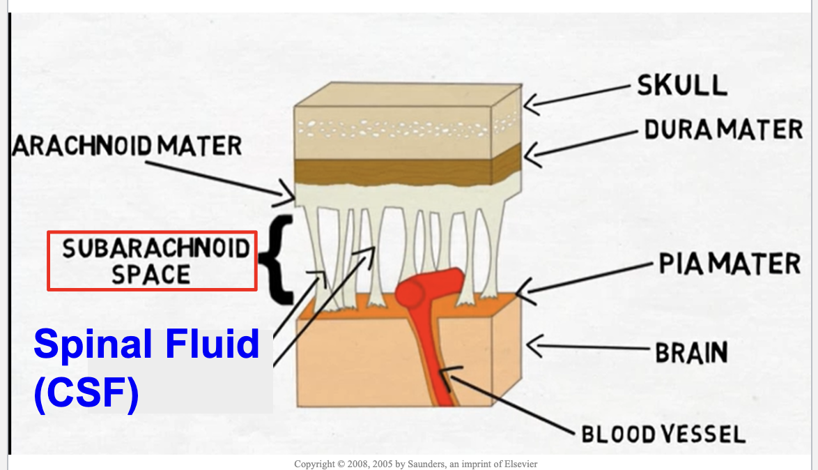
Main Idea:
The brain is protected by three membranes called the meninges and by cerebrospinal fluid (CSF), which cushions and supports the central nervous system.


Meningeal Layers (Outside → Inside) 1. Dura Mater

  • Outermost layer

  • Thick and tough

  • Located directly beneath the skull

  • Provides strong protection for the brain

2. Arachnoid Mater

  • Middle layer

  • Web-like appearance

  • Creates the upper boundary of the subarachnoid space

3. Pia Mater

  • Innermost layer

  • Very thin and delicate

  • Closely attached to the surface of the brain

  • Contains blood vessels that nourish nervous tissue


Subarachnoid Space

Location:

  • Between the arachnoid mater and pia mater

Contains:

  • Cerebrospinal fluid (CSF)

Importance:

  • The CSF-filled space acts as a protective cushion around the brain and spinal cord.


Cerebrospinal Fluid (CSF) Functions:

✅ Cushions the brain and spinal cord from injury

✅ Absorbs shock during movement

✅ Supplies nutrients to nervous tissue

✅ Removes waste products

✅ Helps maintain a stable environment for neurons


Structures Shown in the Diagram

Skull

  • Hard outer protection

Dura Mater

  • Tough outer meningeal layer

Arachnoid Mater

  • Middle, web-like layer

Subarachnoid Space

  • Contains CSF

Pia Mater

  • Thin inner layer attached to the brain

Blood Vessels

  • Carry oxygen and nutrients to brain tissue

Brain

  • Main organ of the central nervous system


Order to Memorize

Skull → Dura Mater → Arachnoid Mater → Subarachnoid Space (CSF) → Pia Mater → Brain


Memory Trick "DAP"

  • Dura = Durable

  • Arachnoid = Arachnid (spider-web)

  • Pia = Personal (sticks directly to the brain)


Exam Tip

The most commonly tested fact is:

👉 CSF is located in the subarachnoid space between the arachnoid mater and pia mater.
<p><strong>Main Idea:</strong><br>The brain is protected by three membranes called the <strong>meninges</strong> and by <strong>cerebrospinal fluid (CSF)</strong>, which cushions and supports the central nervous system.</p><div data-type="horizontalRule"><hr></div><p> Meningeal Layers (Outside → Inside) 1. Dura Mater</p><ul><li><p>Outermost layer</p></li><li><p>Thick and tough</p></li><li><p>Located directly beneath the skull</p></li><li><p>Provides strong protection for the brain</p></li></ul><p> 2. Arachnoid Mater</p><ul><li><p>Middle layer</p></li><li><p>Web-like appearance</p></li><li><p>Creates the upper boundary of the subarachnoid space</p></li></ul><p> 3. Pia Mater</p><ul><li><p>Innermost layer</p></li><li><p>Very thin and delicate</p></li><li><p>Closely attached to the surface of the brain</p></li><li><p>Contains blood vessels that nourish nervous tissue</p></li></ul><div data-type="horizontalRule"><hr></div><p> Subarachnoid Space</p><p><strong>Location:</strong></p><ul><li><p>Between the arachnoid mater and pia mater</p></li></ul><p><strong>Contains:</strong></p><ul><li><p>Cerebrospinal fluid (CSF)</p></li></ul><p><strong>Importance:</strong></p><ul><li><p>The CSF-filled space acts as a protective cushion around the brain and spinal cord.</p></li></ul><div data-type="horizontalRule"><hr></div><p> Cerebrospinal Fluid (CSF) Functions:</p><p><span data-name="check_mark_button" data-type="emoji">✅</span> Cushions the brain and spinal cord from injury</p><p><span data-name="check_mark_button" data-type="emoji">✅</span> Absorbs shock during movement</p><p><span data-name="check_mark_button" data-type="emoji">✅</span> Supplies nutrients to nervous tissue</p><p><span data-name="check_mark_button" data-type="emoji">✅</span> Removes waste products</p><p><span data-name="check_mark_button" data-type="emoji">✅</span> Helps maintain a stable environment for neurons</p><div data-type="horizontalRule"><hr></div><p> Structures Shown in the Diagram</p><p><strong>Skull</strong></p><ul><li><p>Hard outer protection</p></li></ul><p><strong>Dura Mater</strong></p><ul><li><p>Tough outer meningeal layer</p></li></ul><p><strong>Arachnoid Mater</strong></p><ul><li><p>Middle, web-like layer</p></li></ul><p><strong>Subarachnoid Space</strong></p><ul><li><p>Contains CSF</p></li></ul><p><strong>Pia Mater</strong></p><ul><li><p>Thin inner layer attached to the brain</p></li></ul><p><strong>Blood Vessels</strong></p><ul><li><p>Carry oxygen and nutrients to brain tissue</p></li></ul><p><strong>Brain</strong></p><ul><li><p>Main organ of the central nervous system</p></li></ul><div data-type="horizontalRule"><hr></div><p> Order to Memorize</p><p><strong>Skull → Dura Mater → Arachnoid Mater → Subarachnoid Space (CSF) → Pia Mater → Brain</strong></p><div data-type="horizontalRule"><hr></div><p> Memory Trick "DAP"</p><ul><li><p><strong>D</strong>ura = Durable</p></li><li><p><strong>A</strong>rachnoid = Arachnid (spider-web)</p></li><li><p><strong>P</strong>ia = Personal (sticks directly to the brain)</p></li></ul><div data-type="horizontalRule"><hr></div><p> Exam Tip</p><p>The <strong>most commonly tested fact</strong> is:</p><p><span data-name="point_right" data-type="emoji">👉</span> <strong>CSF is located in the subarachnoid space between the arachnoid mater and pia mater.</strong></p>
20
New cards

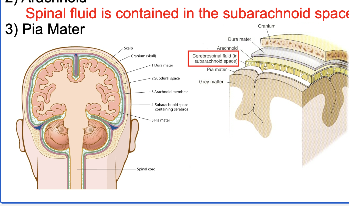
meninges (from outside to inside)

  • dura mater

  • arachnoid (spinal fluid is contained in the subarachnoid space)

  • pia mater

  • Main Idea:
    The meninges are three protective membranes that surround the brain and spinal cord. They provide protection, support, and a space for cerebrospinal fluid (CSF).


    Meningeal Layers (Outside → Inside) 1. Dura Mater

    • Outermost layer

    • Tough, thick, fibrous membrane

    • Located directly beneath the skull

    • Provides the greatest protection

    2. Arachnoid Mater

    • Middle layer

    • Thin, web-like membrane

    • Named after its spiderweb appearance

    • Creates the upper boundary of the subarachnoid space

    Subarachnoid Space

    • Located between the arachnoid mater and pia mater

    • Contains cerebrospinal fluid (CSF)

    Important Fact: 👉 CSF is contained in the subarachnoid space.

    3. Pia Mater

    • Innermost meningeal layer

    • Thin and delicate

    • Closely adheres to the surface of the brain and spinal cord

    • Contains blood vessels that nourish nervous tissue


    Cerebrospinal Fluid (CSF) Functions:

    ✅ Cushions the brain and spinal cord

    ✅ Protects against injury and shock

    ✅ Delivers nutrients

    ✅ Removes waste products

    ✅ Helps maintain a stable environment for neurons


    Layer Order to Memorize

    Skull → Dura Mater → Arachnoid Mater → Subarachnoid Space (CSF) → Pia Mater → Brain


    High-Yield Exam Facts

    • Dura mater = outermost layer

    • Pia mater = innermost layer

    • CSF is located in the subarachnoid space

    • The meninges surround both the brain and spinal cord


    Memory Trick

    "DAP"

    • Dura = Durable

    • Arachnoid = Arachnid (spider-web layer)

    • Pia = Personal (stuck directly to the brain)

    One-Sentence Summary

    The meninges consist of the dura mater, arachnoid mater, and pia mater, and cerebrospinal fluid (CSF) is found in the subarachnoid space between the arachnoid and pia mater. 🧠📚

<ul><li><p>dura mater</p></li><li><p>arachnoid (spinal fluid is contained in the subarachnoid space)</p></li><li><p>pia mater</p></li><li><p><strong>Main Idea:</strong><br>The <strong>meninges</strong> are three protective membranes that surround the brain and spinal cord. They provide protection, support, and a space for <strong>cerebrospinal fluid (CSF)</strong>.</p><div data-type="horizontalRule"><hr></div><p> Meningeal Layers (Outside → Inside) 1. Dura Mater</p><ul><li><p>Outermost layer</p></li><li><p>Tough, thick, fibrous membrane</p></li><li><p>Located directly beneath the skull</p></li><li><p>Provides the greatest protection</p></li></ul><p> 2. Arachnoid Mater</p><ul><li><p>Middle layer</p></li><li><p>Thin, web-like membrane</p></li><li><p>Named after its spiderweb appearance</p></li><li><p>Creates the upper boundary of the subarachnoid space</p></li></ul><p> Subarachnoid Space</p><ul><li><p>Located between the arachnoid mater and pia mater</p></li><li><p>Contains <strong>cerebrospinal fluid (CSF)</strong></p></li></ul><p><strong>Important Fact:</strong> <span data-name="point_right" data-type="emoji">👉</span> <strong>CSF is contained in the subarachnoid space.</strong></p><p> 3. Pia Mater</p><ul><li><p>Innermost meningeal layer</p></li><li><p>Thin and delicate</p></li><li><p>Closely adheres to the surface of the brain and spinal cord</p></li><li><p>Contains blood vessels that nourish nervous tissue</p></li></ul><div data-type="horizontalRule"><hr></div><p> Cerebrospinal Fluid (CSF) Functions:</p><p><span data-name="check_mark_button" data-type="emoji">✅</span> Cushions the brain and spinal cord</p><p><span data-name="check_mark_button" data-type="emoji">✅</span> Protects against injury and shock</p><p><span data-name="check_mark_button" data-type="emoji">✅</span> Delivers nutrients</p><p><span data-name="check_mark_button" data-type="emoji">✅</span> Removes waste products</p><p><span data-name="check_mark_button" data-type="emoji">✅</span> Helps maintain a stable environment for neurons</p><div data-type="horizontalRule"><hr></div><p> Layer Order to Memorize</p><p><strong>Skull → Dura Mater → Arachnoid Mater → Subarachnoid Space (CSF) → Pia Mater → Brain</strong></p><div data-type="horizontalRule"><hr></div><p> High-Yield Exam Facts</p><ul><li><p><strong>Dura mater = outermost layer</strong></p></li><li><p><strong>Pia mater = innermost layer</strong></p></li><li><p><strong>CSF is located in the subarachnoid space</strong></p></li><li><p><strong>The meninges surround both the brain and spinal cord</strong></p></li></ul><div data-type="horizontalRule"><hr></div><p> Memory Trick</p><p><strong>"DAP"</strong></p><ul><li><p><strong>D</strong>ura = Durable</p></li><li><p><strong>A</strong>rachnoid = Arachnid (spider-web layer)</p></li><li><p><strong>P</strong>ia = Personal (stuck directly to the brain)</p></li></ul><p> One-Sentence Summary</p><p><strong>The meninges consist of the dura mater, arachnoid mater, and pia mater, and cerebrospinal fluid (CSF) is found in the subarachnoid space between the arachnoid and pia mater.</strong> <span data-name="brain" data-type="emoji">🧠</span><span data-name="books" data-type="emoji">📚</span></p></li></ul><p></p>
21
New cards
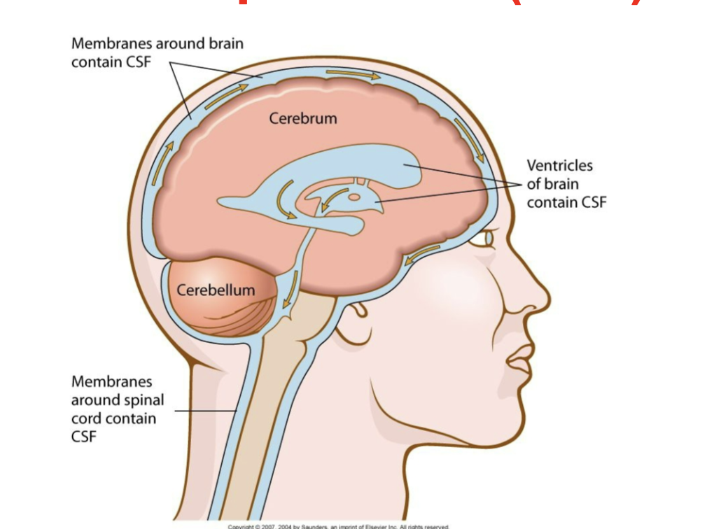
<p>cerebrospinal fluid (CSF)</p>

cerebrospinal fluid (CSF)

ultrafiltrate of plasma (water, glucose, sodium, chloride, protein

20 mL/hr made reasborbed by arachnoid villin

adult volume: 160 mL

infant volume: 60 mL

Main Idea:
Cerebrospinal fluid (CSF) is a clear, colorless fluid that circulates around and through the brain and spinal cord, providing protection, support, and nourishment to the central nervous system (CNS).


Where is CSF Found? Around the Brain and Spinal Cord

  • In the subarachnoid space between the arachnoid mater and pia mater.

  • Surrounds the entire brain and spinal cord.

Inside the Brain

  • Found within the ventricles (fluid-filled chambers of the brain).

  • Flows through connected ventricles and around the CNS.


Functions of CSF 1. Protection

  • Cushions the brain and spinal cord.

  • Acts as a shock absorber against injury.

2. Buoyancy

  • Helps support the weight of the brain.

  • Prevents the brain from pressing heavily against the skull.

3. Nutrient Transport

  • Delivers nutrients and essential substances to nervous tissue.

4. Waste Removal

  • Carries away metabolic waste products from the brain and spinal cord.

5. Homeostasis

  • Maintains a stable chemical environment for neurons to function properly.


Structures Shown in the Diagram Cerebrum

  • Largest part of the brain

  • Responsible for thinking, memory, sensation, and voluntary movement

Cerebellum

  • Coordinates balance, posture, and movement

Ventricles

  • Internal cavities within the brain containing CSF

Membranes Around the Brain and Spinal Cord

  • Meninges that contain and protect the CSF


Flow of CSF (Simple Version)

  1. Produced in the brain's ventricles

  2. Circulates through the ventricles

  3. Flows into the subarachnoid space

  4. Surrounds the brain and spinal cord

  5. Is reabsorbed into the bloodstream


High-Yield Exam Facts

✅ CSF is found in the ventricles of the brain

✅ CSF is found in the subarachnoid space

✅ CSF cushions and protects the brain and spinal cord

✅ CSF helps transport nutrients and remove waste


Memory Trick

CSF = "Cushion, Support, and Flow"

  • Cushions the CNS

  • Supports the brain

  • Flows around the brain and spinal cord

One-Sentence Summary

Cerebrospinal fluid (CSF) circulates through the ventricles and subarachnoid space, where it cushions, protects, nourishes, and supports the brain and spinal cord. 🧠💧📚
<p>ultrafiltrate of plasma (water, glucose, sodium, chloride, protein</p><p>20 mL/hr made reasborbed by arachnoid villin</p><p>adult volume: 160 mL</p><p>infant volume: 60 mL</p><p><strong>Main Idea:</strong><br><strong>Cerebrospinal fluid (CSF)</strong> is a clear, colorless fluid that circulates around and through the brain and spinal cord, providing protection, support, and nourishment to the central nervous system (CNS).</p><div data-type="horizontalRule"><hr></div><p>Where is CSF Found? Around the Brain and Spinal Cord</p><ul><li><p>In the <strong>subarachnoid space</strong> between the arachnoid mater and pia mater.</p></li><li><p>Surrounds the entire brain and spinal cord.</p></li></ul><p>Inside the Brain</p><ul><li><p>Found within the <strong>ventricles</strong> (fluid-filled chambers of the brain).</p></li><li><p>Flows through connected ventricles and around the CNS.</p></li></ul><div data-type="horizontalRule"><hr></div><p>Functions of CSF 1. Protection</p><ul><li><p>Cushions the brain and spinal cord.</p></li><li><p>Acts as a shock absorber against injury.</p></li></ul><p>2. Buoyancy</p><ul><li><p>Helps support the weight of the brain.</p></li><li><p>Prevents the brain from pressing heavily against the skull.</p></li></ul><p>3. Nutrient Transport</p><ul><li><p>Delivers nutrients and essential substances to nervous tissue.</p></li></ul><p>4. Waste Removal</p><ul><li><p>Carries away metabolic waste products from the brain and spinal cord.</p></li></ul><p>5. Homeostasis</p><ul><li><p>Maintains a stable chemical environment for neurons to function properly.</p></li></ul><div data-type="horizontalRule"><hr></div><p>Structures Shown in the Diagram Cerebrum</p><ul><li><p>Largest part of the brain</p></li><li><p>Responsible for thinking, memory, sensation, and voluntary movement</p></li></ul><p>Cerebellum</p><ul><li><p>Coordinates balance, posture, and movement</p></li></ul><p>Ventricles</p><ul><li><p>Internal cavities within the brain containing CSF</p></li></ul><p>Membranes Around the Brain and Spinal Cord</p><ul><li><p>Meninges that contain and protect the CSF</p></li></ul><div data-type="horizontalRule"><hr></div><p>Flow of CSF (Simple Version)</p><ol><li><p>Produced in the brain's ventricles</p></li><li><p>Circulates through the ventricles</p></li><li><p>Flows into the subarachnoid space</p></li><li><p>Surrounds the brain and spinal cord</p></li><li><p>Is reabsorbed into the bloodstream</p></li></ol><div data-type="horizontalRule"><hr></div><p>High-Yield Exam Facts</p><p><span data-name="check_mark_button" data-type="emoji">✅</span> CSF is found in the <strong>ventricles of the brain</strong></p><p><span data-name="check_mark_button" data-type="emoji">✅</span> CSF is found in the <strong>subarachnoid space</strong></p><p><span data-name="check_mark_button" data-type="emoji">✅</span> CSF cushions and protects the brain and spinal cord</p><p><span data-name="check_mark_button" data-type="emoji">✅</span> CSF helps transport nutrients and remove waste</p><div data-type="horizontalRule"><hr></div><p>Memory Trick</p><p><strong>CSF = "Cushion, Support, and Flow"</strong></p><ul><li><p><strong>C</strong>ushions the CNS</p></li><li><p><strong>S</strong>upports the brain</p></li><li><p><strong>F</strong>lows around the brain and spinal cord</p></li></ul><p>One-Sentence Summary</p><p><strong>Cerebrospinal fluid (CSF) circulates through the ventricles and subarachnoid space, where it cushions, protects, nourishes, and supports the brain and spinal cord.</strong> <span data-name="brain" data-type="emoji">🧠</span><span data-name="droplet" data-type="emoji">💧</span><span data-name="books" data-type="emoji">📚</span></p>
22
New cards

spinal tap (lumbar puncture) to obtain cerebrospinal fluid (CSF)

Main Idea:
A lumbar puncture (spinal tap) is a procedure in which a needle is inserted into the subarachnoid space of the lower back to collect or measure cerebrospinal fluid (CSF).


What the Diagram Shows

  • Needle inserted between the L3 and L4 vertebrae.

  • Needle passes through the meninges into the subarachnoid space.

  • The subarachnoid space contains cerebrospinal fluid (CSF).

  • The spinal cord has already ended above this area, making the procedure safer.

  • The needle enters near the cauda equina (bundle of spinal nerve roots).


Important Structures Cauda Equina

  • Means "horse's tail"

  • Bundle of spinal nerve roots below the end of the spinal cord

  • Located in the lower vertebral canal

Subarachnoid Space

  • Located between the arachnoid mater and pia mater

  • Contains CSF

Distal End of the Spinal Cord

  • Adult spinal cord ends around L1-L2

  • Lumbar punctures are performed below this level to reduce the risk of spinal cord injury


Why is a Lumbar Puncture Performed? Diagnostic Uses

  • Test for meningitis

  • Detect infections

  • Detect bleeding around the brain

  • Diagnose certain neurological diseases

Therapeutic Uses

  • Administer medications

  • Deliver anesthesia (spinal anesthesia)

  • Measure CSF pressure


Key Exam Facts

✅ Lumbar puncture collects cerebrospinal fluid (CSF)

✅ CSF is obtained from the subarachnoid space

✅ Procedure is commonly performed between L3-L4 or L4-L5

✅ The spinal cord ends near L1-L2, making lower insertion sites safer

✅ The cauda equina is found below the spinal cord


Memory Trick "LP = Lower Puncture"

  • Lumbar puncture

  • Lower back

  • L3-L4 or L4-L5

Remember:

Spinal Cord Ends → L1-L2
Lumbar Puncture → L3-L4 or L4-L5
CSF → Subarachnoid Space


One-Sentence Summary

A lumbar puncture is performed in the lower back (usually between L3-L4 or L4-L5) to access cerebrospinal fluid located in the subarachnoid space surrounding the cauda equina. 🧠💉📚

<p><strong>Main Idea:</strong><br>A <strong>lumbar puncture (spinal tap)</strong> is a procedure in which a needle is inserted into the <strong>subarachnoid space</strong> of the lower back to collect or measure <strong>cerebrospinal fluid (CSF)</strong>.</p><div data-type="horizontalRule"><hr></div><p> What the Diagram Shows</p><ul><li><p>Needle inserted between the <strong>L3 and L4 vertebrae</strong>.</p></li><li><p>Needle passes through the meninges into the <strong>subarachnoid space</strong>.</p></li><li><p>The subarachnoid space contains <strong>cerebrospinal fluid (CSF)</strong>.</p></li><li><p>The spinal cord has already ended above this area, making the procedure safer.</p></li><li><p>The needle enters near the <strong>cauda equina</strong> (bundle of spinal nerve roots).</p></li></ul><div data-type="horizontalRule"><hr></div><p> Important Structures Cauda Equina</p><ul><li><p>Means "horse's tail"</p></li><li><p>Bundle of spinal nerve roots below the end of the spinal cord</p></li><li><p>Located in the lower vertebral canal</p></li></ul><p> Subarachnoid Space</p><ul><li><p>Located between the arachnoid mater and pia mater</p></li><li><p>Contains CSF</p></li></ul><p> Distal End of the Spinal Cord</p><ul><li><p>Adult spinal cord ends around <strong>L1-L2</strong></p></li><li><p>Lumbar punctures are performed below this level to reduce the risk of spinal cord injury</p></li></ul><div data-type="horizontalRule"><hr></div><p> Why is a Lumbar Puncture Performed? Diagnostic Uses</p><ul><li><p>Test for <strong>meningitis</strong></p></li><li><p>Detect infections</p></li><li><p>Detect bleeding around the brain</p></li><li><p>Diagnose certain neurological diseases</p></li></ul><p> Therapeutic Uses</p><ul><li><p>Administer medications</p></li><li><p>Deliver anesthesia (spinal anesthesia)</p></li><li><p>Measure CSF pressure</p></li></ul><div data-type="horizontalRule"><hr></div><p> Key Exam Facts</p><p><span data-name="check_mark_button" data-type="emoji">✅</span> <strong>Lumbar puncture collects cerebrospinal fluid (CSF)</strong></p><p><span data-name="check_mark_button" data-type="emoji">✅</span> <strong>CSF is obtained from the subarachnoid space</strong></p><p><span data-name="check_mark_button" data-type="emoji">✅</span> <strong>Procedure is commonly performed between L3-L4 or L4-L5</strong></p><p><span data-name="check_mark_button" data-type="emoji">✅</span> <strong>The spinal cord ends near L1-L2, making lower insertion sites safer</strong></p><p><span data-name="check_mark_button" data-type="emoji">✅</span> <strong>The cauda equina is found below the spinal cord</strong></p><div data-type="horizontalRule"><hr></div><p> Memory Trick "LP = Lower Puncture"</p><ul><li><p><strong>L</strong>umbar puncture</p></li><li><p><strong>L</strong>ower back</p></li><li><p><strong>L</strong>3-L4 or L4-L5</p></li></ul><p> Remember:</p><p><strong>Spinal Cord Ends → L1-L2</strong><br><strong>Lumbar Puncture → L3-L4 or L4-L5</strong><br><strong>CSF → Subarachnoid Space</strong></p><div data-type="horizontalRule"><hr></div><p> One-Sentence Summary</p><p><strong>A lumbar puncture is performed in the lower back (usually between L3-L4 or L4-L5) to access cerebrospinal fluid located in the subarachnoid space surrounding the cauda equina.</strong> <span data-name="brain" data-type="emoji">🧠</span><span data-name="syringe" data-type="emoji">💉</span><span data-name="books" data-type="emoji">📚</span></p>
23
New cards

neurons vs glial cells

neurons = parenchymal cells degenerate (die) as aging

glial = stromal cells may become cancer
<p>neurons = parenchymal cells degenerate (die) as aging</p><p>glial = stromal cells may become cancer</p>
24
New cards

four types of neuroglia (glia cells)

To maintain health of nervous system. They don’t transmit impulse, but are classified into stromal cells

1) Astrocytes (astroglial cells): transport water and salts between

capillaries and neurons. Regulate chemical compositions in neurons.

2) Microglia (microglial cells): phagocytic cells protecting neurons from

infections and disposing debris.

3) Oligodendroglia (oligodendroglial cells): produce and form the myelin sheath around nerve fibers.

4) Ependymal cells

a) GK: upper garment

b) Line the membraneswithin the brain and spinal cord where CSF circulates

<p>To maintain health of nervous system. They don’t transmit impulse, but are classified into stromal cells</p><p>1) Astrocytes (astroglial cells): transport water and salts between</p><p>capillaries and neurons. Regulate chemical compositions in neurons.</p><p>2) Microglia (microglial cells): phagocytic cells protecting neurons from</p><p>infections and disposing debris.</p><p>3) Oligodendroglia (oligodendroglial cells): produce and form the myelin sheath around nerve fibers.</p><p>4) Ependymal cells</p><p>a) GK: upper garment</p><p>b) Line the membraneswithin the brain and spinal cord where CSF circulates</p>
25
New cards

glioblastoma multiforme (GBM)

Most common and most aggressive malignant primary brain tumor.

• It involves glial cells and accounting for 52% of all brain tissue tumor cases.

• Common symptoms of the disease include seizure, nausea and vomiting, headache, memory loss, and hemiparesis.

• The single most prevalent symptom is a progressive memory, personality, or neurological deficit due to temporal and frontal lobe involvement. The kind of symptoms produced depends highly on the location of the tumor, more so than on its pathological properties.

• About 50% of the people diagnosed with GBM die within one year, while 90% within three years

26
New cards

brain

Definition: Three protective layers (dura mater, arachnoid mater, and pia mater) that surround and protect the brain and spinal cord.

Function: Protect the CNS and contain cerebrospinal fluid (CSF).


Thalamus

Definition: A relay center located near the middle of the brain.

Function: Receives sensory information (except smell) and sends it to the appropriate areas of the cerebral cortex.

Think: "Sensory relay station."


Hypothalamus

Definition: A small region located below the thalamus.

Function: Maintains homeostasis by regulating:

  • Body temperature

  • Hunger

  • Thirst

  • Sleep

  • Emotions

  • Hormone production (through the pituitary gland)

Think: "Body's thermostat and control center."


Pituitary Gland

Definition: Small endocrine gland attached to the hypothalamus.

Function: Produces hormones that regulate:

  • Growth

  • Metabolism

  • Reproduction

  • Other endocrine glands

Nickname: "Master gland."


Frontal Lobe

Definition: Front portion of the cerebrum.

Function:

  • Voluntary movement

  • Planning and decision-making

  • Problem solving

  • Personality

  • Speech production

Think: "Movement and personality."


Parietal Lobe

Definition: Upper middle region of the cerebrum.

Function:

  • Processes touch, pressure, pain, and temperature

  • Interprets sensory information from the body

Think: "Sensory lobe."


Occipital Lobe

Definition: Rear portion of the cerebrum.

Function:

  • Processes visual information

Think: "Vision center."


Temporal Lobe

Definition: Side portion of the cerebrum near the ears.

Function:

  • Hearing

  • Smell

  • Memory

  • Language comprehension

Think: "Hearing and memory."


Pons

Definition: Part of the brainstem located above the medulla.

Function:

  • Relays information between brain regions

  • Helps regulate breathing

  • Involved in sleep and arousal

Think: "Bridge" between brain areas.


Cerebellum

Definition: Structure located at the back of the brain beneath the cerebrum.

Function:

  • Balance

  • Coordination

  • Posture

  • Fine motor control

Think: "Balance and coordination center."


Medulla (Medulla Oblongata)

Definition: Lowest portion of the brainstem, connecting the brain to the spinal cord.

Function: Controls vital involuntary functions:

  • Heart rate

  • Blood pressure

  • Breathing

  • Swallowing

Think: "Keeps you alive."


Super Quick Memory Chart

Structure

Main Function

Thalamus

Sensory relay

Hypothalamus

Homeostasis

Pituitary Gland

Hormone control

Frontal Lobe

Movement, personality

Parietal Lobe

Touch and sensation

Temporal Lobe

Hearing and memory

Occipital Lobe

Vision

Cerebellum

Balance and coordination

Pons

Breathing and communication

Medulla

Heart rate and breathing

Meninges

Protect brain and spinal cord

Easy Mnemonic for the Lobes

"F-POT"

  • Frontal = Functions (thinking, personality)

  • Parietal = Pressure/touch

  • Occipital = Optical (vision)

  • Temporal = Tunes (hearing) and memory


<p><strong>Definition:</strong> Three protective layers (dura mater, arachnoid mater, and pia mater) that surround and protect the brain and spinal cord.</p><p><strong>Function:</strong> Protect the CNS and contain cerebrospinal fluid (CSF).</p><div data-type="horizontalRule"><hr></div><p><strong>Thalamus</strong></p><p><strong>Definition:</strong> A relay center located near the middle of the brain.</p><p><strong>Function:</strong> Receives sensory information (except smell) and sends it to the appropriate areas of the cerebral cortex.</p><p><strong>Think:</strong> "Sensory relay station."</p><div data-type="horizontalRule"><hr></div><p><strong>Hypothalamus</strong></p><p><strong>Definition:</strong> A small region located below the thalamus.</p><p><strong>Function:</strong> Maintains homeostasis by regulating:</p><ul><li><p>Body temperature</p></li><li><p>Hunger</p></li><li><p>Thirst</p></li><li><p>Sleep</p></li><li><p>Emotions</p></li><li><p>Hormone production (through the pituitary gland)</p></li></ul><p><strong>Think:</strong> "Body's thermostat and control center."</p><div data-type="horizontalRule"><hr></div><p><strong>Pituitary Gland</strong></p><p><strong>Definition:</strong> Small endocrine gland attached to the hypothalamus.</p><p><strong>Function:</strong> Produces hormones that regulate:</p><ul><li><p>Growth</p></li><li><p>Metabolism</p></li><li><p>Reproduction</p></li><li><p>Other endocrine glands</p></li></ul><p><strong>Nickname:</strong> "Master gland."</p><div data-type="horizontalRule"><hr></div><p><strong>Frontal Lobe</strong></p><p><strong>Definition:</strong> Front portion of the cerebrum.</p><p><strong>Function:</strong></p><ul><li><p>Voluntary movement</p></li><li><p>Planning and decision-making</p></li><li><p>Problem solving</p></li><li><p>Personality</p></li><li><p>Speech production</p></li></ul><p><strong>Think:</strong> "Movement and personality."</p><div data-type="horizontalRule"><hr></div><p><strong>Parietal Lobe</strong></p><p><strong>Definition:</strong> Upper middle region of the cerebrum.</p><p><strong>Function:</strong></p><ul><li><p>Processes touch, pressure, pain, and temperature</p></li><li><p>Interprets sensory information from the body</p></li></ul><p><strong>Think:</strong> "Sensory lobe."</p><div data-type="horizontalRule"><hr></div><p><strong>Occipital Lobe</strong></p><p><strong>Definition:</strong> Rear portion of the cerebrum.</p><p><strong>Function:</strong></p><ul><li><p>Processes visual information</p></li></ul><p><strong>Think:</strong> "Vision center."</p><div data-type="horizontalRule"><hr></div><p><strong>Temporal Lobe</strong></p><p><strong>Definition:</strong> Side portion of the cerebrum near the ears.</p><p><strong>Function:</strong></p><ul><li><p>Hearing</p></li><li><p>Smell</p></li><li><p>Memory</p></li><li><p>Language comprehension</p></li></ul><p><strong>Think:</strong> "Hearing and memory."</p><div data-type="horizontalRule"><hr></div><p><strong>Pons</strong></p><p><strong>Definition:</strong> Part of the brainstem located above the medulla.</p><p><strong>Function:</strong></p><ul><li><p>Relays information between brain regions</p></li><li><p>Helps regulate breathing</p></li><li><p>Involved in sleep and arousal</p></li></ul><p><strong>Think:</strong> "Bridge" between brain areas.</p><div data-type="horizontalRule"><hr></div><p><strong>Cerebellum</strong></p><p><strong>Definition:</strong> Structure located at the back of the brain beneath the cerebrum.</p><p><strong>Function:</strong></p><ul><li><p>Balance</p></li><li><p>Coordination</p></li><li><p>Posture</p></li><li><p>Fine motor control</p></li></ul><p><strong>Think:</strong> "Balance and coordination center."</p><div data-type="horizontalRule"><hr></div><p><strong>Medulla (Medulla Oblongata)</strong></p><p><strong>Definition:</strong> Lowest portion of the brainstem, connecting the brain to the spinal cord.</p><p><strong>Function:</strong> Controls vital involuntary functions:</p><ul><li><p>Heart rate</p></li><li><p>Blood pressure</p></li><li><p>Breathing</p></li><li><p>Swallowing</p></li></ul><p><strong>Think:</strong> "Keeps you alive."</p><div data-type="horizontalRule"><hr></div><p> Super Quick Memory Chart</p><table style="min-width: 50px;"><colgroup><col style="min-width: 25px;"><col style="min-width: 25px;"></colgroup><tbody><tr><th colspan="1" rowspan="1"><p>Structure</p></th><th colspan="1" rowspan="1"><p>Main Function</p></th></tr><tr><td colspan="1" rowspan="1"><p>Thalamus</p></td><td colspan="1" rowspan="1"><p>Sensory relay</p></td></tr><tr><td colspan="1" rowspan="1"><p>Hypothalamus</p></td><td colspan="1" rowspan="1"><p>Homeostasis</p></td></tr><tr><td colspan="1" rowspan="1"><p>Pituitary Gland</p></td><td colspan="1" rowspan="1"><p>Hormone control</p></td></tr><tr><td colspan="1" rowspan="1"><p>Frontal Lobe</p></td><td colspan="1" rowspan="1"><p>Movement, personality</p></td></tr><tr><td colspan="1" rowspan="1"><p>Parietal Lobe</p></td><td colspan="1" rowspan="1"><p>Touch and sensation</p></td></tr><tr><td colspan="1" rowspan="1"><p>Temporal Lobe</p></td><td colspan="1" rowspan="1"><p>Hearing and memory</p></td></tr><tr><td colspan="1" rowspan="1"><p>Occipital Lobe</p></td><td colspan="1" rowspan="1"><p>Vision</p></td></tr><tr><td colspan="1" rowspan="1"><p>Cerebellum</p></td><td colspan="1" rowspan="1"><p>Balance and coordination</p></td></tr><tr><td colspan="1" rowspan="1"><p>Pons</p></td><td colspan="1" rowspan="1"><p>Breathing and communication</p></td></tr><tr><td colspan="1" rowspan="1"><p>Medulla</p></td><td colspan="1" rowspan="1"><p>Heart rate and breathing</p></td></tr><tr><td colspan="1" rowspan="1"><p>Meninges</p></td><td colspan="1" rowspan="1"><p>Protect brain and spinal cord</p></td></tr></tbody></table><p> Easy Mnemonic for the Lobes</p><p><strong>"F-POT"</strong></p><ul><li><p><strong>F</strong>rontal = <strong>F</strong>unctions (thinking, personality)</p></li><li><p><strong>P</strong>arietal = <strong>P</strong>ressure/touch</p></li><li><p><strong>O</strong>ccipital = <strong>O</strong>ptical (vision)</p></li><li><p><strong>T</strong>emporal = <strong>T</strong>unes (hearing) and memory</p></li></ul><p></p>
27
New cards

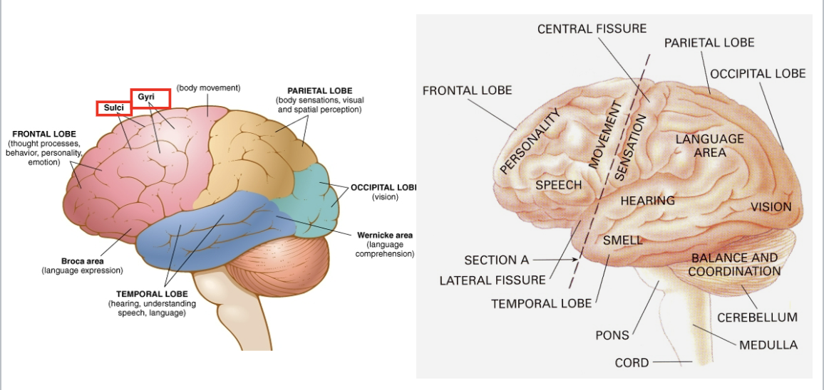
cerebrum

  • sulcus: depression, groove in the surface of the cerebral cortex

  • gyrus: sheet of nerve cells producing a rounded fold on the surface of the cerebrym

<ul><li><p>sulcus: depression, groove in the surface of the cerebral cortex</p></li><li><p>gyrus: sheet of nerve cells producing a rounded fold on the surface of the cerebrym</p></li></ul><p></p>
28
New cards

gyri and sulci lay on cerebral cortex

Main Idea:
The surface of the brain is highly folded. These folds increase the surface area of the cerebral cortex, allowing for more neurons and greater brain function.


Gyrus (plural: Gyri)

Definition:
A gyrus is a raised ridge or fold on the surface of the brain.

Function:

  • Increases the surface area of the cerebral cortex.

  • Allows more neurons to fit within the skull.

  • Helps improve information processing.

Think: A "hill" or "ridge" on the brain's surface.


Sulcus (plural: Sulci)

Definition:
A sulcus is a shallow groove or furrow between gyri.

Function:

  • Separates adjacent gyri.

  • Helps organize different functional regions of the brain.

Think: A "valley" between brain folds.


Why Are Gyri and Sulci Important?

✅ Increase the surface area of the cerebral cortex

✅ Allow more neurons to fit into the brain

✅ Improve the brain's processing capacity

✅ Help separate different functional brain regions


Visualizing It

  • Gyrus = Raised fold (hill)

  • Sulcus = Groove (valley)

In the diagram:

  • The rounded raised area is the gyrus.

  • The indentation between folds is the sulcus.


Memory Trick

"Gyrus = Grows Up" ⛰

  • Gyri stick up.

"Sulcus = Sink" ⬇

  • Sulci go down.


Exam Tip

Professors often ask:

Q: What is the difference between a gyrus and a sulcus?

A: A gyrus is a raised fold of the cerebral cortex, while a sulcus is a groove or depression between gyri.

One-Sentence Summary

Gyri are the raised folds of the cerebral cortex, and sulci are the grooves between them; together they increase the brain's surface area and allow for greater processing power. 🧠📚

<p><strong>Main Idea:</strong><br>The surface of the brain is highly folded. These folds increase the surface area of the cerebral cortex, allowing for more neurons and greater brain function.</p><div data-type="horizontalRule"><hr></div><p> Gyrus (plural: Gyri)</p><p><strong>Definition:</strong><br>A <strong>gyrus</strong> is a raised ridge or fold on the surface of the brain.</p><p><strong>Function:</strong></p><ul><li><p>Increases the surface area of the cerebral cortex.</p></li><li><p>Allows more neurons to fit within the skull.</p></li><li><p>Helps improve information processing.</p></li></ul><p><strong>Think:</strong> A "hill" or "ridge" on the brain's surface.</p><div data-type="horizontalRule"><hr></div><p> Sulcus (plural: Sulci)</p><p><strong>Definition:</strong><br>A <strong>sulcus</strong> is a shallow groove or furrow between gyri.</p><p><strong>Function:</strong></p><ul><li><p>Separates adjacent gyri.</p></li><li><p>Helps organize different functional regions of the brain.</p></li></ul><p><strong>Think:</strong> A "valley" between brain folds.</p><div data-type="horizontalRule"><hr></div><p> Why Are Gyri and Sulci Important?</p><p><span data-name="check_mark_button" data-type="emoji">✅</span> Increase the surface area of the cerebral cortex</p><p><span data-name="check_mark_button" data-type="emoji">✅</span> Allow more neurons to fit into the brain</p><p><span data-name="check_mark_button" data-type="emoji">✅</span> Improve the brain's processing capacity</p><p><span data-name="check_mark_button" data-type="emoji">✅</span> Help separate different functional brain regions</p><div data-type="horizontalRule"><hr></div><p> Visualizing It</p><ul><li><p><strong>Gyrus = Raised fold (hill)</strong></p></li><li><p><strong>Sulcus = Groove (valley)</strong></p></li></ul><p>In the diagram:</p><ul><li><p>The rounded raised area is the <strong>gyrus</strong>.</p></li><li><p>The indentation between folds is the <strong>sulcus</strong>.</p></li></ul><div data-type="horizontalRule"><hr></div><p> Memory Trick</p><p><strong>"Gyrus = Grows Up"</strong> <span data-name="mountain" data-type="emoji">⛰</span></p><ul><li><p>Gyri stick up.</p></li></ul><p><strong>"Sulcus = Sink"</strong> <span data-name="arrow_down" data-type="emoji">⬇</span></p><ul><li><p>Sulci go down.</p></li></ul><div data-type="horizontalRule"><hr></div><p> Exam Tip</p><p>Professors often ask:</p><p><strong>Q:</strong> What is the difference between a gyrus and a sulcus?</p><p><strong>A:</strong> A <strong>gyrus</strong> is a raised fold of the cerebral cortex, while a <strong>sulcus</strong> is a groove or depression between gyri.</p><p> One-Sentence Summary</p><p><strong>Gyri are the raised folds of the cerebral cortex, and sulci are the grooves between them; together they increase the brain's surface area and allow for greater processing power.</strong> <span data-name="brain" data-type="emoji">🧠</span><span data-name="books" data-type="emoji">📚</span></p>
29
New cards

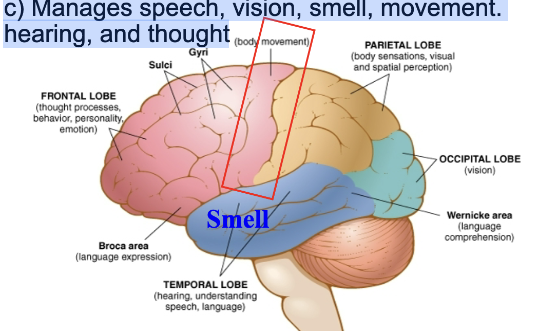
cerebrum

a) Largest section of brain

b) Surface area is called cerebral cortex

c) Manages speech, vision, smell, movement. hearing, and thought
<p>a) Largest section of brain</p><p>b) Surface area is called cerebral cortex</p><p>c) Manages speech, vision, smell, movement. hearing, and thought</p>
30
New cards

afferent and efferent nerve fibers

Cranial and spinal nerves

a) Sensory nerves (Afferent Nerve Fibers) carry messages

toward the brain

b) Motor nerves (Efferent Nerve Fibers) carry messages

from the brain.

• Sensory receptors in organs and glands

a) Sight

b) Hearing and balance

c) Smell

31
New cards

sensory and motor areas of cerebral cortex

Main Idea:
The cerebral cortex contains specialized areas that control movement (motor cortex) and process sensations (sensory cortex). Different body parts are represented in different regions of these cortices.


Motor Cortex (Precentral Gyrus) Definition:

The motor cortex is located in the frontal lobe and controls voluntary skeletal muscle movements.

Function:

  • Initiates movement

  • Sends signals to muscles throughout the body

Important Fact:

  • The left motor cortex controls the right side of the body

  • The right motor cortex controls the left side of the body


Somatic Sensory Cortex (Postcentral Gyrus) Definition:

The somatic sensory cortex is located in the parietal lobe and receives sensory information from the body.

Function:

Processes:

  • Touch

  • Pressure

  • Pain

  • Temperature

  • Vibration

  • Body position (proprioception)

Important Fact:

  • The left sensory cortex receives information from the right side of the body

  • The right sensory cortex receives information from the left side of the body


Precentral Gyrus Definition:

A gyrus located immediately anterior (in front of) the central sulcus.

Function:

Contains the primary motor cortex.

Think: Pre = Before the central sulcus = Movement


Postcentral Gyrus Definition:

A gyrus located immediately posterior (behind) the central sulcus.

Function:

Contains the primary somatic sensory cortex.

Think: Post = After the central sulcus = Sensation


Cortical Homunculus Definition:

A map showing how different body parts are represented in the motor and sensory cortices.

Key Concept:

The amount of cortex devoted to a body part depends on:

  • Fine motor control (motor cortex)

  • Sensory sensitivity (sensory cortex)


Why Are the Hands and Face So Large?

The hands, lips, tongue, and face take up a large area because they:

  • Require precise movements

  • Have many sensory receptors

  • Need greater cortical processing

Not because they are physically larger.


Body Arrangement on the Cortex Top of Cortex

  • Toes

  • Feet

  • Legs

Middle

  • Trunk

  • Arms

  • Hands

Lower Side

  • Face

  • Lips

  • Tongue


High-Yield Exam Facts

✅ Motor cortex = Precentral gyrus

✅ Sensory cortex = Postcentral gyrus

✅ Motor cortex is in the frontal lobe

✅ Sensory cortex is in the parietal lobe

✅ Left hemisphere controls/receives input from the right side of the body

✅ Hands and face occupy large cortical areas


Memory Tricks

Motor = Move

  • Precentral gyrus = Motor cortex

Sensory = Sense

  • Postcentral gyrus = Sensory cortex

"PRE moves, POST feels."


One-Sentence Summary

The precentral gyrus contains the primary motor cortex responsible for voluntary movement, while the postcentral gyrus contains the primary somatic sensory cortex responsible for processing touch, pain, temperature, and other sensations from the body. 🧠📚

32
New cards

spinal cord (afferent and efferent nerves)

Gray matter: nerve cell bodies

White matter: Axons covered with myelin sheets

Main Idea:
Sensory information travels into the spinal cord through the dorsal root, while motor commands travel out of the spinal cord through the ventral root.


Dorsal Root (Sensory Root) Definition:

The dorsal root is the posterior (back) root of a spinal nerve that carries sensory (afferent) information into the spinal cord.

Function:

  • Brings sensory signals from the skin, muscles, and organs to the CNS.

  • Carries sensations such as:

    • Touch

    • Pain

    • Temperature

    • Pressure

Remember:
Dorsal = Sensory = Afferent


Dorsal Root Ganglion Definition:

A cluster of sensory neuron cell bodies located on the dorsal root.

Function:

  • Houses the cell bodies of sensory neurons before they enter the spinal cord.

Exam Tip:
The dorsal root ganglion contains sensory neuron cell bodies.


Afferent Neuron (Sensory Neuron) Definition:

A neuron that carries information toward the CNS.

Function:

  • Transmits sensory information from receptors to the brain and spinal cord.

Pathway:

Receptor → Afferent Neuron → Dorsal Root → Spinal Cord

Memory Trick:
Afferent = Arrives at the CNS


Ventral Root (Motor Root) Definition:

The anterior (front) root of a spinal nerve that carries motor (efferent) signals out of the spinal cord.

Function:

  • Sends commands from the CNS to muscles and glands.

Remember:
Ventral = Motor = Efferent


Efferent Neuron (Motor Neuron) Definition:

A neuron that carries information away from the CNS.

Function:

  • Sends motor commands to skeletal muscles.

  • Produces movement.

Pathway:

Spinal Cord → Efferent Neuron → Muscle

Memory Trick:
Efferent = Exits the CNS


Gray Matter Definition:

The inner butterfly-shaped region of the spinal cord.

Function:

  • Contains neuron cell bodies.

  • Site where information is processed.


White Matter Definition:

The outer region of the spinal cord.

Function:

  • Contains myelinated axons.

  • Carries information up and down the spinal cord.


Central Canal Definition:

A small canal running through the center of the spinal cord.

Function:

  • Contains cerebrospinal fluid (CSF).


High-Yield Exam Facts

✅ Dorsal root = sensory (afferent)

✅ Ventral root = motor (efferent)

✅ Dorsal root ganglion contains sensory neuron cell bodies

✅ Afferent neurons carry information TO the CNS

✅ Efferent neurons carry information AWAY from the CNS


Memory Trick

SAME DAVE

  • Sensory = Afferent

  • Motor = Efferent

Dorsal = Afferent
Ventral = Efferent


One-Sentence Summary

Sensory (afferent) neurons enter the spinal cord through the dorsal root and dorsal root ganglion, while motor (efferent) neurons exit the spinal cord through the ventral root to control muscles. 🧠➡💪

<p>Gray matter: nerve cell bodies</p><p>White matter: Axons covered with myelin sheets</p><p><strong>Main Idea:</strong><br>Sensory information travels <strong>into</strong> the spinal cord through the <strong>dorsal root</strong>, while motor commands travel <strong>out of</strong> the spinal cord through the <strong>ventral root</strong>.</p><div data-type="horizontalRule"><hr></div><p> Dorsal Root (Sensory Root) Definition:</p><p>The <strong>dorsal root</strong> is the posterior (back) root of a spinal nerve that carries <strong>sensory (afferent)</strong> information into the spinal cord.</p><p> Function:</p><ul><li><p>Brings sensory signals from the skin, muscles, and organs to the CNS.</p></li><li><p>Carries sensations such as:</p><ul><li><p>Touch</p></li><li><p>Pain</p></li><li><p>Temperature</p></li><li><p>Pressure</p></li></ul></li></ul><p><strong>Remember:</strong><br><strong>Dorsal = Sensory = Afferent</strong></p><div data-type="horizontalRule"><hr></div><p> Dorsal Root Ganglion Definition:</p><p>A cluster of sensory neuron cell bodies located on the dorsal root.</p><p> Function:</p><ul><li><p>Houses the cell bodies of sensory neurons before they enter the spinal cord.</p></li></ul><p><strong>Exam Tip:</strong><br>The dorsal root ganglion contains <strong>sensory neuron cell bodies</strong>.</p><div data-type="horizontalRule"><hr></div><p> Afferent Neuron (Sensory Neuron) Definition:</p><p>A neuron that carries information <strong>toward the CNS</strong>.</p><p> Function:</p><ul><li><p>Transmits sensory information from receptors to the brain and spinal cord.</p></li></ul><p> Pathway:</p><p>Receptor → Afferent Neuron → Dorsal Root → Spinal Cord</p><p><strong>Memory Trick:</strong><br><strong>Afferent = Arrives at the CNS</strong></p><div data-type="horizontalRule"><hr></div><p> Ventral Root (Motor Root) Definition:</p><p>The anterior (front) root of a spinal nerve that carries <strong>motor (efferent)</strong> signals out of the spinal cord.</p><p> Function:</p><ul><li><p>Sends commands from the CNS to muscles and glands.</p></li></ul><p><strong>Remember:</strong><br><strong>Ventral = Motor = Efferent</strong></p><div data-type="horizontalRule"><hr></div><p> Efferent Neuron (Motor Neuron) Definition:</p><p>A neuron that carries information <strong>away from the CNS</strong>.</p><p> Function:</p><ul><li><p>Sends motor commands to skeletal muscles.</p></li><li><p>Produces movement.</p></li></ul><p> Pathway:</p><p>Spinal Cord → Efferent Neuron → Muscle</p><p><strong>Memory Trick:</strong><br><strong>Efferent = Exits the CNS</strong></p><div data-type="horizontalRule"><hr></div><p> Gray Matter Definition:</p><p>The inner butterfly-shaped region of the spinal cord.</p><p> Function:</p><ul><li><p>Contains neuron cell bodies.</p></li><li><p>Site where information is processed.</p></li></ul><div data-type="horizontalRule"><hr></div><p> White Matter Definition:</p><p>The outer region of the spinal cord.</p><p> Function:</p><ul><li><p>Contains myelinated axons.</p></li><li><p>Carries information up and down the spinal cord.</p></li></ul><div data-type="horizontalRule"><hr></div><p> Central Canal Definition:</p><p>A small canal running through the center of the spinal cord.</p><p> Function:</p><ul><li><p>Contains cerebrospinal fluid (CSF).</p></li></ul><div data-type="horizontalRule"><hr></div><p> High-Yield Exam Facts</p><p><span data-name="check_mark_button" data-type="emoji">✅</span> <strong>Dorsal root = sensory (afferent)</strong></p><p><span data-name="check_mark_button" data-type="emoji">✅</span> <strong>Ventral root = motor (efferent)</strong></p><p><span data-name="check_mark_button" data-type="emoji">✅</span> <strong>Dorsal root ganglion contains sensory neuron cell bodies</strong></p><p><span data-name="check_mark_button" data-type="emoji">✅</span> <strong>Afferent neurons carry information TO the CNS</strong></p><p><span data-name="check_mark_button" data-type="emoji">✅</span> <strong>Efferent neurons carry information AWAY from the CNS</strong></p><div data-type="horizontalRule"><hr></div><p> Memory Trick</p><p><strong>SAME DAVE</strong></p><ul><li><p><strong>S</strong>ensory = <strong>A</strong>fferent</p></li><li><p><strong>M</strong>otor = <strong>E</strong>fferent</p></li></ul><p><strong>D</strong>orsal = <strong>A</strong>fferent<br><strong>V</strong>entral = <strong>E</strong>fferent</p><div data-type="horizontalRule"><hr></div><p> One-Sentence Summary</p><p><strong>Sensory (afferent) neurons enter the spinal cord through the dorsal root and dorsal root ganglion, while motor (efferent) neurons exit the spinal cord through the ventral root to control muscles.</strong> <span data-name="brain" data-type="emoji">🧠</span><span data-name="arrow_right" data-type="emoji">➡</span><span data-name="muscle" data-type="emoji">💪</span></p>
33
New cards

brain

Cerebellum (“little brain”)

a) Coordinates voluntary movements

b) Maintains balance coordination

Thalamus (Gk: chamber)

1) Relaying & processing sensory signals prior to sending them to cerebral cortex.

2) Regulation of alertness & consciousness.

<p>Cerebellum (“little brain”)</p><p>a) Coordinates voluntary movements</p><p>b) Maintains balance coordination</p><p>Thalamus (Gk: chamber)</p><p>1) Relaying &amp; processing sensory signals prior to sending them to cerebral cortex.</p><p>2) Regulation of alertness &amp; consciousness.</p>
34
New cards

hypothalamus

Controls body temperature, sleep, appetite, sexual

desire, and emotions

•Supervises the release of hormones from pituitary

gland.

•Integrates activities of sympathetic and

parasympathetic nervous systems

<p>Controls body temperature, sleep, appetite, sexual</p><p>desire, and emotions</p><p>•Supervises the release of hormones from pituitary</p><p>gland.</p><p>•Integrates activities of sympathetic and</p><p>parasympathetic nervous systems</p>
35
New cards

brain stem pons

Bridges cerebrum with cerebellum and medulla

•Passes on pulses from cerebellum and houses

nerves for face and eyes, including hearing, tasting,

eye movement, facial expression, chewing, swallowing and secretion of saliva.

•Lower part interacts with

Cranial nerve

<p>Bridges cerebrum with cerebellum and medulla</p><p>•Passes on pulses from cerebellum and houses</p><p>nerves for face and eyes, including hearing, tasting,</p><p>eye movement, facial expression, chewing, swallowing and secretion of saliva.</p><p>•Lower part interacts with</p><p>Cranial nerve</p>
36
New cards

cranial nerves are autonomic nerve system

Main Idea:
There are 12 pairs of cranial nerves that arise directly from the brain and brainstem. They control sensory functions, motor functions, or both in the head, neck, and some internal organs.


I. Olfactory Nerve

Function: Smell

Memory Aid: "Olfaction = Odor"


II. Optic Nerve

Function: Vision

Word Parts:

  • Opt/o = eye, vision

  • Optic/o = eye, sight


III. Oculomotor Nerve

Function: Most eye movements and pupil constriction

Word Part:

  • Ocul/o = eye


IV. Trochlear Nerve

Function: Eye movement

Memory Aid: "Trochlea" means pulley, referring to how the eye muscle moves.


V. Trigeminal Nerve

Function:

  • Facial sensation

  • Chewing

Memory Aid: "Tri" = three branches

Three Branches:

  1. Ophthalmic

  2. Maxillary

  3. Mandibular

Think: "Born in threes"


VI. Abducens Nerve

Function: Moves the eye laterally (away from the nose)

Memory Aid: "Abduct = move away"


VII. Facial Nerve

Function:

  • Facial expressions

  • Taste (anterior tongue)

  • Some ear sensation

Think: "Makes the face move"


VIII. Vestibulocochlear Nerve

Function:

  • Hearing

  • Balance

Memory Aid:

  • Vestibule = balance

  • Cochlea = hearing ("snail shell")


IX. Glossopharyngeal Nerve

Function:

  • Taste

  • Tongue sensation

  • Swallowing

  • Throat sensation

Word Parts:

  • Gloss/o = tongue

  • Pharyng/o = throat


X. Vagus Nerve

Function:

  • Controls many autonomic functions

  • Heart rate

  • Blood pressure

  • Digestion

  • Breathing support

Memory Aid:
Vagus means "wandering nerve" because it travels throughout the body.

Very Important Exam Fact: The vagus nerve is the major nerve of the parasympathetic nervous system.


XI. Accessory Nerve

Function:

  • Head movement

  • Shoulder shrugging

  • Neck muscles


XII. Hypoglossal Nerve

Function:

  • Tongue movement

  • Speech

  • Swallowing

Word Part:

  • Hypo = under

  • Glossal = tongue


Brainstem Cranial Nerves

Many cranial nerves emerge from the:

  • Pons

  • Medulla Oblongata


Cranial Nerves in Order

Number

Name

Function

I

Olfactory

Smell

II

Optic

Vision

III

Oculomotor

Eye movement

IV

Trochlear

Eye movement

V

Trigeminal

Facial sensation, chewing

VI

Abducens

Eye movement

VII

Facial

Facial expression, taste

VIII

Vestibulocochlear

Hearing, balance

IX

Glossopharyngeal

Taste, swallowing

X

Vagus

Parasympathetic control

XI

Accessory

Head and shoulder movement

XII

Hypoglossal

Tongue movement


Easy Mnemonic (Names)

"Oh, Oh, Oh, To Touch And Feel Very Good Velvet, AH!"

  1. Olfactory

  2. Optic

  3. Oculomotor

  4. Trochlear

  5. Trigeminal

  6. Abducens

  7. Facial

  8. Vestibulocochlear

  9. Glossopharyngeal

  10. Vagus

  11. Accessory

  12. Hypoglossal

One-Sentence Summary

The 12 cranial nerves arise from the brain and brainstem and control smell, vision, eye movements, facial sensation and expression, hearing, balance, swallowing, autonomic functions, and tongue movement. 🧠📚

<p><strong>Main Idea:</strong><br>There are <strong>12 pairs of cranial nerves</strong> that arise directly from the brain and brainstem. They control sensory functions, motor functions, or both in the head, neck, and some internal organs.</p><div data-type="horizontalRule"><hr></div><p> I. Olfactory Nerve</p><p><strong>Function:</strong> Smell</p><p><strong>Memory Aid:</strong> "Olfaction = Odor"</p><div data-type="horizontalRule"><hr></div><p> II. Optic Nerve</p><p><strong>Function:</strong> Vision</p><p><strong>Word Parts:</strong></p><ul><li><p><strong>Opt/o</strong> = eye, vision</p></li><li><p><strong>Optic/o</strong> = eye, sight</p></li></ul><div data-type="horizontalRule"><hr></div><p> III. Oculomotor Nerve</p><p><strong>Function:</strong> Most eye movements and pupil constriction</p><p><strong>Word Part:</strong></p><ul><li><p><strong>Ocul/o</strong> = eye</p></li></ul><div data-type="horizontalRule"><hr></div><p> IV. Trochlear Nerve</p><p><strong>Function:</strong> Eye movement</p><p><strong>Memory Aid:</strong> "Trochlea" means <strong>pulley</strong>, referring to how the eye muscle moves.</p><div data-type="horizontalRule"><hr></div><p> V. Trigeminal Nerve</p><p><strong>Function:</strong></p><ul><li><p>Facial sensation</p></li><li><p>Chewing</p></li></ul><p><strong>Memory Aid:</strong> "Tri" = three branches</p><p> Three Branches:</p><ol><li><p>Ophthalmic</p></li><li><p>Maxillary</p></li><li><p>Mandibular</p></li></ol><p><strong>Think:</strong> "Born in threes"</p><div data-type="horizontalRule"><hr></div><p> VI. Abducens Nerve</p><p><strong>Function:</strong> Moves the eye laterally (away from the nose)</p><p><strong>Memory Aid:</strong> "Abduct = move away"</p><div data-type="horizontalRule"><hr></div><p> VII. Facial Nerve</p><p><strong>Function:</strong></p><ul><li><p>Facial expressions</p></li><li><p>Taste (anterior tongue)</p></li><li><p>Some ear sensation</p></li></ul><p><strong>Think:</strong> "Makes the face move"</p><div data-type="horizontalRule"><hr></div><p> VIII. Vestibulocochlear Nerve</p><p><strong>Function:</strong></p><ul><li><p>Hearing</p></li><li><p>Balance</p></li></ul><p><strong>Memory Aid:</strong></p><ul><li><p>Vestibule = balance</p></li><li><p>Cochlea = hearing ("snail shell")</p></li></ul><div data-type="horizontalRule"><hr></div><p> IX. Glossopharyngeal Nerve</p><p><strong>Function:</strong></p><ul><li><p>Taste</p></li><li><p>Tongue sensation</p></li><li><p>Swallowing</p></li><li><p>Throat sensation</p></li></ul><p><strong>Word Parts:</strong></p><ul><li><p>Gloss/o = tongue</p></li><li><p>Pharyng/o = throat</p></li></ul><div data-type="horizontalRule"><hr></div><p> X. Vagus Nerve</p><p><strong>Function:</strong></p><ul><li><p>Controls many autonomic functions</p></li><li><p>Heart rate</p></li><li><p>Blood pressure</p></li><li><p>Digestion</p></li><li><p>Breathing support</p></li></ul><p><strong>Memory Aid:</strong><br>Vagus means "<strong>wandering nerve</strong>" because it travels throughout the body.</p><p><strong>Very Important Exam Fact:</strong> The vagus nerve is the major nerve of the <strong>parasympathetic nervous system</strong>.</p><div data-type="horizontalRule"><hr></div><p> XI. Accessory Nerve</p><p><strong>Function:</strong></p><ul><li><p>Head movement</p></li><li><p>Shoulder shrugging</p></li><li><p>Neck muscles</p></li></ul><div data-type="horizontalRule"><hr></div><p> XII. Hypoglossal Nerve</p><p><strong>Function:</strong></p><ul><li><p>Tongue movement</p></li><li><p>Speech</p></li><li><p>Swallowing</p></li></ul><p><strong>Word Part:</strong></p><ul><li><p>Hypo = under</p></li><li><p>Glossal = tongue</p></li></ul><div data-type="horizontalRule"><hr></div><p> Brainstem Cranial Nerves</p><p>Many cranial nerves emerge from the:</p><ul><li><p><strong>Pons</strong></p></li><li><p><strong>Medulla Oblongata</strong></p></li></ul><div data-type="horizontalRule"><hr></div><p> Cranial Nerves in Order</p><table style="min-width: 75px;"><colgroup><col style="min-width: 25px;"><col style="min-width: 25px;"><col style="min-width: 25px;"></colgroup><tbody><tr><th colspan="1" rowspan="1"><p>Number</p></th><th colspan="1" rowspan="1"><p>Name</p></th><th colspan="1" rowspan="1"><p>Function</p></th></tr><tr><td colspan="1" rowspan="1"><p>I</p></td><td colspan="1" rowspan="1"><p>Olfactory</p></td><td colspan="1" rowspan="1"><p>Smell</p></td></tr><tr><td colspan="1" rowspan="1"><p>II</p></td><td colspan="1" rowspan="1"><p>Optic</p></td><td colspan="1" rowspan="1"><p>Vision</p></td></tr><tr><td colspan="1" rowspan="1"><p>III</p></td><td colspan="1" rowspan="1"><p>Oculomotor</p></td><td colspan="1" rowspan="1"><p>Eye movement</p></td></tr><tr><td colspan="1" rowspan="1"><p>IV</p></td><td colspan="1" rowspan="1"><p>Trochlear</p></td><td colspan="1" rowspan="1"><p>Eye movement</p></td></tr><tr><td colspan="1" rowspan="1"><p>V</p></td><td colspan="1" rowspan="1"><p>Trigeminal</p></td><td colspan="1" rowspan="1"><p>Facial sensation, chewing</p></td></tr><tr><td colspan="1" rowspan="1"><p>VI</p></td><td colspan="1" rowspan="1"><p>Abducens</p></td><td colspan="1" rowspan="1"><p>Eye movement</p></td></tr><tr><td colspan="1" rowspan="1"><p>VII</p></td><td colspan="1" rowspan="1"><p>Facial</p></td><td colspan="1" rowspan="1"><p>Facial expression, taste</p></td></tr><tr><td colspan="1" rowspan="1"><p>VIII</p></td><td colspan="1" rowspan="1"><p>Vestibulocochlear</p></td><td colspan="1" rowspan="1"><p>Hearing, balance</p></td></tr><tr><td colspan="1" rowspan="1"><p>IX</p></td><td colspan="1" rowspan="1"><p>Glossopharyngeal</p></td><td colspan="1" rowspan="1"><p>Taste, swallowing</p></td></tr><tr><td colspan="1" rowspan="1"><p>X</p></td><td colspan="1" rowspan="1"><p>Vagus</p></td><td colspan="1" rowspan="1"><p>Parasympathetic control</p></td></tr><tr><td colspan="1" rowspan="1"><p>XI</p></td><td colspan="1" rowspan="1"><p>Accessory</p></td><td colspan="1" rowspan="1"><p>Head and shoulder movement</p></td></tr><tr><td colspan="1" rowspan="1"><p>XII</p></td><td colspan="1" rowspan="1"><p>Hypoglossal</p></td><td colspan="1" rowspan="1"><p>Tongue movement</p></td></tr></tbody></table><div data-type="horizontalRule"><hr></div><p> Easy Mnemonic (Names)</p><p><strong>"Oh, Oh, Oh, To Touch And Feel Very Good Velvet, AH!"</strong></p><ol><li><p>Olfactory</p></li><li><p>Optic</p></li><li><p>Oculomotor</p></li><li><p>Trochlear</p></li><li><p>Trigeminal</p></li><li><p>Abducens</p></li><li><p>Facial</p></li><li><p>Vestibulocochlear</p></li><li><p>Glossopharyngeal</p></li><li><p>Vagus</p></li><li><p>Accessory</p></li><li><p>Hypoglossal</p></li></ol><p> One-Sentence Summary</p><p><strong>The 12 cranial nerves arise from the brain and brainstem and control smell, vision, eye movements, facial sensation and expression, hearing, balance, swallowing, autonomic functions, and tongue movement.</strong> <span data-name="brain" data-type="emoji">🧠</span><span data-name="books" data-type="emoji">📚</span></p>
37
New cards

brainstem: medulla oblongata

“Long marrow”

•Below pons

•Connects spinal cord to brain

•Nerve tracts crossover

•Regulates:

a) Blood vessels (vasomotor sensor)

b) Heart rates

c) Respiratory system

<p>“Long marrow”</p><p>•Below pons</p><p>•Connects spinal cord to brain</p><p>•Nerve tracts crossover</p><p>•Regulates:</p><p>a) Blood vessels (vasomotor sensor)</p><p>b) Heart rates</p><p>c) Respiratory system</p>
38
New cards

spina bifida

  • birth defect where there is incomplete closing of the backbone (vertebrae) and membranes around the spinal cord

  • is believed to be due to a combo of genetic and environmental factors. not having enough folate in the diet during pregnancy also plays a significant role. mole cases can be prevented if the mother gets enough folate

    before and during pregnancy. Other risk factors include certain antiseizure

    medications, obesity, and poorly controlled diabetes.

    • Diagnosis may occur either before or after a child is born. Before birth if a blood

    test or amniocentesis finds a high level of alpha-fetoprotein (AFP) there is a

    higher chance of spina bifida. Ultrasound examination may also detect the

    problem.

    • There are three main types: spina bifida occulta, meningocele, and

    myelomeningocele..

    1) Occulta has no or only mild signs. Signs of occulta may include a hairy

    patch, dimple, dark spot, or swelling on the back at the site of the gap in the

    spine.

    2) Cystica with Meningocele typically causes mild problems with a sac of fluid

    present at the gap in the spine.

    3) Cystica with myelomeningocele, also known as open spina bifida, is the

    most severe form. Associated problems include poor ability to walk,

    problems with bladder or bowel control, hydrocephalus, and latex allergy.


<ul><li><p>birth defect where there is incomplete closing of the backbone (vertebrae) and membranes around the spinal cord</p></li><li><p>is believed to be due to a combo of genetic and environmental factors. not having enough folate in the diet during pregnancy also plays a significant role. mole cases can be prevented if the mother gets enough folate</p><p>before and during pregnancy. Other risk factors include certain antiseizure</p><p>medications, obesity, and poorly controlled diabetes.</p><p>• Diagnosis may occur either before or after a child is born. Before birth if a blood</p><p>test or amniocentesis finds a high level of alpha-fetoprotein (AFP) there is a</p><p>higher chance of spina bifida. Ultrasound examination may also detect the</p><p>problem.</p><p>• There are three main types: spina bifida occulta, meningocele, and</p><p>myelomeningocele..</p><p>1) Occulta has no or only mild signs. Signs of occulta may include a hairy</p><p>patch, dimple, dark spot, or swelling on the back at the site of the gap in the</p><p>spine.</p><p>2) Cystica with Meningocele typically causes mild problems with a sac of fluid</p><p>present at the gap in the spine.</p><p>3) Cystica with myelomeningocele, also known as open spina bifida, is the</p><p>most severe form. Associated problems include poor ability to walk,</p><p>problems with bladder or bowel control, hydrocephalus, and latex allergy.</p></li></ul><p></p>
39
New cards

amniocentesis

Definition:
Amniocentesis is a prenatal diagnostic procedure in which a needle is inserted through the mother's abdomen into the uterus to withdraw a sample of amniotic fluid surrounding the fetus.


When is it Performed?

  • Usually between 14-16 weeks of pregnancy (as shown in the diagram).

  • Used when there is concern about possible genetic or developmental abnormalities.


What is Collected? Amniotic Fluid

  • Fluid that surrounds and protects the fetus.

  • Contains fetal cells and chemicals produced by the fetus.


What Happens to the Sample? 1. Fluid Analysis

The amniotic fluid is tested for substances such as:

Alpha-Fetoprotein (AFP)

Definition: A protein produced by the fetus.

Purpose: Used to screen for certain developmental abnormalities, especially neural tube defects.


2. Fetal Cell Analysis

Fetal cells in the fluid are separated and grown in culture.

Karyotyping

Definition: The examination of chromosomes to determine their number and structure.

Purpose: Detects chromosomal abnormalities such as:

  • Down syndrome (Trisomy 21)

  • Trisomy 18

  • Other chromosomal disorders


Structures in the Diagram Placenta

  • Organ that provides nutrients and oxygen to the fetus.

Fetus

  • Developing baby.

Uterus (Womb)

  • Muscular organ where the fetus develops.

Cervix

  • Lower opening of the uterus leading to the vagina.

Amniotic Fluid

  • Protective fluid surrounding the fetus.


Why is Amniocentesis Performed?

✅ Detect genetic disorders

✅ Detect chromosomal abnormalities

✅ Evaluate fetal development

✅ Measure AFP levels

✅ Obtain fetal cells for chromosome analysis


High-Yield Exam Facts

✅ Amniocentesis = removal of amniotic fluid

✅ Performed around 14-16 weeks

✅ AFP is measured from amniotic fluid

✅ Fetal cells can be used for karyotyping

✅ Karyotyping examines chromosomes


Memory Trick

Amnio = Amniotic Fluid

AFP = Alpha-Fetoprotein

Karyotype = Chromosome Picture


One-Sentence Summary

Amniocentesis is a prenatal test performed around 14-16 weeks of pregnancy in which amniotic fluid containing fetal cells is collected to measure AFP levels and perform karyotyping for the detection of genetic and chromosomal abnormalities. 👶🧬📚

<p><strong>Definition:</strong><br><strong>Amniocentesis</strong> is a prenatal diagnostic procedure in which a needle is inserted through the mother's abdomen into the uterus to withdraw a sample of <strong>amniotic fluid</strong> surrounding the fetus.</p><div data-type="horizontalRule"><hr></div><p> When is it Performed?</p><ul><li><p>Usually between <strong>14-16 weeks of pregnancy</strong> (as shown in the diagram).</p></li><li><p>Used when there is concern about possible genetic or developmental abnormalities.</p></li></ul><div data-type="horizontalRule"><hr></div><p> What is Collected? Amniotic Fluid</p><ul><li><p>Fluid that surrounds and protects the fetus.</p></li><li><p>Contains fetal cells and chemicals produced by the fetus.</p></li></ul><div data-type="horizontalRule"><hr></div><p> What Happens to the Sample? 1. Fluid Analysis</p><p>The amniotic fluid is tested for substances such as:</p><p> Alpha-Fetoprotein (AFP)</p><p><strong>Definition:</strong> A protein produced by the fetus.</p><p><strong>Purpose:</strong> Used to screen for certain developmental abnormalities, especially neural tube defects.</p><div data-type="horizontalRule"><hr></div><p> 2. Fetal Cell Analysis</p><p>Fetal cells in the fluid are separated and grown in culture.</p><p> Karyotyping</p><p><strong>Definition:</strong> The examination of chromosomes to determine their number and structure.</p><p><strong>Purpose:</strong> Detects chromosomal abnormalities such as:</p><ul><li><p>Down syndrome (Trisomy 21)</p></li><li><p>Trisomy 18</p></li><li><p>Other chromosomal disorders</p></li></ul><div data-type="horizontalRule"><hr></div><p> Structures in the Diagram Placenta</p><ul><li><p>Organ that provides nutrients and oxygen to the fetus.</p></li></ul><p> Fetus</p><ul><li><p>Developing baby.</p></li></ul><p> Uterus (Womb)</p><ul><li><p>Muscular organ where the fetus develops.</p></li></ul><p> Cervix</p><ul><li><p>Lower opening of the uterus leading to the vagina.</p></li></ul><p> Amniotic Fluid</p><ul><li><p>Protective fluid surrounding the fetus.</p></li></ul><div data-type="horizontalRule"><hr></div><p> Why is Amniocentesis Performed?</p><p><span data-name="check_mark_button" data-type="emoji">✅</span> Detect genetic disorders</p><p><span data-name="check_mark_button" data-type="emoji">✅</span> Detect chromosomal abnormalities</p><p><span data-name="check_mark_button" data-type="emoji">✅</span> Evaluate fetal development</p><p><span data-name="check_mark_button" data-type="emoji">✅</span> Measure AFP levels</p><p><span data-name="check_mark_button" data-type="emoji">✅</span> Obtain fetal cells for chromosome analysis</p><div data-type="horizontalRule"><hr></div><p> High-Yield Exam Facts</p><p><span data-name="check_mark_button" data-type="emoji">✅</span> <strong>Amniocentesis = removal of amniotic fluid</strong></p><p><span data-name="check_mark_button" data-type="emoji">✅</span> <strong>Performed around 14-16 weeks</strong></p><p><span data-name="check_mark_button" data-type="emoji">✅</span> <strong>AFP is measured from amniotic fluid</strong></p><p><span data-name="check_mark_button" data-type="emoji">✅</span> <strong>Fetal cells can be used for karyotyping</strong></p><p><span data-name="check_mark_button" data-type="emoji">✅</span> <strong>Karyotyping examines chromosomes</strong></p><div data-type="horizontalRule"><hr></div><p> Memory Trick</p><p><strong>Amnio = Amniotic Fluid</strong></p><p><strong>AFP = Alpha-Fetoprotein</strong></p><p><strong>Karyotype = Chromosome Picture</strong></p><div data-type="horizontalRule"><hr></div><p> One-Sentence Summary</p><p><strong>Amniocentesis is a prenatal test performed around 14-16 weeks of pregnancy in which amniotic fluid containing fetal cells is collected to measure AFP levels and perform karyotyping for the detection of genetic and chromosomal abnormalities.</strong> <span data-name="baby" data-type="emoji">👶</span><span data-name="dna" data-type="emoji">🧬</span><span data-name="books" data-type="emoji">📚</span></p>
40
New cards

hydrocephalus

water on the brain

41
New cards

ventriculoperitoneal shunt placement

Definition:
A ventriculoperitoneal (VP) shunt is a surgically implanted tube that drains excess cerebrospinal fluid (CSF) from the brain's ventricles into the abdominal (peritoneal) cavity, where the fluid can be absorbed.


Why is a VP Shunt Needed? Hydrocephalus

Definition: A condition in which excess CSF accumulates in the ventricles of the brain.

Effects:

  • Enlarged ventricles

  • Increased intracranial pressure

  • Brain tissue compression

Nickname: "Water on the brain"


Parts of the VP Shunt Ventricular Catheter

  • Inserted into a ventricle of the brain.

  • Collects excess CSF.

Valve

  • Located behind the ear.

  • Controls the rate of CSF drainage.

  • Prevents too much fluid from being removed.

Shunt Tubing

  • Runs underneath the skin from the head to the abdomen.

  • Carries CSF away from the brain.

Peritoneal Cavity

  • Space within the abdomen.

  • Absorbs the drained CSF into the body.


How It Works

  1. Excess CSF builds up in the ventricles.

  2. The shunt catheter collects the CSF.

  3. Fluid passes through the valve.

  4. Tubing carries the fluid under the skin.

  5. CSF drains into the peritoneal cavity.

  6. The body absorbs the excess fluid.


Structures Shown in the Diagram Enlarged Left Ventricle

  • Indicates excess CSF accumulation.

Cranium Entry Site

  • Point where the shunt enters the skull.

Valve (Behind Ear)

  • Regulates CSF flow.

Tubing Under Skin

  • Connects the brain to the abdomen.

Peritoneal Cavity

  • Final drainage site for the fluid.


High-Yield Exam Facts

✅ VP = Ventriculoperitoneal

✅ Used to treat hydrocephalus

✅ Drains CSF from the ventricles

✅ Fluid is transported to the peritoneal cavity

✅ Valve controls drainage rate


Memory Trick

VP Shunt

  • V = Ventricle (where fluid comes from)

  • P = Peritoneal cavity (where fluid goes)

Brain ➜ Tube ➜ Abdomen


One-Sentence Summary

A ventriculoperitoneal (VP) shunt is a device used to treat hydrocephalus by draining excess cerebrospinal fluid from the brain's ventricles to the peritoneal cavity, where it can be safely absorbed. 🧠➡🩺➡🫃📚

<p><strong>Definition:</strong><br>A <strong>ventriculoperitoneal (VP) shunt</strong> is a surgically implanted tube that drains excess <strong>cerebrospinal fluid (CSF)</strong> from the brain's ventricles into the abdominal (peritoneal) cavity, where the fluid can be absorbed.</p><div data-type="horizontalRule"><hr></div><p> Why is a VP Shunt Needed? Hydrocephalus</p><p><strong>Definition:</strong> A condition in which excess CSF accumulates in the ventricles of the brain.</p><p> Effects:</p><ul><li><p>Enlarged ventricles</p></li><li><p>Increased intracranial pressure</p></li><li><p>Brain tissue compression</p></li></ul><p><strong>Nickname:</strong> "Water on the brain"</p><div data-type="horizontalRule"><hr></div><p> Parts of the VP Shunt Ventricular Catheter</p><ul><li><p>Inserted into a ventricle of the brain.</p></li><li><p>Collects excess CSF.</p></li></ul><p> Valve</p><ul><li><p>Located behind the ear.</p></li><li><p>Controls the rate of CSF drainage.</p></li><li><p>Prevents too much fluid from being removed.</p></li></ul><p> Shunt Tubing</p><ul><li><p>Runs underneath the skin from the head to the abdomen.</p></li><li><p>Carries CSF away from the brain.</p></li></ul><p> Peritoneal Cavity</p><ul><li><p>Space within the abdomen.</p></li><li><p>Absorbs the drained CSF into the body.</p></li></ul><div data-type="horizontalRule"><hr></div><p> How It Works</p><ol><li><p>Excess CSF builds up in the ventricles.</p></li><li><p>The shunt catheter collects the CSF.</p></li><li><p>Fluid passes through the valve.</p></li><li><p>Tubing carries the fluid under the skin.</p></li><li><p>CSF drains into the peritoneal cavity.</p></li><li><p>The body absorbs the excess fluid.</p></li></ol><div data-type="horizontalRule"><hr></div><p> Structures Shown in the Diagram Enlarged Left Ventricle</p><ul><li><p>Indicates excess CSF accumulation.</p></li></ul><p> Cranium Entry Site</p><ul><li><p>Point where the shunt enters the skull.</p></li></ul><p> Valve (Behind Ear)</p><ul><li><p>Regulates CSF flow.</p></li></ul><p> Tubing Under Skin</p><ul><li><p>Connects the brain to the abdomen.</p></li></ul><p> Peritoneal Cavity</p><ul><li><p>Final drainage site for the fluid.</p></li></ul><div data-type="horizontalRule"><hr></div><p> High-Yield Exam Facts</p><p><span data-name="check_mark_button" data-type="emoji">✅</span> <strong>VP = Ventriculoperitoneal</strong></p><p><span data-name="check_mark_button" data-type="emoji">✅</span> <strong>Used to treat hydrocephalus</strong></p><p><span data-name="check_mark_button" data-type="emoji">✅</span> <strong>Drains CSF from the ventricles</strong></p><p><span data-name="check_mark_button" data-type="emoji">✅</span> <strong>Fluid is transported to the peritoneal cavity</strong></p><p><span data-name="check_mark_button" data-type="emoji">✅</span> <strong>Valve controls drainage rate</strong></p><div data-type="horizontalRule"><hr></div><p> Memory Trick</p><p><strong>VP Shunt</strong></p><ul><li><p><strong>V</strong> = Ventricle (where fluid comes from)</p></li><li><p><strong>P</strong> = Peritoneal cavity (where fluid goes)</p></li></ul><p><strong>Brain ➜ Tube ➜ Abdomen</strong></p><div data-type="horizontalRule"><hr></div><p> One-Sentence Summary</p><p><strong>A ventriculoperitoneal (VP) shunt is a device used to treat hydrocephalus by draining excess cerebrospinal fluid from the brain's ventricles to the peritoneal cavity, where it can be safely absorbed.</strong> <span data-name="brain" data-type="emoji">🧠</span><span data-name="arrow_right" data-type="emoji">➡</span><span data-name="stethoscope" data-type="emoji">🩺</span><span data-name="arrow_right" data-type="emoji">➡</span><span data-name="pregnant_man" data-type="emoji">🫃</span><span data-name="books" data-type="emoji">📚</span></p>
42
New cards

hydrocephalyus

  • meaning "water", and

    kephalos, meaning "head") is a medical condition in which

    there is an abnormal accumulation of cerebrospinal fluid

    (CSF) in the brain.

    • This causes increased intracranial pressure inside the

    skull and may cause progressive enlargement of the head

    if it occurs in childhood, potentially causing convulsion,

    tunnel vision, and mental disability. It was once informally

    called “water on the brain”.

    • Hydrocephalus can be caused by congenital or acquired

    factors. Congenital causes include Spina Bifida, Arnold–

    Chiari malformation, craniosynostosis, Dandy–Walker

    syndrome, and Vein of Galen malformations.

    • Acquired causes include hemorrhage, meningitis, head

    trauma, tumors, and cysts.


43
New cards

Alzheimer Disease (AD)

  • Accounts for 60% to 70% of cases of dementia (mental inability).

    • It is a chronic neurodegenerative disease that usually starts slowly

    and gets worse over time. The disease process is associated with

    plaques and tangles in the brain, as well as loss of neurons and

    synapses in cerebral cortex . Examination of brain tissue is

    needed for a definite diagnosis.

    • The most common early symptom is difficulty in remembering

    recent events (short-term memory loss). As the disease advances,

    symptoms can include problems with language, disorientation

    (including easily getting lost), mood swings, loss of motivation, not

    managing self care, and behavioral issues. Gradually, bodily

    functions are lost, ultimately leading to death. The average life

    expectancy following diagnosis is three to nine years.

    • The cause of Alzheimer's disease is poorly understood. About

    70% of the risk is believed to be genetic with many genes usually

    involved. Mental and physical exercise, and avoiding obesity may

    decrease the risk, There are no medications or supplements that

    decrease risk. 26.6 million people worldwide had Alzheimer's in

    2006; this number may quadruple by 2050. It is becoming one of

    the most financially costly disease.


<ul><li><p>Accounts for 60% to 70% of cases of dementia (mental inability).</p><p>• It is a chronic neurodegenerative disease that usually starts slowly</p><p>and gets worse over time. The disease process is associated with</p><p>plaques and tangles in the brain, as well as loss of neurons and</p><p>synapses in cerebral cortex . Examination of brain tissue is</p><p>needed for a definite diagnosis.</p><p>• The most common early symptom is difficulty in remembering</p><p>recent events (short-term memory loss). As the disease advances,</p><p>symptoms can include problems with language, disorientation</p><p>(including easily getting lost), mood swings, loss of motivation, not</p><p>managing self care, and behavioral issues. Gradually, bodily</p><p>functions are lost, ultimately leading to death. The average life</p><p>expectancy following diagnosis is three to nine years.</p><p>• The cause of Alzheimer's disease is poorly understood. About</p><p>70% of the risk is believed to be genetic with many genes usually</p><p>involved. Mental and physical exercise, and avoiding obesity may</p><p>decrease the risk, There are no medications or supplements that</p><p>decrease risk. 26.6 million people worldwide had Alzheimer's in</p><p>2006; this number may quadruple by 2050. It is becoming one of</p><p>the most financially costly disease.</p></li></ul><p></p>
44
New cards

amyotrophic lateral sclerosis (ALS)

  • also known as Lou Gehrig's disease

    •no + muscle+ nourishment, indicating muscle

    wasting from degeneration of motor neurons.

    •It involves the death of neurons, characterized by

    stiff muscles, muscle twitching, and gradually

    worsening weakness due to muscle wasting. This

    results in difficulty speaking, swallowing, and

    eventually breathing (causing death). The average

    survival from onset to death is three to four years

    •The cause is not known. About 5–10% of cases

    are inherited. About half of these genetic cases

    are due to one of two specific genes.


45
New cards

epilepsy

  • greek for "to seize, possess, or afflict“.

    Symptoms include unconscious and uncontrolled

    movement.

    B. Patients suffer from chronic recurrent seizures.

    Epileptic seizures are the result of excessive and

    abnormal cortical nerve cell activity in the brain.

    Seizures are episodes that can vary from brief and

    nearly undetectable to long periods of vigorous

    shaking.

    C. 65 million individuals affected (1 % people worldwide).

    a) Younger patients: genetic, congenital, and

    developmental conditions

    b) Over 40: tumors are more likely.

    c) At any age, some people develop epilepsy as the

    result of brain injury, stroke, brain tumor, and


46
New cards

huntington disease

  • can be passed on to children in family. A child of an

    affected parent typically has a 50% chance of inheriting the disease

    (dominant autosomal inheritance).

     Huntington's disease is an incurable neurodegenerative genetic disorder

    that affects muscle coordination and leads to mental decline (dementia),

    behavioral symptoms, uncontrollable movements of arms, legs, and

    face.

     Complications include pneumonia, and heart disease. Life expectancy to

    around twenty years from the point at which symptoms begin


<ul><li><p>can be passed on to children in family. A child of an</p><p>affected parent typically has a 50% chance of inheriting the disease</p><p>(dominant autosomal inheritance).</p><p> Huntington's disease is an incurable neurodegenerative genetic disorder</p><p>that affects muscle coordination and leads to mental decline (dementia),</p><p>behavioral symptoms, uncontrollable movements of arms, legs, and</p><p>face.</p><p> Complications include pneumonia, and heart disease. Life expectancy to</p><p>around twenty years from the point at which symptoms begin</p></li></ul><p></p>
47
New cards

parkinson disease

a disorder of the central nervous

system that results from the loss of cells in a basal ganglia

that normally produce dopamine, a chemical messenger

responsible for transmitting signals within the brain and allows

for coordination of movement.

2) Loss of dopamine causes neurons to fire without normal

control, leaving patients less able to direct or control their

movement. Common symptoms include cognitive impairment,

muscle rigidity, tremors, shuffling gait, changes in speech,

mild memory loss, dementia, mood disorders, depression,

anxiety, sleep difficulties, loss of sense of smell, constipation,

speech and swallowing problems, unexplained pains, and

drooling.

3) The exact cause of Parkinson's disease is unknown, although

research points to a combination of genetic and environmental

<p>a disorder of the central nervous</p><p>system that results from the loss of cells in a basal ganglia</p><p>that normally produce dopamine, a chemical messenger</p><p>responsible for transmitting signals within the brain and allows</p><p>for coordination of movement.</p><p>2) Loss of dopamine causes neurons to fire without normal</p><p>control, leaving patients less able to direct or control their</p><p>movement. Common symptoms include cognitive impairment,</p><p>muscle rigidity, tremors, shuffling gait, changes in speech,</p><p>mild memory loss, dementia, mood disorders, depression,</p><p>anxiety, sleep difficulties, loss of sense of smell, constipation,</p><p>speech and swallowing problems, unexplained pains, and</p><p>drooling.</p><p>3) The exact cause of Parkinson's disease is unknown, although</p><p>research points to a combination of genetic and environmental</p>
48
New cards

tourette’s syndrome

Associated with excessive dopamine,

Tourette’s syndrome is an inherited

neuropsychiatric disorder, with onset

in childhood, characterized by multiple

physical (motor) tics including

provisional, transient and persistent

(chronic).

• Common tics of eye blinking, coughing,

throat clearing, sniffing, and facial

movements. Extreme Tourette's in

adulthood is a rarity, and Tourette's

does not adversely affect intelligence or

life expectancy.

• Genetic and environmental factors play

a role in the etiology of Tourette's, but

the exact causes are unknown.

49
New cards

bell palsy

Nerve that controls muscles on one side of the face is

damaged by inflammation.

2) The cause of Bell palsy is not clear. Most cases are thought

to be caused by the herpes virus .

3) The nerve damage may also affect the sense of taste and

the way tears and saliva are made. This condition comes on

suddenly, often overnight, and usually self-resolve on its

own within a few months

<p>Nerve that controls muscles on one side of the face is</p><p>damaged by inflammation.</p><p>2) The cause of Bell palsy is not clear. Most cases are thought</p><p>to be caused by the herpes virus .</p><p>3) The nerve damage may also affect the sense of taste and</p><p>the way tears and saliva are made. This condition comes on</p><p>suddenly, often overnight, and usually self-resolve on its</p><p>own within a few months</p>
50
New cards

traumatic disorders

  • Cerebral concussion (“shake together”):

    • Temporary brain dysfunction after injury

    There may be no evidence of structural damage

    to brain tissue or loss of consciousness

    Rest is very important to allow the brain to heal

    • Usually clears within 24 hours

    • Cerebral contusion (“break into pieces”):

    • Bruising of brain tissue through direct trauma to

    head.

    • Usually associated with skull fracture as well as

    edema and an increase in intracranial pressure.

    • Neurological deficits persist longer than 24 hours


51
New cards

cerebrovascular accident (CVA) commonly called:

  • stroke

  • is a disruption of normal

    blood supply to brain.

    Hemorrhagic — A blood vessel, such as the cerebral artery, breaks

    and bleeding occurs

    Embolic — A dislodged clot that travels to the cerebral arteries.

    Thrombotic — A blood clot in an artery heading to the brain


<ul><li><p>stroke</p></li><li><p>is a disruption of normal</p><p>blood supply to brain.</p><p>Hemorrhagic — A blood vessel, such as the cerebral artery, breaks</p><p>and bleeding occurs</p><p>Embolic — A dislodged clot that travels to the cerebral arteries.</p><p>Thrombotic — A blood clot in an artery heading to the brain</p></li></ul><p></p>
52
New cards

cerebral angiography

X-ray Tests

Cerebral angiography.

a) Provides images of blood vessels

and detects abnormalities such as

arteriovenous malformations and

aneurysms.

b) a contrast dye is delivered to carotid

artery. A series of radiographs are

taken as the contrast agent spreads

through the brain's arterial system,

then a second series as it reaches the

venous system.

c) This method may yield better images

than non-invasive methods such as

computed tomography angiography

and magnetic resonance angiography

53
New cards

computed tomography (CT)

X-ray Tests

• Cerebral angiography

• Computed tomography (CT) of

the brain. It is also called CT

scan.

a) Uses a series of x-rays of the

head taken from many different

directions.

b) It quickly evaluates swelling,

injuries, bleeding, and in

assessment of ventricle size

c) It uses a computer program

that performs a integral

calculation on the measured x-

ray series to estimate how

much of an x-ray beam is

absorbed in brain.

<p>X-ray Tests</p><p>• Cerebral angiography</p><p>• Computed tomography (CT) of</p><p>the brain. It is also called CT</p><p>scan.</p><p>a) Uses a series of x-rays of the</p><p>head taken from many different</p><p>directions.</p><p>b) It quickly evaluates swelling,</p><p>injuries, bleeding, and in</p><p>assessment of ventricle size</p><p>c) It uses a computer program</p><p>that performs a integral</p><p>calculation on the measured x-</p><p>ray series to estimate how</p><p>much of an x-ray beam is</p><p>absorbed in brain.</p>
54
New cards

myelography


a) A type of radiographic examination

that uses a contrast medium to detect

pathology of the spinal cord,

including the location of a spinal cord

injury, cysts, and tumors.

b) The procedure often involves

injection of contrast dye into the

cervical or lumbar spine, followed by

several X-ray projections.

c) A myelogram may help to find the

cause of pain not found by an MRI or

CT.

55
New cards

MRI—Magnetic resonance Imaging


a) Magnetic resonance imaging (MRI) is

a noninvasive medical test that

physicians use to diagnose medical

conditions.

b) MRI uses a powerful magnetic field,

radio frequency pulses and a

computer to produce detailed

pictures of organs, soft tissues, bone

and virtually all other internal body

structures.

c) MRI can show tissue damage or

disease, such as infection or

inflammation, a tumor, stroke or seizure

56
New cards

PET - positron emission scan


Post seizure

1) Red/yellow = more activity

2) Green/blue = less activity

Use biologically active molecule (like

glucose analog) to produce 3-

dimensional image reflecting the

functional processes (activities) of brain

tissues.

57
New cards

EEG-Electroencephalography

most often used to

diagnose epilepsy. It records

electrical activity/waves of the

brain and detect seizure.

• For detecting sleep disorders,

coma, encephalopathies, brain

death, tumors, stroke and other

focal brain disorders.

58
New cards

acetylecholine

Neurotransmitter chemical released at ends

of nerve cells

59
New cards

afferent nerve

Carries messages toward the brain and

spinal cord

60
New cards

arachnoid membrane

Middle layer of the three membranes that

surround the brain and spinal cord

61
New cards

astrocyte

Glial cell that transports water and

salts from capillaries

62
New cards

autonomic nervous

system

Nerves that control involuntary body

functions of muscles, glands, and

internal organs

63
New cards

axon

Microscopic fiber that carries the

nervous impulse along a nerve cell

64
New cards

blood-brain barrier

Protective separation between the

blood and brain cells that keeps

substances (such as anticancer drugs)

from penetrating capillary walls and

entering the brain

65
New cards

brainstem

Posterior portion of the brain that

connects the cerebrum with the spinal

cord

66
New cards

cauda equina

collection of spinal nerves below the

end of the spinal cord

67
New cards

cell body

the part of a nerve cell that contains the

nucleus

68
New cards

central nervous

system (CNS)

the brain and spinal cord

69
New cards

cerebellum

the posterior part of the brain that

coordinates muscle movements and

maintains balance

70
New cards

cerebral cortex

Outer region of the cerebrum; contains

sheets of nerve cells; gray matter

71
New cards

cerebrospinal fluid

Circulates throughout the brain and spinal

cord

72
New cards

cerebrum

the largest part of the brain; responsible for

voluntary muscle activity, vision, speech,

taste, hearing, thought, and memory

73
New cards

cranial nerves

Twelve pairs; carry messages to and from

the brain with regard to the head and neck

(except the vagus nerve)

74
New cards

dendrite

Microscopic branching fiber of a nerve

cell; the first part to receive the nervous

impulse

75
New cards

dura mater

The thick, outermost layer of the

meninges surrounding and protecting the

brain and spinal cord

76
New cards

efferent nerve

Carries messages away from the brain

and spinal cord; motor nerve

77
New cards

ependymal cell

glial cell that lines the membranes within

the brain and spinal cord; helps form

cerebrospinal fluid

78
New cards

ganglion (plural:

ganglia)

Collection of nerve cell bodies in the

peripheral nervous system

79
New cards

glial cell (neuroglial

cell)

Supportive and connective nerve cell that

does not carry nervous impulses; can

reproduce itself

80
New cards

gyrus (plural: gyri

Sheet of nerve cells that produces a

rounded ridge on the surface of the

cerebral cortex; convolution

81
New cards

hypothalamus

The portion of the brain beneath the

thalamus; controls sleep, appetite, body

temperature, and pituitary gland secretions

82
New cards

medulla oblongata

the part of the brain just above the

spinal cord; controls breathing,

heartbeat, and size of blood vessels

83
New cards

meninges

Three protective membranes that

surround the brain and spinal cor

84
New cards

microglial cell

Phagocytic glial cell that removes waste

products from the central nervous

system

85
New cards

motor nerve

Carries messages away from the brain and

spinal cord to muscles and organs; efferent

nerve

86
New cards

myelin sheath

Covering of white fatty tissue that surrounds

and insulates the axon of a nerve cell;

speeds impulse conduction

87
New cards

nerve

Macroscopic cord-like collection of fibers

(axons and dendrites) that carry electrical

impulses

88
New cards

neuron

Nerve cell that carries impulses

throughout body; parenchyma of the

nervous system

89
New cards

neurotransmitter

Chemical messenger released at the

end of a nerve cell; stimulates or

inhibits another cell

90
New cards

oligodendroglial cell

Glial cell that forms the myelin sheath

covering axons

91
New cards

parasympathetic

nerves

Involuntary, autonomic nerves that

regulate normal body functions such as

heart rate, breathing, and muscles of the

gastrointestinal tract

92
New cards

parenchyma

Essential, distinguishing tissue of any

organ or system; for the nervous system,

neurons and nerves that carry impulses

93
New cards

peripheral nervous

system

Nerves outside the brain and spinal cord;

cranial, spinal, and autonomic nerves

94
New cards

pia mater

The thin, delicate inner membrane of the

meninges

95
New cards

plexus (plural:

plexuses)

A large, interlacing network of nerves

96
New cards

pons

The part of the brain anterior to the

cerebellum and between the medulla and

the rest of the midbrain

97
New cards

receptor

An organ that receives a nervous stimulus

and passes it on to afferent nerves; the

skin, ears, eyes, and taste buds are

receptors

98
New cards

sciatic nerve

The nerve extending from the base of the

spine down the thigh, lower leg, and foot

99
New cards

sensory nerve

Carries messages toward the brain and

spinal cord from receptor; afferent nerve

100
New cards

spinal nerves

Thirty-one pairs arising from the spinal

cord