Lesser tubercle
 Call Kai
 Learn
 Practice Test
 Spaced Repetition
 Match
 Flashcards
 Knowt Play
1/269

There's no tags or description
Looks like no tags are added yet.
270 Terms
Cervical Vertebrae is located
In the neck c1-c7
Cervical Vertebrae body is shaped
small,oval,curved face (Shape c1-c7)
Cervical Vertebrae foramen
Large hole in the medial section C1-C7
Cervical Vertebrae spinous process is
long split tip external c1-c7
Cervical Vertebrae function is
support the skull, stabilize relative position of brain and spinal cord and allow controlled head movement
Cervical Vertebrae looks like

Thoracic Vertebrae location is
chest
Thoracic Vertebrae shape is
medium, heart-shaped flat faces, facets for rib articulations
Thoracic vertebral foramen is
medium shaped
Thoracic Vertebrae function is
support weight of head, neck, upper limbs and chest, articulate with ribs to allow
Thoracic vertebrae looks like
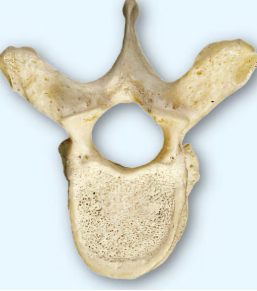
Lumbar Vertebrae is located in
the inferior portion of the back
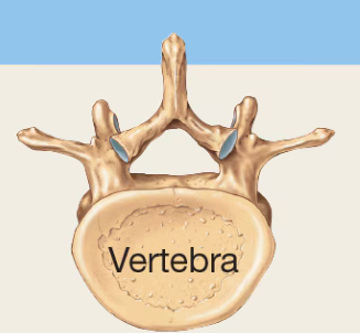
Lumbar Vertebrae shape is
Massive, oval, flat faces
Lumbar Vertebrae vertebral foramen
size is small
Lumbar Vertebrae spinous Process
Blunt,broad; points, posteriorly
Lumbar Vertebrae supports
weight of head, neck, upper limbs, and trunk
Lumbar Vertebrae

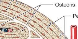
Concentric Lamellae is
a transverse section of lamellae

Osteon (Haversian Systems)
A ring of concentric thing plates (lamellae)
Central (Haversian Canals)
Central channel containing blood vessels, lymphatics and nerves
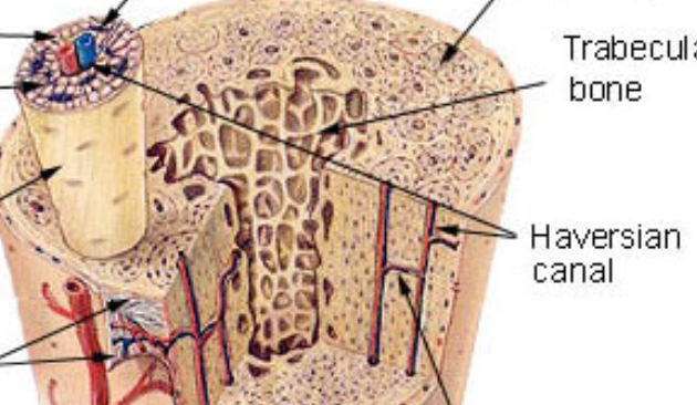
Lacunae
small spaces between lamellae

Perforating (Volkmann’) canals
Channels that pierce the central canals horizontally. Containing neurovascular bundles

Interstitial Lamellae (system)
Remnants of lamellae not circling the central canal

Canaliculi
Small diffusion channels for nutrients and gas exchange

Sutural Bones
Small, flat, oddly shaped bones found between the flat bones of the skull.

Flat bones
Have thin, parallel surfaces. Form roof of the skull, sternum, ribs, and the scapulae. Provides protection for underlying soft tissue and muscle. ( also where muscle can attach)

Irregular Bones
Have short, flat notched or ridged surfaces. Exp vertebrae, pelvis bones, some skull bones
Long bones
Long and slender
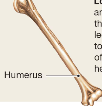
Short bones
Boxlike in appearance.

Sesamoid Bones
Small, round and flat. Near hands, knees , and feet.
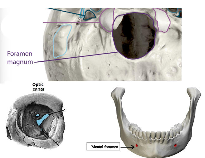
Foramen
Optic Foramen, foramen magnum, and mental foramina
Fissure
Superior and inferior orbital fissure

Meatus / Canal
External auditory Meatus

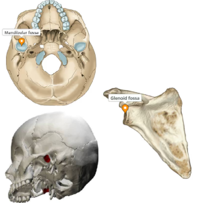
Fossa
Mandibular Fossa; glenoid fossa
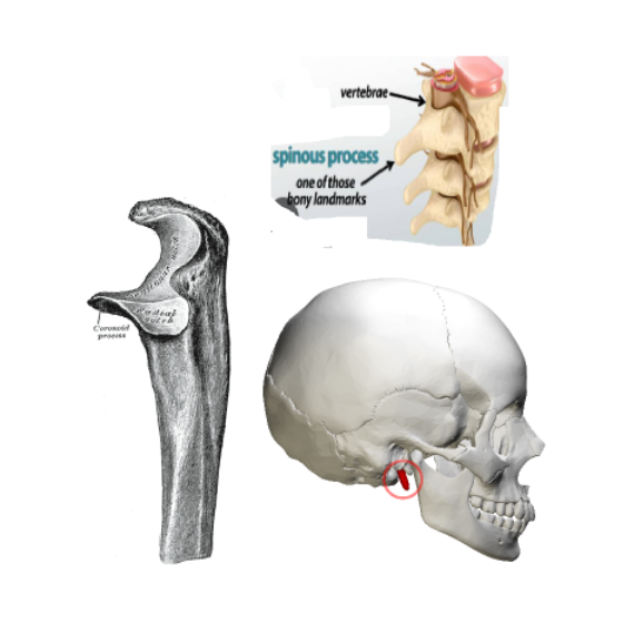
Process
Coronoid process (mandible); styloid process (temporal bone) ; spinous process (vertebra)
Tubercle
Rib tubercle; conoid tubercle of clavicle

Facet
Articular facet (vertebra)

Angle
Mandibular angle; scapular angles

Condyle
Occipital condyles; mandibular condyle (condylar process)

Fossa
Mandibular fossa of temporal bone; glenoid fossa

Spine
Anterior superior iliac spine (ASIS); Anterior inferior iliac spine (AIIS)
Anterior superior iliac spine (ASIS); Anterior inferior iliac spine (AIIS)

tuberosity
Ischial tuberosity; iliac tuberosity

Structure 1
Skull

The Cranial Bone has
Eight bones
Occipital Bone
Posterior hear, base of the skull
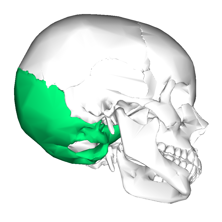
External Occipital Protuberance
The bump on the posterior side
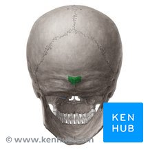
Foramen Magnum
Large hole for the spinal cord to pass through

Frontal Bone
Forehead
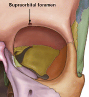
Supraorbital margin
Ridge of bones above each eye

Supraorbital foramen
Hole above the supraorbital margin
Parietal Bones
Form part of the sidewall and roof of the cranium
Coronal Suture
Junction between frontal bone and 2 parietal bones

Sagittal Suture
Junction between 2 parietal bones

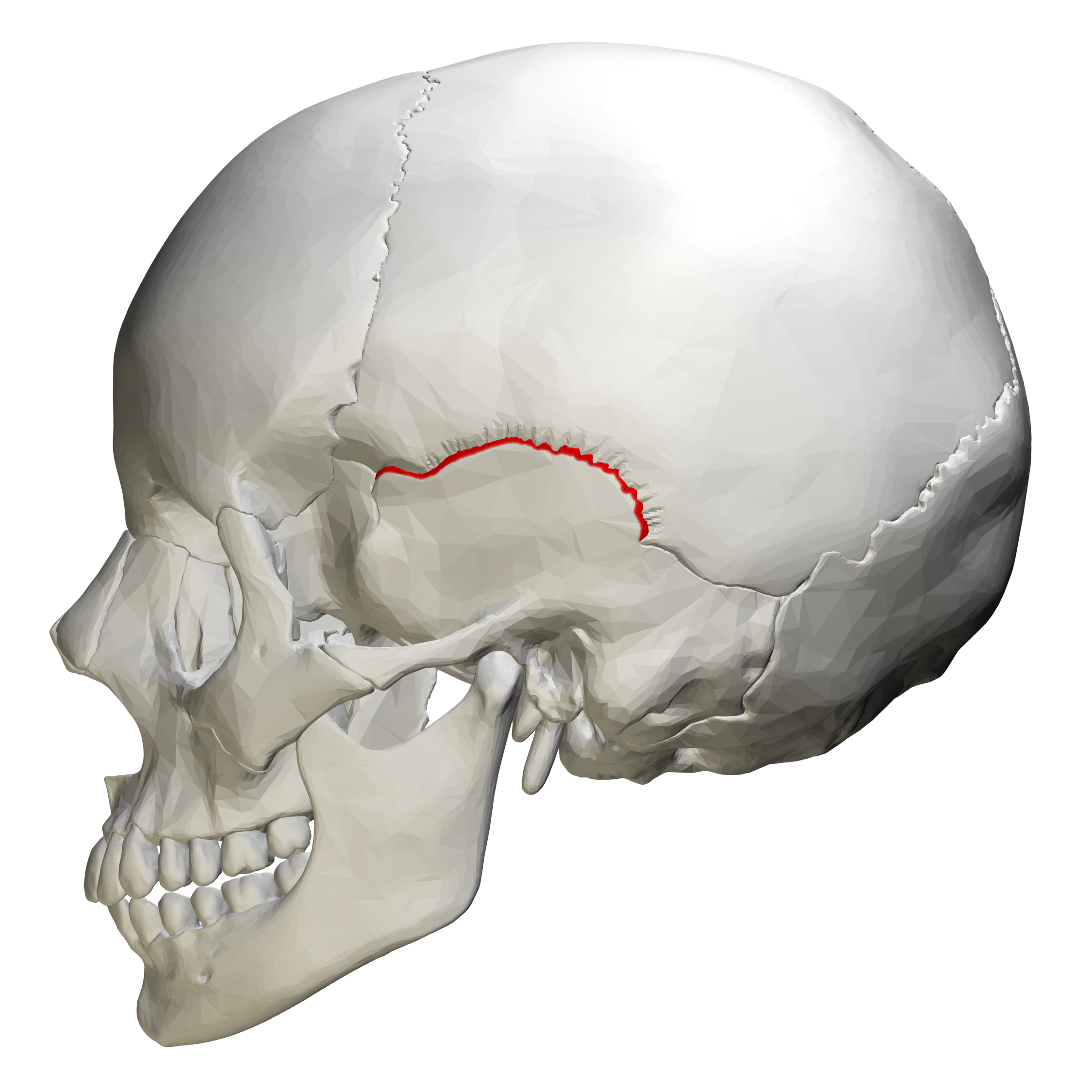
Squamous Suture
Junction between temporal bone and parietal bone
Lambdoidal Suture
Junction between 2 parietal bones and the occipital bone

Temporal Bones
Form part of the sidewall of the cranium, the temple
External Auditory Meatus
Ear canal

Styloid Process
Elongated, pin-lake process on inferior side

Mastoid Process
Large bony mass inferior and posterior to ear

Mandibular Fossa
Articulates with the mandibular condyles on mandible

Zygomatic Process
Projection of temporal bone
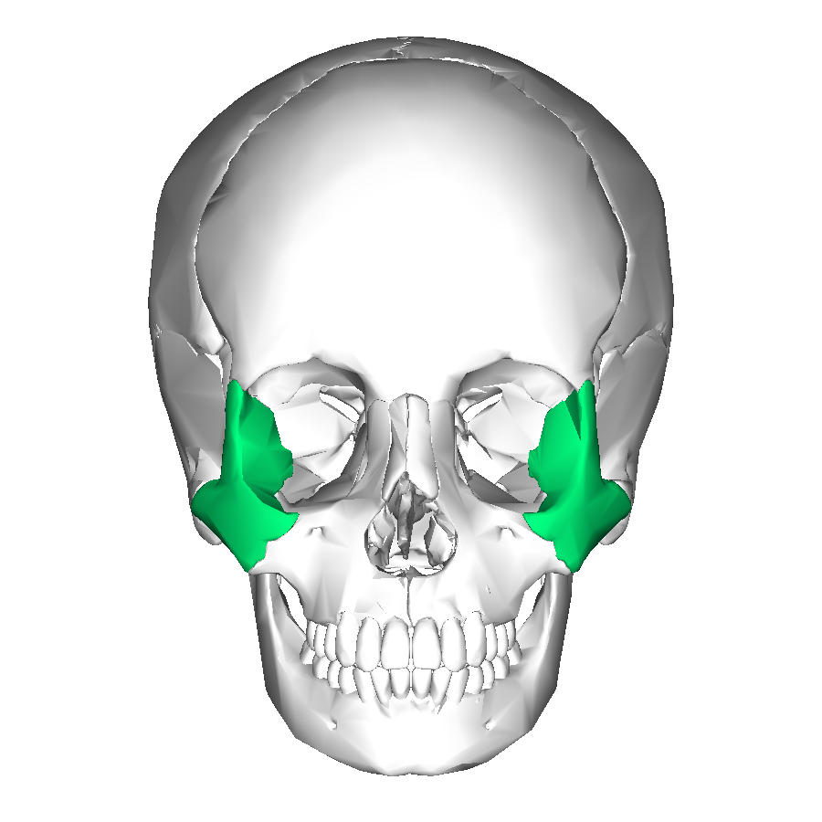
Sphenoid
Irregular bone on the floor of the cranium, looks like a “Bat”

Optic Foramen (Canal)
Hole for the optic nerve to pass through towards each eye

Sella turcica
Saddle like structure on superior surface (pituitary gland sits here)

Greater and Lesser Wings
Large and small wing like structures which help form the floor of the cranium

Superior orbital fissure
Large slits which can be seen in the eye sockets, it is between th greater and lesser wings
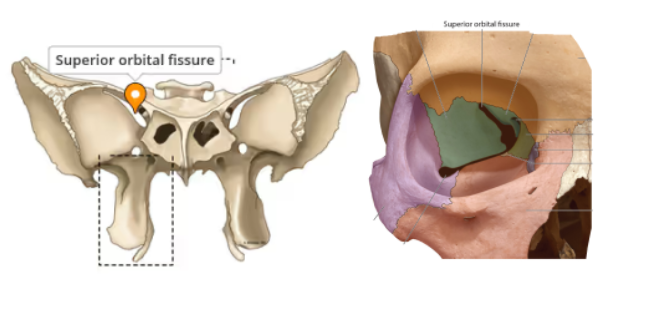
Inferior orbital fissure
Smaller slits, also seen in the eye sockets just inferior to the superior orbital fissures

Ethmoid
Irregular bone anterior to sphenoid and between the eyes
Crista galli
Bony projection on superior surface

Cribriform plate
Horizontal plate on the superior surface that is perforated with many small holes for the olfactory nerve

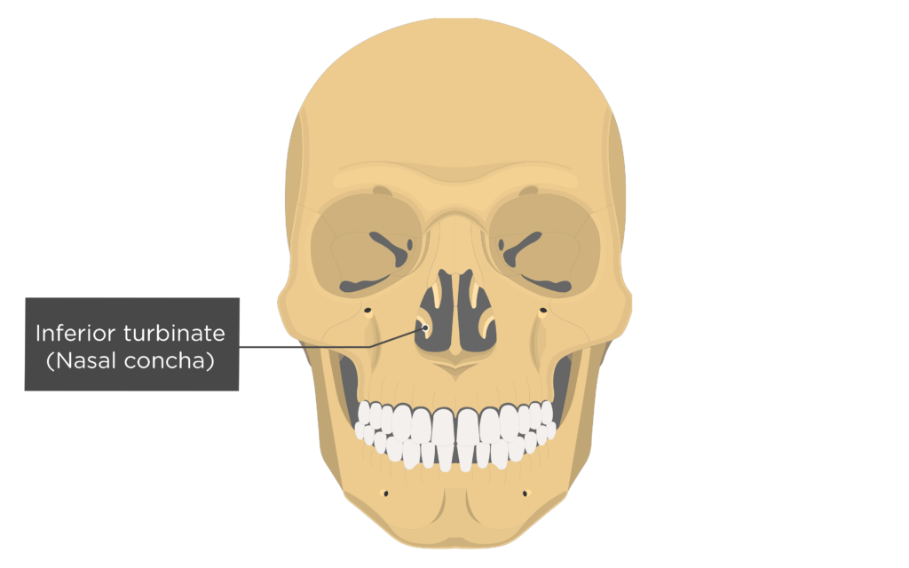
Middle nasal concha
Not a named bone, but a part of the ethmoid bone. Above inferior nasal concha
Superior nasal concha
Not a named bone, but part of the ethmoid bone. Above
middle nasal conch

Perpendicular plate
Inferior plate that divides the nasal cavity

Orbital plate
Smooth plates which form the medial walls of the eye sockets

How many bones does the facial bone have
14
Mandible
Lower Jaw

Ramus
Vertical projection on each side
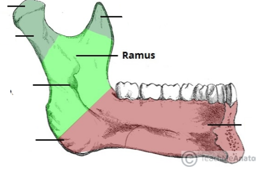
Condylar process
Rounded projections on the superior surface of the rami

Coronoid process
Flat process on the superior surface of the rami

Body
Chin area
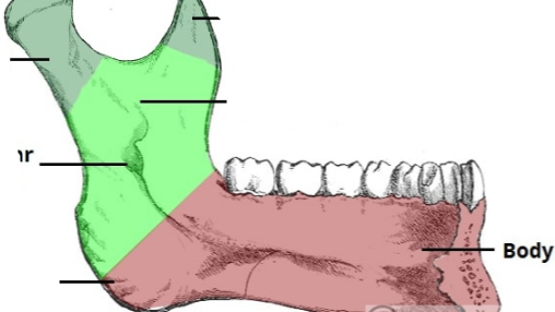
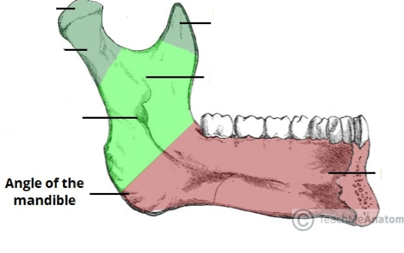
Mandibular Angle
Where body and rami meet
Mental Foramen (foramina)
Hole(s) penetration body on each side of chin

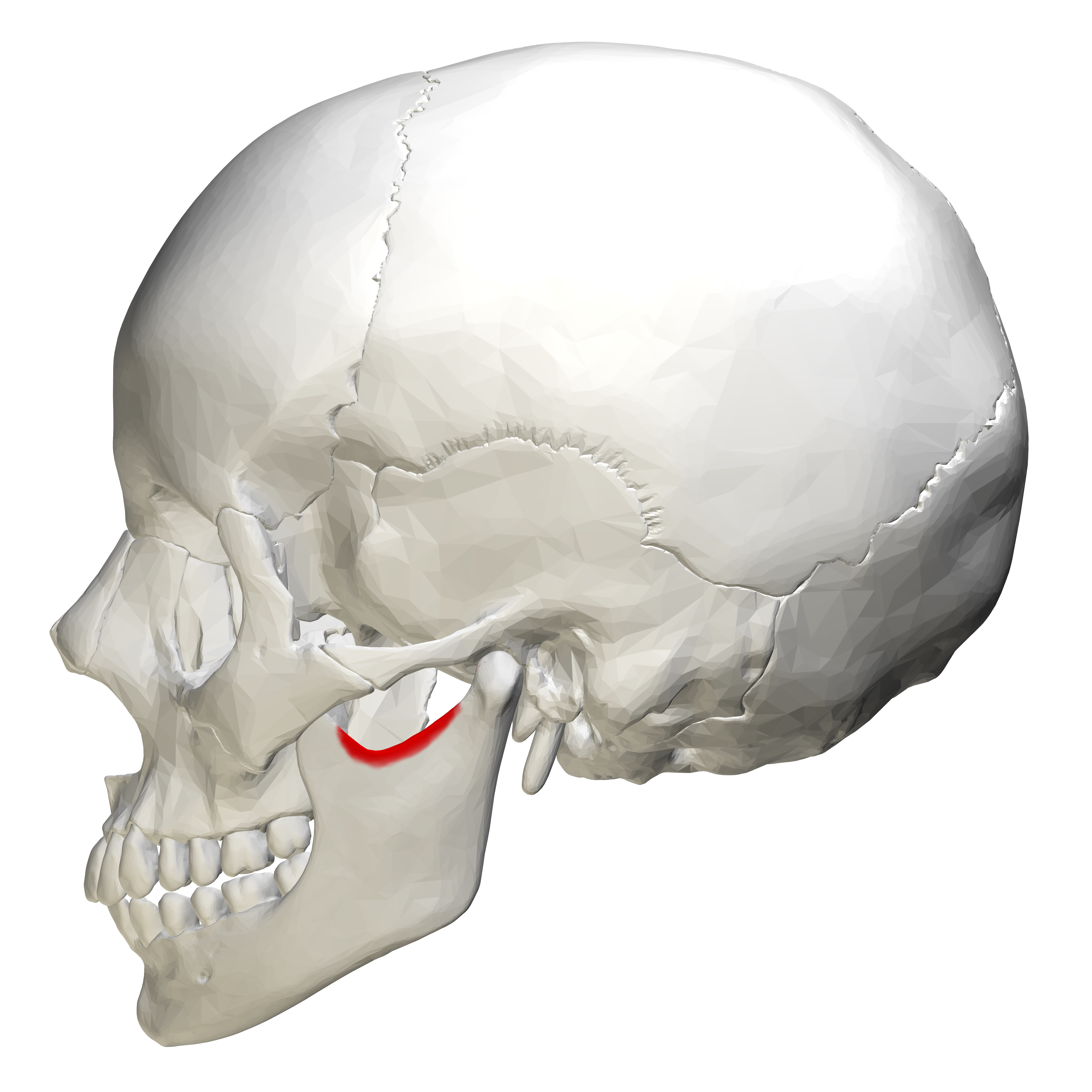
Mandibular notch
Depression that lies between condylar and coronoid processes
Maxilla
Upper Jaw

Infraorbital foramen
Hole that penetrates the orbital rim

Zygomatic bones
Cheek bones

Temporal Process

Nasal Bone
Forms bridge of the nose

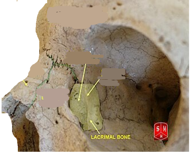
Lacrimal bones
Located on medial wall of socket
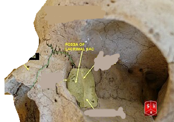
Lacrimal fosaa
Deep groove on anterior/medial surface (tear duct or lacrimal duct)
Palatine Bones
Form roof of mouth and wall of nasal cavity

Vomer
In nasal cavity, forms lower portion of nasal septum

Inferior Nasal Concha
Hyoid
Horse-shoe bone only facial bone not connected to the skull

how many thoracic joints are there
twelve
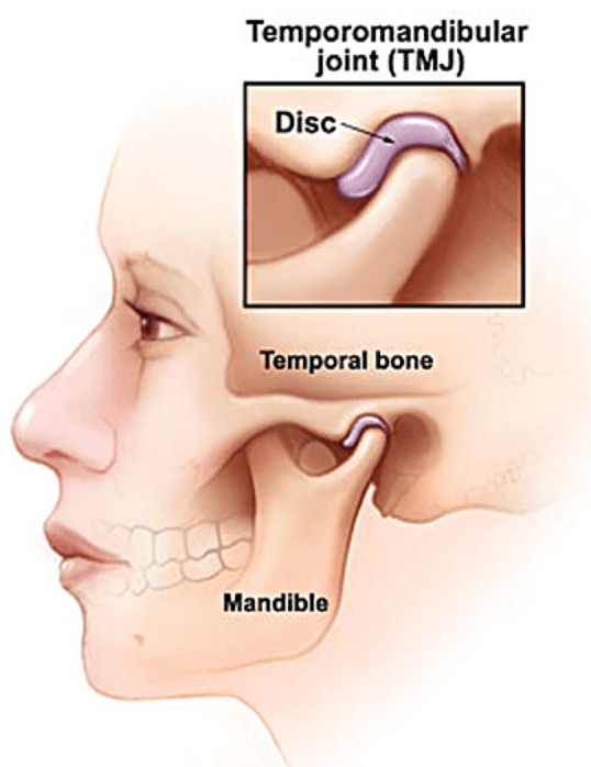
Temporomandibular joint
TMJ combines plane joint and hinge diarthorsis
Atlanto-occipital joint
Flexion-extension

Atlanto-axial joint
Pivots joint only permits rotation
Intervertebral joint
A fibrocartilage pad between the bodies of successive vertebrae that absorbs shocks

Sternoclavicular joint
Protracration/reaction/evelation/ depression, slight rotation
