BSCI202 Lab Practical 1
 Call Kai
 Learn
 Practice Test
 Spaced Repetition
 Match
 Flashcards
 Knowt Play
1/230

There's no tags or description
Looks like no tags are added yet.
231 Terms

Where is the heart located?
Between the lungs in inferior mediastinum
What is the size of a heart?
about the size of a human fist and it weighs less than 1 lb
What is an artery?
vessel that carries blood AWAY from heart
What is a vein?
vessel that carries blood TO heart
Which chambers are filled with deoxygenated blood and which with oxygenated blood?
The right atrium & ventricle are filled with deoxygenated blood.
The left atrium & ventricle are filled with oxygenated blood.
What is the difference between arteries and veins?
Arteries transport blood TO heart, while veins transport blood AWAY from heart.
Arteries have thick walls & small lumens, cuasing high pressure, while veins have thin walls & big lumens, causing lower pressure.
What is the systemic circulation & what is its path?
carries blood to all tissues for nourishment, then transports deoxygenated blood back to heart
Left ventricle → aorta → other arteries → arterioles → capillaries (within tissues) → venules → veins → venae cavae → right atrium
What is the pulmonary circulation & what is its path?
carries deoxygenated blood to the lungs for gas exchange, the brings oxygenated blood back to the heart
Right ventricle → pulmonary trunk → pulmonary arteries → capillaries within the lungs → pulmonary vein → left atrium
Why are the atria are structurally different from ventricles?
The atria recieve blood from the vena cavae and pulmonary vein to pump into the ventricles via just pressure
The ventricles recieve blood from the atria and use cardiac myscle to pump the blood into the aorta and pulmonary artery, so they have thicker walls.
Why is the left ventricle structurally different from the right ventricle?
The left ventricle has thicker layer of myocardium than the right one becasue it needs to pump harder to travel farther since it sends blood to the top and bottom of the body and is part of systemic circulation.
But the right sends blood to the lungs which is of closer distance, so less pressure is needed.
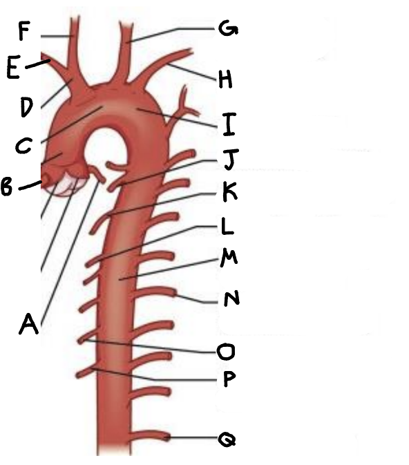
Identify major arteries arising from the aorta and indicate the body region supplied by each.
A. left coronary artery → heart
B. right coronary artery → heart
C. arch of aorta
D. Brachiocephalic trunk
E. right subclavian artery
F. right common carotid artery
G. Left common carotid artery
H. Left subclavian artery
I. Ascending aorta:
J. bronchial branches → lungs
K. mediastinal branches
L. esophageal branches - esophagus
M. Descending aorta: thoracic arteries
N. posterior intercostal arteries → muscles of thorax wall
O. pericardial branches
P. superior phrenic arteries → diaphragm
Q. subcostal artery

Identify major arteries arising from the abdominal aorta and indicate the body region supplied by each.
A. Abdominal aorta
B. right gastric artery
C. gastroduodenal artery
D. hepatic artery proper
E. common hepatic artery → liver
F. Celiac trunk → stomach, spleen, liver
G. left gastric artery → stomach
H. splenic artery → spleen
I. left gastroepiploic artery
J. right gastroepiploic artery
K. superior mesentric artery
L. median sacral artery
M. lumbar arteries
N. inferior phrenic artery
O. middle suprarenal artery
P. renal artery → kidneys
Q. gonadal artery - ovaries & testicles
R. inferior mesentric artery → 2nd half of large intestine
S. common iliac artery → pelvic organs
Identify the major veins draining into the superior vena cavae, and indicate the body regions drained
arms,
axillary vein, brachial vein, medial cubital vein
head,
vertebral vein
brain,
internal jugular vein
brachiocephalic veins
thorax,
azygous vein
Identify the major veins draining into the inferior vena cavae, and indicate the body regions drained
legs
anterior & posterior tibial veins
anterior & posterior fibial veins
external iliac vein
great saphenous veins
common iliac veins
Ovaries & testicles
right gonadal vein
abdomen
renal veins
liver
hepatic portal vein
hepatic veins
What is the circle of Willis (cerebral arterial circle) and discuss its importance in the body?
It is the anterior and posterior blood supplies(containing internal carotid and vertebral arteries) which are united by small communicating arterial branches. They form a complete circle of connecting blood vessels.
It’s important becasue the circle shape allows so that you always have an alternate path to the brain if there is a blockage.


What structures of the fetal circulation system are absent in newborns? What do the fetal structures become in the newborn circulatory system? Identify them in the diagram and say what they do.
Ductus arteriosus (passes to aorta) → ligamentum arteriosum,
foramen ovale (bypasses lungs to enter left atrium) → fossa ovalis,
ductus venosus (bypasses liver to enter inferior vena cava) → ligamentus venosum,
umbilical vein (carry O2 and nutrient-rich blood to fetus) → ligamentis teres,
umbilial arteries (carry CO2 & debris blood from fetus to placenta) → median umbilical ligaments
What organs are associated with the hepatic portal circulation system? What drains them
(superior mesenteric vein) → small intestine, stomach, large intestine
(inferior mesenteric vein) → distal large intestine
(splenic vein) → spleen
(gastric veins) → stomach
(hepatic portal vein, hepatic vein) → liver
pancreas
What is coronary circulation & what does it do?
It is the heart’s own nourishing circulatory system that nourishes the myocardium
The coronary arteries branch from the aorta to supply the heart muscle with oxygenated blood
The cardiac veins drain the myocardium of blood and deliver blood to the coronary sinus, a large vein on the posterior of the heart that empties into rigth atrium.
What is pulmonary circulation & what does it do?
It is the system that carries blood through the lungs and oxygenates the blood supply.
It involves the pulmonary trunk, pulmonary arteries, alveoli, and bronchial arteries.
What is the hepatic portal system & what does it do?
It is the system that drains the digesive organs and carries the blood to the liver. It absorbs nutrients and releases toxins.
It invloves the superior mesentric vein, inferior mesentric vein, splenic vein, gastric veins, and hepatic vein, and hepatic portal vein.
What is fetal circulation & what does it do?
Fetal circulation recieves exchanges of gases, nutrients, and wastes through the placenta.
It bypasses the liver and lungs.
What are the layers of the heart wall?
epicardium, myocardium, endocardium
What is the pericardium & what does it do?
It is a double-walled sac around the heart that protects the heart.
It contains an outer wall (fibrous pericardium) & an inner wall (parietal epericardium & visceral pericardium).
What is the epicardium?
The outermost layer of heart wall
What is the myocardium?
Is the muccle layer of heart wall. Is striated, has intercalated discs, is under involuntary control
Has fibrous skeleton composed of a dense network of fibrous connective tissue that supports the cardiac muscle fibers and heart valves.
What is the endocardium?
Is the innermost layer. Composed of simple squamous epithelium cells.
What is the path of blood-flow of blood?
Venae cavae → right atrium → right ventricle → pulmonary trunk → pulmonary arteries → capillaries within the lungs → pulmonary vein → left atrium → left ventricle → aorta → other arteries → arterioles → capillaries (within tissues) → venules → veins → venae cavae → …
What are the 3 main mechanisms that fascilitate venous blood return to the heart?
skeletal muscle pump, valves, respiratory pump
How does the skeletal muscle pump work?
skeletal muscle contractions from normal movement squeeze veins and push blood through them
How does the respiratory pump work?
during inspiration, a decrease in intra-thoracic pressure and increase in intra-abdominal pressure in lung cavity causes blood to flow from veins in the high abdominal region to veins in the low thoracic region
How do valves work?
prevent backflow of blood using pressure and 1-way flaps.
What is the Tunica intima?
Is the deepest layer of artery/vein that forms a friction-reducing lining to the lumen. Composed of an endothelium of simple squamous cells.
What is the Tunica media?
Is the middle layer of artery/vein that controls lumen diameter composed of circularly arranged smooth muscle and elastic CT.
What is the Tunica externa?
Is the outermostlayer of artery/vein that protects and anchors blood vessels and is composed of areolar or fibrous CT.

What is a capillary?
A blood vessel that provide for exchange of materials in tissues. Has narrow vessels with very thin walls only 1 cell layer thick and made of simple squamous cells in endothelial layer.
Name the types of blood vessels and what is their order is most to least pressure?
elastic arteries
muscular arteries
arteriole
Capilaries
Venules
Veins
What are elastic arteries?
arge, highly expandable arteries close to the heart
What are muscular arteries
medium-sized arteries that distribute blood around the body
What are arterioles?
the smallest arteries, with very thin tunica externa and tunica media
What are venules?
(smallest veins) that drain capillary beds and then merge to form larger vein
What is atherosclerosis?
A condition where the arteries ar eblocked by fat and cholesterol deposits → higher pressure in arteries

What happens during a myocardial infarction (heart attack)?
During myocardial infarction, blood flow to heart tissue is impaired and cells die through necrosis. After necrosis, neutrophils infiltrate the dead tissue. The arrows on the images point out areas of this neutrophilic infiltrate.
Immediately after (within 12 hrs) → loss of striations & nuclei in cardiac tissue.
During healing (next 1-4 days) → necrosis of cardiac tissue & invasion of neutrophils, which clean debris
In healed tissue (6+ mo) → lot of collagen deposits (scarring) & scar tissue is noncontractile


What type of cell is this?
cardiac muscle

What pathology occured here?
acute myocardial infarction
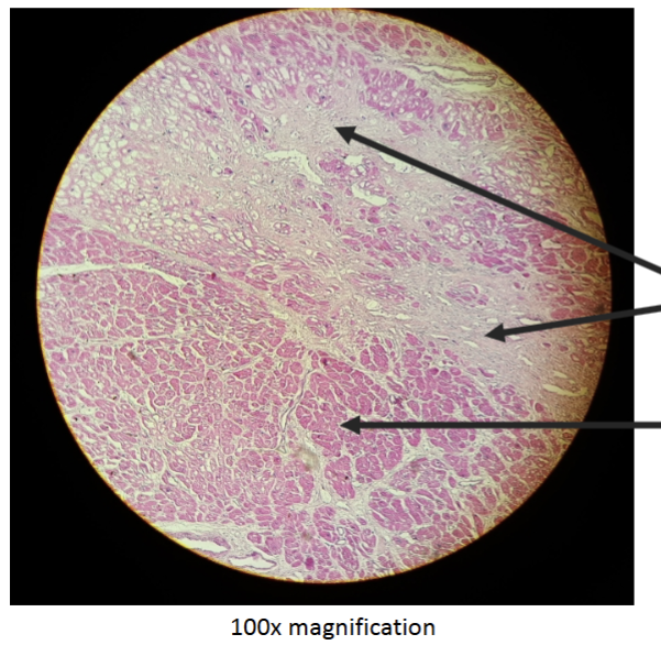
What pathology happened here ?
Old Myocardial Infarction
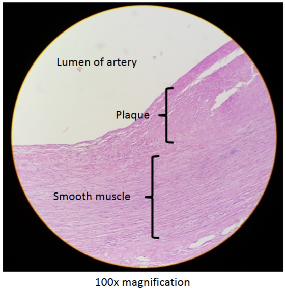
What pathology is shown here?
Atherosclerosis

What is shown here (3 vessel types)
A. Artery
B. Vein
C. Capillary

WHat is shown here? What are the layers?
Vena cava
A. tunica intima
B. tunica media
C. tunica externa
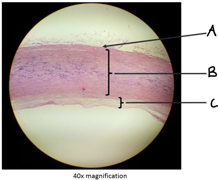
WHat is shown here? What are the layers?
Aorta
A. tunica intima
B. tunica media
C. tunica externa

What pathology is shown here?
dissecting aneurism

What is a dissecting aneurism?
When bleeding into the aortic wall splits the artery longitudinally, forcing the layers of the wall apart.
What is the blood path of the hepatic portal system?
Capillary beds of the digestive system absorb nutrients and toxins
Nutrient-rich blood drains into veins of the hepatic portal circulatory system
These veins empty into the hepatic portal vein, which carries this blood to the liver
Liver helps maintain proper glucose, fat, and protein concentrations in blood
Liver also filters toxins from the blood
Blood drains into the hepatic vein as it leaves the liver


Identify the major vessels of the hepatic portal circulation and what they drain.
hepatic portal vein
Gastric veins
Splenic vein
Superior mesenteric vein
hepatic veins
Splenic vein
Inferior mesenteric vein

Identify the parts of the heart of the heart.
superior vena cava
right pulmonary artery
a
right atrium
right pulmonary veins
a
a
right ventricle
a
a
aorta
left pulmonary artery
a
left pulmonary veins
bicuspid valve
aortic semilunar valve
pulmonary semilunar valve
left ventricle
interventricular septum
myocardium
visceral pericardium
apex
base
interatrial septum

Name the parts of the heart wall.
A. fibrous cardium
B.parieta layer of serous pericardium
C. pericardial cavity
D. visceral layer of serous pericardium
E. epicardium
F. myocardium
G. endocardium
H. heart chamber
I. pericardium
J. heart wall
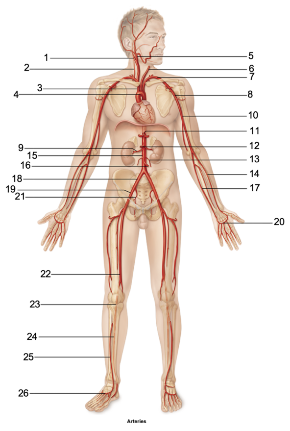
Name the arteries.
internal carotid artery
vertebral artery
brachiocephalic trunk
aortic arch
external carotid artery
common carotid arteries
subclavian artery
axillary artery
renal artery
brachial artery
thoracic aorta
superior mesentric artery
gonadal artery
radial artery
abdominal aorta
inferior mesentric artery
ulnar artery
common iliac artery
external iliac artery
superficial palmar arch / digital arteries
internal iliac artery
femoral artery
popliteal artery
posterior tibial artery
anterior tibial artery
arcuate artery

Name the blood supplies of brain.
middle cerebral artery
anterior communicating artery
anterior cerebral artery
posterior communicating artery
posterior cerebral artery
basilar artery
vertebral artery
internal carotid artery
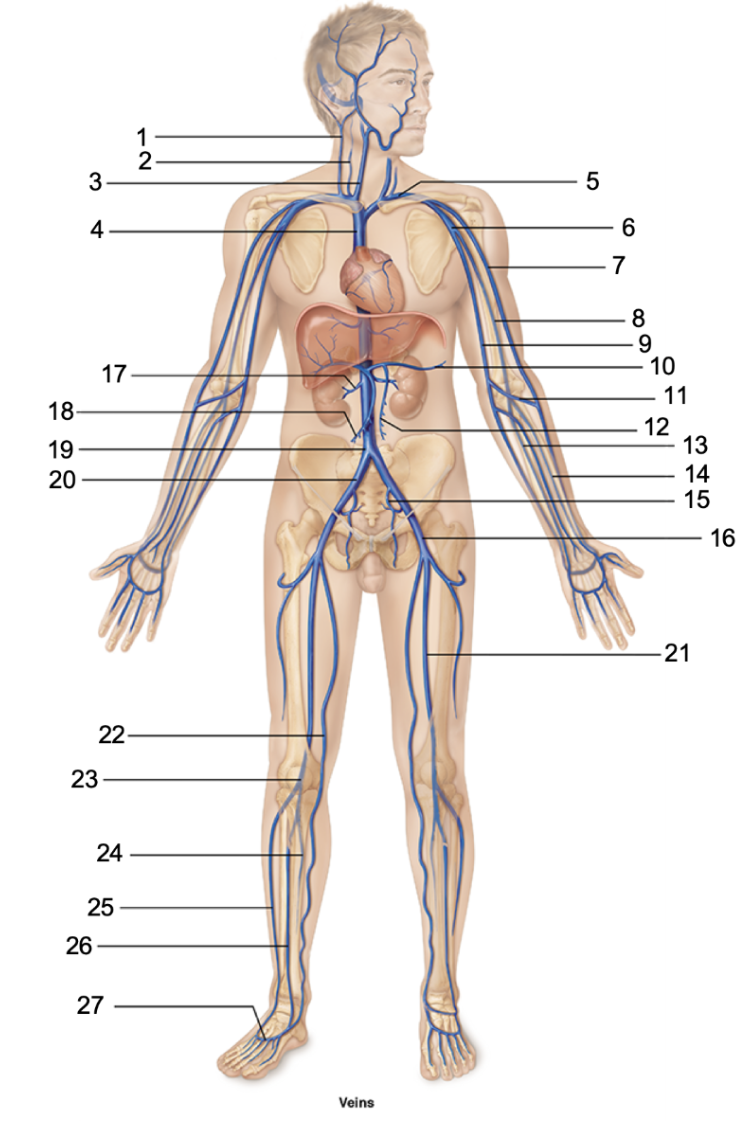
Name the veins.
external jugular vein
vertebral vein
internal jugular vein
superior vena cava
subclavian vein
axillary vein
cephalic vein
brachial vein
basilic vein
splenic vein
medial cubital vein
inferior mesenteric vein
ulnar vein
radial vein
internal iliac vein
external iliac vein
renal vein
superior mesenteric vein
inferior vena cava
common iliac vein
femoral vein
great saphenous vein
popliteal vein
posterior tibial vein
small saphenous vein
anterior tibial vein
dorsal metatarsal veins

Name the arteries in head & neck.
internal carotid artery
superficial temporal artery
maxillary artery?
external carotid artery
common carotid artery
occipital artery?
opthalmic artery?
facial artery
lingual artery?
superior thyroid artery
vertebral artery
basilar artery
What causes the “lub dub” sounds of a heartbeat?
The closure the the heart valves
What causes Lub (S1)?
the closure of the AV valves at the start of ventricular systole
What causes Dub (S2)?
The closur eof the SL valves ar the end of the ventricular systole
What is systole?
contraction
What is diastole
relaxation
What is the normal heart rate?
60-100 bpm at rest
(Avg resting hr is 70-76 bpm)
What is arrhythmia?
irregular heartbeat or inconsistent pattern
Can describe badycardia or tachycardia
What is bradycardia?
a low heart rate <60 bpm
is considered abnormal EXCEPT in well-conditioned athletes
What is tachycardia?
a fast heart rate >100 bpm
is abnormal when at rest
if untreated, may lead to fibrillation
What is fibrillation?
Is rapid unccordinated contractions
This renders heart useless as a pump
What type of cells are involved in heart conduction?
atrial cells and ventricular cells
How often do atrial cells beat per minute? How often do ventricular cells beat per minute?
Atrial cells beat 60 times per min.
Ventricular cells beat 20-40 time per min
What is the role of the nervous system on regulating heart rate and conduction velocity?
Autonomic nervous system helps regulate heart rate (but does not initiate contractions)
• Sympathetic nervous system (SNS) accelerates heart rate
• Parasympathetic nervous system (PNS) decelerates heart rate
What does the intrisic conduction system do?
It coordinates spontaneous contractions of the heart & ensures the heart beats as a coordinated unit. It enforces a hr of 75 bpm
How is the signal to contract transmitted through the heart?
Through the intrinsic conduction system
What is the intrisnsic conduction system composed of ?
specialized nervous tissue embedded in the myocardium:
Sinoatrial node
Atrioventricular node
Atrioventricular bundle
Purkinje fibers
Explain the order of impulse transmission through the intrisnic conduction system.
The SA node starts each heartbeat → Impulse spreads through the atria to the AV node
• Atria contract; blood pushed into ventricles
• At the AV node, the impulse is delayed briefly
• Impulse travels through the AV bundle, bundle branches, and subendocardial network
• Ventricles contract; blood is ejected from the heart
What is an electrocardiogram and what does it do?
It measures the heart’s electrical activity from the body surface. It combines all of the cardiac events into 1 diagram
What does the PR interval tell you?
It tells you the time it takes for the electrical impulse to travel from the SA node to the AV node
How long should a normal PR interval be? What does a prolonged PR interval mean for a patient?
It should normally be 0.12 - 0.2 sec.
partial heart block (problem with communication between SA and AV)
What does the QRS complex tell you?
Ventricular depolarization
Atrial repolarization also happens here, at same time with ventricular contraction
What does a prolonged QRS complex mean for a patient?
partial blockage of right or left bundle branch (first degree block)
What does the QT interval mean?
Ventricular depolarization to repolarizaation
What does a shorter QT interval mean for the patient?
faster heart rate
What does a prolonged QT interval mean for the patient?
risk of arrhythmia (irregular ventricular contractions)
What does the P wave mean?
depolarization of the atria
What does T wave tell you?
repolarization of ventricles
What does the S-T segment tell you?
the interval between the end of ventricular depolarization (contraction) and the beginning of ventricular repolarization (relaxation), corresponding to the plateau phase when the heart muscle contracts to pump blood out to the body
What does the T-P segment tell you?
on an EKG (electrocardiogram) is the flat line that runs from the end of the T wave to the beginning of the next P wave
shows the time when the heart muscle cells are electrically silent and resting between heartbeats
What does the R-R interval tell you?
the time elapsed between the peak of one R wave and the peak of the next R wave on an electrocardiogram (ECG). It represents a single complete ventricular cardiac cycle

Identify the parts of this ECG segment.
P wave
QRS complex
R wave
T wave
P-R Interval
S-T Interval
Q-T Interval

How do you calculate the heart rate from an EKG?
(60 bpm) / (R-R interval time in sec)
Is this EKG normal? If not, what is the pathology?
Normal sinus rhythm
Is this EKG normal? If not, what is the pathology?
Junctional rhythm
P wave is absent (or inverted)
SA node is not acting as the pacemaker, leading to the AV node pacing the heart
Is this EKG normal? If not, what is the pathology?
second degree heart block
Not all P waves are followed by a QRS complex
Indicates damage to the AV node
Is this EKG normal? If not, what is the pathology?
Ventricular fibrillation
Impulses generated in the atria do not pace ventricular contractions
Uncoordinated contractions of the myocardium
Typical in acute myocardial infarction
What is atrial fibrillation?
An irregular and often very rapid heart rhythm. It can lead to blood clots in the heart. The condition also increases the risk of stroke, heart failure and other heart-related complications.
What is ventricular fibrillation?
It is a type of irregular heart rhythm (arrhythmia). The lower heart chambers contract in a very rapid and uncoordinated manner. As a result, the heart doesn't pump blood to the rest of the body.
It is an emergency that requires immediate medical attention. It's the most frequent cause of sudden cardiac death.
What is the Sinoatrial node?
It’s located in right atrium & serves as heart’s pacemaker.
Starts the impulse

What is the Atrioventricular node?
Is at the junction of atria & ventricles.

What is the atrioventricular bundle (Bundle of His)?
This & bundle branches are in interventricular septum
