Perio II, Lecture 8 - Osseous Resective Surgery (Dr. Ho)
 Call Kai
 Learn
 Practice Test
 Spaced Repetition
 Match
 Flashcards
 Knowt Play
1/93

There's no tags or description
Looks like no tags are added yet.
94 Terms
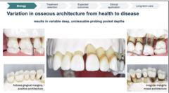
T/F: Variation in osseous architecture from health to disease results in variable deep, uncleasable probing pocket depths
True

How would you describe the architecture?
- Follows gingival margins
Positive architecture

How would you describe the architecture?
- Irregular margins
Mixed architecture
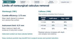
What is the mean depth at which curettes can achieve a plaque- and calculus-free root surface?
3.73 mm

What is the mean maximum depth at which instrument contact can be detected during scaling and root planing?
6.21 mm
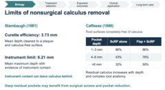
T/F: Instrument contact can leave calculus behind
True

_______ residual pockets may benefit from surgical access and pocket reduction
Deep
At which pocket depth is scaling and root planing (ScRP) alone most effective at achieving calculus-free root surfaces?
1-3 mm (86%)

What percentage of root surfaces are completely calculus-free following ScRP alone in 4-6 mm pockets?
43%
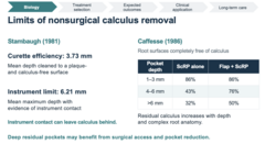
What percentage of root surfaces are completely calculus-free following flap surgery + ScRP in 4-6 mm pockets?
76%

In pockets greater than 6 mm, what percentage of root surfaces are completely calculus-free following ScRP alone versus flap surgery + ScRP?
32% vs. 50%

Which of the following patients would most likely benefit from surgical access for calculus removal?
A) Patient with 2 mm pockets and minimal calculus
B) Patient with 3 mm pockets and healthy gingiva
C) Patient with persistent 7 mm pockets following ScRP
D) Patient with 2 mm pockets following prophylaxis
C) Patient with persistent 7 mm pockets following ScRP


ID the type of architecture:
Positive architecture

ID the type of architecture:
Negative architecture

ID the type of architecture:
Flat architecture
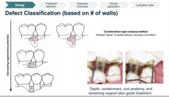
What is the defect classification?
- "Funnel Shaped"
- Appear Moat-like
- Often noted: distal 2nd molars
3-wall

What is the defect classification?
- Common
- Bone loss ~ equal
- Cup or bowl-shaped
2-wall

What is the defect classification?
- Usually remaining bone wall exists interdentally
1-wall defect
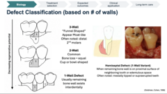
What is the defect classification?
- When remaining bone wall is on proximal surface of neighboring tooth or edentulous space
- Often noted: mesially tipped or supraerupted teeth
Hemiseptal defect (1-wall variant)
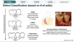
What clinical conditions are most commonly associated with a hemiseptal defect?
- Mesially tipped teeth
- Supraerupted teeth

What wall-variant is a hemiseptal defect?
1-wall defect

Which periodontal defect has the greatest regenerative potential?
3-wall defect

As the number of remaining bony walls decreases, what happens to the regenerative potential of a periodontal defect?
Decreases

Vrotsos et al. directly observed 981 osseous defects during surgery in 286 patients with moderate-to-advanced periodontitis. Craters accounted for _______ of all defects, were more prevalent in posterior segments
Almost half

Periodontal crater defects are most commonly found in which region?
Posterior region

A periodontal crater defect is classified as which type of osseous defect?
2-wall defect

Define the following:
An interdental concavity with remaining buccal and lingual bony walls
Periodontal crater defect

Define the following:
A defect with multiple wall configurations at different depths
Combination-type osseous defect
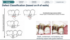
A periodontal defect has three remaining bony walls at its deepest portion and two remaining walls coronally. How is this defect classified?
Combination-type defect
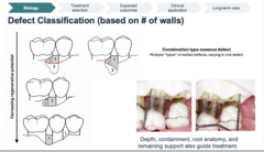
Which of the following factors influence treatment selection for periodontal osseous defects?
A) Defect depth
B) Defect containment
C) Root anatomy
D) Remaining periodontal support
E) All of the above
E) All of the above
In periodontitis staging, defect depth, containment, and root anatomy are considered under which category?
Complexity

Indication or contraindication for osseous resection surgery?
- "Early and moderate" periodontal defects (~Stage 2 Periodontitis)
Indication

Indication or contraindication for osseous resection surgery?
- Intrabony and hemiseptal defects
Indication

Indication or contraindication for osseous resection surgery?
- Shallow to Medium depth Craters
Indication

Indication or contraindication for osseous resection surgery?
- Incipient furcation involvement
Indication

Indication or contraindication for osseous resection surgery?
- Bony ledges and tori
Indication

Indication or contraindication for osseous resection surgery?
- Negative architecture
Indication

Indication or contraindication for osseous resection surgery?
- Deep osseous craters (>3 mm)
Contraindication

Indication or contraindication for osseous resection surgery?
- Deep three-wall osseous defects
Contraindication

Indication or contraindication for osseous resection surgery?
- Moderate-deep circumferential defects
Contraindication

Indication or contraindication for osseous resection surgery?
- Buccal defects on terminal mandibular molars
Contraindication

Indication or contraindication for osseous resection surgery?
- Shorter root trunk lengths (furcation exposure risk)
Contraindication
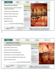
Indication or contraindication for osseous resection surgery?
- Regions of esthetic concern
Contraindication

What treatment is most appropriate for a deep, wide intrabony defect?
Flap curettage with barrier membrane (GTR) and bone graft

What treatment is most appropriate for a deep, narrow intrabony defect according to the diagram?
Flap curettage with or without bone graft
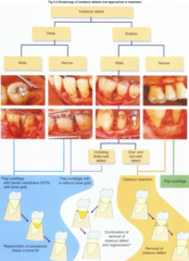
What treatment is most appropriate for a shallow, wide intrabony defect with a 3-wall configuration?
Flap curettage with or without bone graft

What treatment is most appropriate for a shallow, wide intrabony defect with a 1- or 2-wall configuration?
Osseous resection
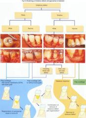
What treatment is most appropriate for a shallow, narrow intrabony defect according to the diagram?
Flap curettage

What is the primary goal of guided tissue regeneration (GTR) in the treatment of intrabony defects?
Promote regeneration of periodontal supporting tissues
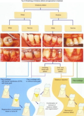
What is the primary goal of osseous resective surgery in the treatment of intrabony defects?
Removal of osseous defect

Define the following:
- Reshapes (cuts) alveolar bone and places the gingival margin at a more apical level
- Re-establish physiologic contour: positive, maintainable architecture with a shallow sulcus and a short epithelial seal at a new apical level.
Osseous resective surgery

What is the goal of osseous resective surgery?
Health or stability with "closed pockets"
- BOP
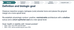
In a successfully treated, stable periodontitis patient, which probing depth may be considered clinically healthy if there is no bleeding on probing (BOP)?
≤4 mm

A patient with a history of successfully treated periodontitis has a 4 mm probing depth without BOP. How is this site classified?
Clinical health on a reduced periodontium
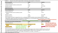
What percentage of bleeding on probing (BOP) is consistent with clinical gingival health on a reduced periodontium?

In a successfully treated periodontitis patient, gingival inflammation on a reduced periodontium is characterized by a probing depth and BOP of what?
- PD ≤3 mm
- BOP ≥10%

What phase of osseous resective surgery (ORS) includes the following?
- Establish plaque control, reduce modifiable risks
(systemic and local contributors)
- Perform subgingival instrumentation, instruct and motivate - Periodontal Re-evaluation (6-8 weeks*):
- - 4-5mm with BOP: may benefit from repeated subgingival instrumentation and reassessment
- - Residual >6mm PPD: Consider surgery after adequate initial therapy.
- - - Select access, resective, or regenerative treatment by anatomy.
Phase I

What phase of osseous resective surgery (ORS) includes the following?
- Surgical intervention using a site-specific approach (e.g. ORS vs regeneration)
Phase II

What phase of osseous resective surgery (ORS) includes the following?
- Provide continuing supportive periodontal care (q3mo)
Phase III

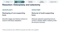
Define the following:
Reshaping of non-supporting bone
Osteoplasty

Define the following:
Smooths ledges and blends contours to improve soft-tissue adaptation
Osteoplasty

Define the following:
Removal of tooth-supporting bone
Ostectomy

Define the following:
Reduces selected supporting bone to correct a shallow defect. Anatomy limits the amount removed
Ostectomy
________ exposes more root surface and can alter appearance. ________ exposure may produce transient or persistent hypersensitivity.
Recession, Dentin

During osseous resective surgery, some supporting bone and clinical attachment may be sacrificed. Increased mobility is expected and may take up to _______ to stabilize
1 year

What is resection necessary at these sites?
- Persistent posterior pockets remained after nonsurgical treatment.
- The chart shows residual 5-6 mm sites with bleeding.
- The next decision depends on defect anatomy and plaque control.
- Shallow, two-wall crater defects.
- The target was a maintainable posterior contour with reduced pockets.
- Furcation position does limit the amount of supporting bone that could be removed.
- Different sites in the same mouth can require different treatment approaches.

What is the surgical sequence of osseous resectitive surgery?
1. Evaluate the crest and plan incisions
2. Reflect the flap, remove granulation tissue, and debride roots
3. Perform osteoplasty and selective ostectomy
4. Adapt and stabilize the flap at the planned apical level
ID the type of incision:
A resective incision angled in an apical-to-coronal direction which is intended to reduce the thickness of amount of mucogingival tissue and allow for healing by secondary intention. Often used during gingivectomy and gingivoplasty
Submarginal external bevel

ID the type of incision:
An acute or oblique surgical incision angled toward the tooth surface in a coronal-to-apical direction which is intended to reduce the thickness or amount of mucogingival tissue.
Submarginal internal bevel (inverse, reverse, inverted)

ID the type of incision:
Made to increase access to alveolar bone, enhance flap mobility, facilitate lateral or coronal flap advancement, limit the inclusion of non-diseased sites in the surgical field, and/or decrease tension on retracted flaps.
Releasing (vertical)


ID the type of incision:
External bevel

ID the type of incision:
Internal bevel
What is the primary consideration when planning a periodontal flap incision?
Apicocoronal dimension of keratinized tissue

When adequate keratinized tissue is present, what should be determined before selecting the incision position?
Position of the alveolar crest

Which techniques can be used to determine the position of the alveolar crest before periodontal flap surgery?
- Bone sound
- Crestal anticipation

If bone sounding measures 6 mm from the gingival margin, approximately how far from the gingival margin should the incision be positioned to anticipate the alveolar crest, according to the slide?
3 mm
[6 mm − 3 mm (approximately 2 mm supracrestal tissue attachment + 1 mm sulcus) = 3 mm]
During periodontal flap suturing, where should the flap margin ideally coincide according to the incision-planning approach shown?
Alveolar crest
When the apicocoronal dimension of keratinized tissue is minimal, which incisions are generally preferred?
- Intrasulcular incision
- Releasing incision (possible)
- Require portion of split thickness for periosteal anchorage

What type of flap exposes root surfaces and bony defects?
Mucoperiosteal (Full thickness) Flap reflection

During osteoplasty, what is the purpose of grooving?
Establish the width and volume of interdental bone
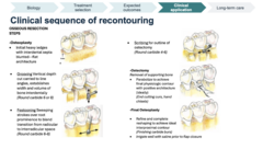
During osteoplasty, _________ strokes over root prominence to blend transition from radicular to interradicular space
Festooning Sweeping
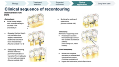
Which step of osseous recontouring involves outlining the areas where supporting bone will be removed?
Scribing

During ostectomy, what is the purpose of parabolizing the alveolar bone?
Achieve physiologic contour with positive architecture

What is the primary goal of final osteoplasty?
Refine the interproximal contour to achieve ideal architecture

When treating a deep periodontal crater defect, which direction is ideal for osseous ramping?
Palatal/lingual

What approach: Palatal or lingual?
- Avoids surgical exposure of buccal furcations and possible root fenestrations or dehiscences
- Avoids dealing with a shallow vestibule
- More cancellous bone
- All keratinized tissue
- Wider interdental space
- Natural cleansing of the tongue
Palatal approach

What approach: Palatal or lingual?
- Base of defects commonly located lingually (due to tipping of molars 20º 1st molar, 25° 2nd molar-thus, lingual incision will not req scalloping)
- Avoids buccal furcation exposure Avoids the shallow vestibule
- Thicker bone
- Slightly wider embrasures
- Tongue cleanses area
Lingual appraoch

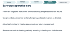
What are some instructions for early postoperative care with osseous resective surgery?
- Follow the surgeon's instructions for local cleaning and protection of the wound
- Use prescribed pain control and any temporary antiseptic regimen as directed
- Attend early review for healing assessment and suture management
- Resume mechanical cleaning gradually according to healing and clinical advice
What are some important guidelines for referring a patient to a periodontist?
- Recognize residual disease after adequate initial treatment
- Refer complex defects and patients whose needs exceed your training
- Send a complete chart, suitable radiographs, risk history, and treatment response
- Coordinate restorations with the periodontist and the patient's maintenance needs

What are three things osseous resective surgery case selection depends on?
- Residual disease
- Defect anatomy
- Patient risk

Pocket elimination trades some tissue and support for ________ maintainability
Greater
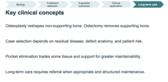
T/F: Long-term care requires referral when appropriate and structured maintenance
True

An osseous crater is an example of what type of bone loss?
A) 2-wall defect
B) 3-wall defect
C) Horizontal bone loss
D) Suprabony defect
A) 2-wall defect
Osetoplasty is __________?
A) Supporting bone is removed to create sufficient supracrestal attachment (biologic width)
B) No bone was removed
C) Supporting bone is removed, but non-pathological bone was not touched
D) Non-supporting bone is removed for better flap adaptation
D) Non-supporting bone is removed for better flap adaptation