nervous system week 3
 Call Kai
 Learn
Learn Practice Test
 Spaced Repetition
 Match
Match Flashcards
 Knowt Play
1/124

There's no tags or description
Looks like no tags are added yet.
Name | Mastery | Learn | Test | Matching | Spaced | Call with Kai | Chat |
|---|
No analytics yet
Send a link to your students to track their progress
125 Terms
usually 3-4
how many cervical ganglia are there
usually 12
thoracic ganglia
usually 4
lumbar ganglia
usually 4
sacral ganglia
CN III, ciliary ganglion, smooth m. in eye, constrict pupil
Pathway of CN III
CN VII, pterygopalatine ganglion, lacrimal gland, inc. tear production
1st pathway of CN VII
CN VII, sublingual ganglion, submandibular gland, inc. salvation
2nd pathway of CN VII
CN IX, otic ganglion, parotid gland, inc. salvation
pathway for CN IX
CN X, intramural ganglia, cardiac m., dec. heart rate
1st pathway for CN X
CN X, intramural ganglia, smooth m. in digestive tract, inc. blood flow
2nd pathway for CN X
sacral spinal cord, intramural ganglion, smooth m. in pelvic organs, inc. urine excretion
Pathway for sacral spinal cord
upper thoracic spinal cord, superior cervical ganglion, smooth m. in eye, dilate pupil
1st Pathway for upper thoracic spinal cord (head)
upper thoracic spinal cord, upper thoracic chain ganglion, cardiac m., inc. heart rate
2nd pathway upper thoracic spinal cord (thorax)
lower thoracic spinal cord, celiac ganglion, smooth m. in stomach or intestines, dec. blood flow
pathway for lower thoracic spinal cord
pre g= long, post g = short
what is the difference between pre ganglion and post ganglion for parasympathetic division of ANS
pre g=short, post g= long
what is the difference between pre ganglion and post ganglion for sympathetic division of ANS
Fibrous layer of the eye
collective. white part and the clear part

sclera
region. just the white part

cornea
structure. outermost clear part

Vascular layer of the eye
entire red line running inside eye

choroid of the eye
region. continuous width at the back of the eye

ciliary body
collective. where it wides out, anterior part of the eye

ciliary muscle
backing of love seat

ciliary processes
white lines on seat

iris
colored part

pupil
space inside iris

constrictor pupillae m.
lines inside of iris

dilator pupillae m.
lines outside of iris

retina
internal structure. purple and orange

optic part of the retina
portion: orange part

nonvisual part of the retina
portion: purple part

ora serrata
serrated edge between orange and purple

macula
structure. yellow disc lateral to opening at back

fovea centralis:dense with cones so it is the area of sharpest vision
depression. probe inside yellow disc

optic disc
yellow disc that has vascularization that aligns with opening

optic nerve
corn cob structure at the back of the eye
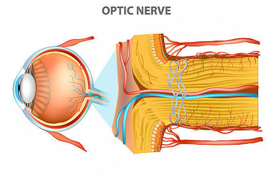
central artery of the retina
red line in optic nerve

lens
bubbled 2 clear parts

suspensory ligament of the lens
bq only: attaches to ciliary body
anterior segment of the eye
region: entire segment from cornea to lens

aqueous humor of the eye
fluid: fluid that fills the anterior segment

anterior chamber of the eye
space: cornea to iris

posterior chamber of the eye
space: iris to the lens

posterior segment of the eye
region. lens to optic nerve

vitreous body of the eye
fluid. fluid that fills posterior segment

1) cornea
2) aqueous humor
3) lens
4) vitreous body
what is the summary of refractive media or the pathway of light
superior rectus m.
12 o clock

inferior rectus m.
6 oclock

medial rectus m.
3 oclock

lateral rectus m.
9 oclock

superior oblique m.
2 oclock. connected to blue thing

trochlea of the eye
sits medially; blue thing attached to superior oblique m.

inferior oblique m.
cradles under the eye and runs transverse

levator palpebrae superioris m.
attached to eye brain, most superior

superior eyelids
top eyelid

inferior eyelids
bottom eyelid

palpebral fissure
space between eyelids

eyelashes
microstructure. probe brush up on eyelashes

bulbar conjunctiva: its many blood vessels can become irritated producing bloodshot eyes
covers eye itself. same showing as palpebral fissure

palpebral conjunctiva
probe inside inferior eyelid

lacrimal caruncle
structure: tear duct inner corner of eye

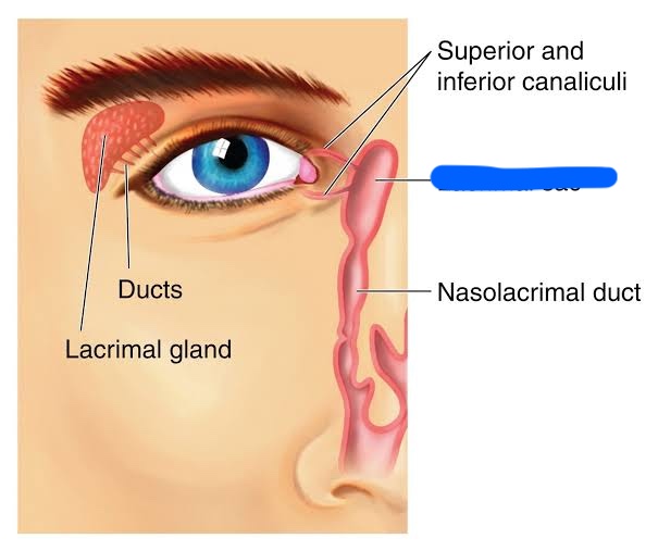
lacrimal gland: makes tears
structure: brain looking thing, most superior “eye brain”

lacrimal sac
pink sac on medial side of eye
auricle
collective: entire ear

lobule of the auricle
lobe of the ear. pinch

helix
structure outer ridge

tragus
structure. medial to earhole

concha
depression. divit in the ear

external acoustic meatus
passageway. probe through earhole inside earhole
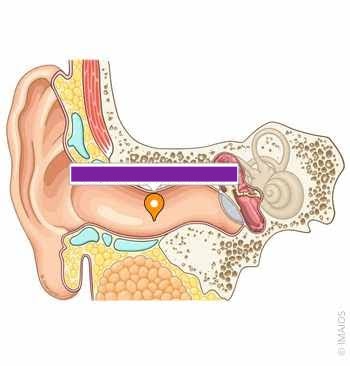
tympanic membrane
structure. small little eardrum

lateral wall
probe in external acoustic meatus

medial wall
inside of tympanic membrane

oval window
opening: oval hole

round window
opening: round hole

posterior wall
take tympanic membrane out and behind it

mastoid cells
pink golf ball like structure

anterior wall
hand in front of tympanic membrane

eustachian tube
passage. behind tympanic membrane in canal

malleus b.,incus b., stapes b.
bq only: what structues make up the auditory ossicles
malleus b.
golf club on tympanic membrane

head of the malleus b.
bottom part not directly attached

handle of the malleus b.
directly attached to tympanic membrane

incus b.
not touching membrane

stapes b.
cow bell

head of the stapes b.
little numb

footplate of the stapes b.
flat part underneath

bony labyrinth
bq only: filled with fluid called perilymph
semicircular canals
all of the loops

anterior semicircular canal
back of the chair

posterior semicircular canal
arm of the chair

lateral semicircular canal
seat of the chair

vestibule of the bony labyrinth
junction. where all arches meet

cochlea
snail shell

modiolus of the cochlea
structure. yellow divits

osseous spiral lamina
bq only: partially divides the cochlea into two channels
scala vestibuli
spaces. upper space. V/T

scala tympani
spaces. lower space. V/T

helicotrema
feature. tip of snail shell

membranous labyrinth
bq only: filled with a fluid called endolymph
semicircular ducts:within the semicircular canals
clear tubes
