8. respiration and circulation
 Call Kai
 Learn
 Practice Test
 Spaced Repetition
 Match
 Flashcards
 Knowt Play
1/61

There's no tags or description
Looks like no tags are added yet.
62 Terms
5 functions of circulatory system
nutrient delivery
hydrostatic pressure
immune response
clotting
thermoregulation
hemolymph
blood
hemocoel
body cavity
describe the open circulatory system
hemolymph (blood) is released into the open hemocoel (body cavity) to circulate

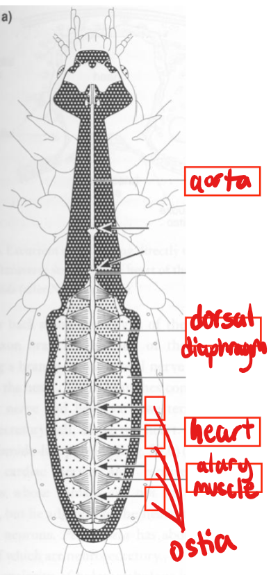
function of tubular dorsal vessel
pumps hemolymph anteriorly into the pulsatile heart (abdomen) and aorta (thorax and head)

parts of pulsatile heart
allary muscles
ostia
alary muscles
at every chamber/segment of pulsatile heart, create rhythmic pumping

ostia
paired one-way valves at each chamber

incurrent ostia vs. excurrent ostia
incurrent ostia along heart
excurrent ostia at ends
heartbeat is __ but gets __ and __ signaling
myogenic, neuronal, endocrine
contractile wave mostly moves…
anteriorly
can contractile waves of the heart reverse?
yes — can reverse in pupae in response to anterior pressure
2 functions of alary muscles for heartbeat
dilate heart chambers
draw hemolymph through incurrent ostia
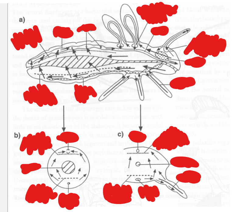
ventral and dorsal diaphragms
guide hemolymph into sinuses by forming channels and pulsating

accessory hearts
circulate hemolymph thru appendages (legs, wings, antennae, ovipositor)

hematopoietic organ/lymph gland
houses stem cells and releases mature hemocytes
usually in close association w dorsal vessel




6 substances that make up the hemolymph
ions
circulating sugars and AAs
hemocytes
circulating metabolic compounds (yolk proteins, lipid chaperones, etc.)
defensive chemicals (synthesized or sequestered)
endocrine signals
trehalose
disaccharide w antifreeze function
necessary because we need glucose to diffuse to extremities. however, that would mean [glucose] would be so high that it would COUNTER its gradient from gut to hemolymph
to resolve this, we rapidly convert glucose to trehalose for nutrient delivery

fat bodies + location and functions
lobed ribbon-like sheets of storage cells distributed throughout the hemocoel
close proximity to the heart and midgut (no capillaries)
functions: metabolic regulation, secretions into hemolymph
describe fat bodies’ involvement in metabolic regulation
fat bodies synthesize trehalose from glucose for immediate circulation
triglycerides and glycogen are stored in lipid droplets for induced energy release
4 things fat bodies secrete into the hemolymph
lipophorins - lipid chaperones
vitellogenins - yolk protein for provisioning eggs
hormones involved in molting
antimicrobial proteins
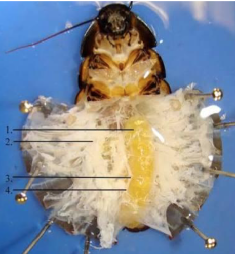
which number is the fat body?
2
(1 is ootheca, 2 is fat body, 3 is nerve ganglion, 4 is ventral nerve cord)
eclosion
process of hatching from an egg/cocoon
ex:
fly “ptilinum”
inflating wings
molting from exuviae at suture lines
what do caterpillars’ prolegs rely on to function?
hydrostatic pressure
what are male moth “coremata” used for?
phermone release
what must humoral compounds recognize to induce cellular responses?
“universal” molecular signatures
prior exposure sensitizes subsequent responses (but not adaptive immunity)
less expensive than adaptive immunity
less flexible
pattern recognition receptors
involved in immune response
bind specific molecular signatures associated w microbes
trigger signaling cascades to induce cellular and humeral responses
3 types of hemocytes
prohemocytes
phagocytes
enzyme producing (oenocytoids, crystal cells)
prohemocytes
undifferentiated precursors to all other hemocytes
phagocytes
consume cellular debris
encapsulating: flatten to engulf microorganisms
enzyme producing
oenocytoids, crystal cells
store enzymes that induce formation of melanin and toxic compounds
3 cellular immune responses
phagocytosis
encapsulation
nodulation
phagocytosis
singular target, phagocyte engulfs that one pathogen into itself
encapsulation
target is a small number of large pathogens
nodulation
target is a large number of small pathogens
cellular encapsulation and nodulation
involves phagocytes surrounding the pathogens
melanization in encapsulation and nodulation
oenocytoids release enzymes to melanize
polyDNA viruses
parasitic wasps inject polyDNA viruses along w eggs
they don’t kill host cells — instead, they cause production of proteins that suppress;
molting and growth
production of antimicrobial compounds
encapsulation
melanization
… which prevent the wasp egg from being killed
2 steps to clotting
hemolymph thickens when exposed to air (forms a gel)
hemocytes aggregate and melanize (similar to encapsulation response)
insects are generally (exothermic/endothermic)?
exothermic
antifreeze agents
polypeptides that bind ice nuclei as they form and lower the freezing point → often parallel secondary structures
an exception to insects being Mostly exothermic
muscle-based thermogenesis
contracting flight muscles heats up blood which is then circulated
another exception to insects being Mostly exothermic
tracheal system
primary respiratory system of insects. a complex system of hollow vessels where O2 and CO2 are passively exchanged

tracheoles
large trachea narrow into these, which innervate tissue (muscles, brain, gut, etc.)

spiracles
valves to the tracheal system
muscular — can be opened or closed
can also prevent water loss

when do spiracles open?
during “energetic” behavior
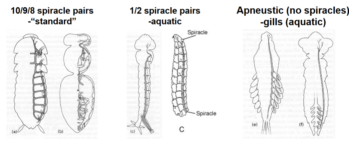
spiracle pairs for standard vs. aquatic vs. gills
“standard” - 10/9/8 spiracle pairs
aquatic - 1/2 spiracle pairs
gills (aquatic) - no spiracles
intima
cuticular layer formed by tracheal epithelium secreting chitin

taenidium
cuticular spirals within intima
provides support but also allows some compression

are trachea or tracheoles shed during molting?
tracheoles
describe wing expansion
as fluid is pumped into the wing veins, trachea compress to enable wing expansion
air sacs
used for “active” respiration
these are enlarged trachea which form sacs
sacs are vented to increase air exchange
diving beetles may also use sace for buoyancy
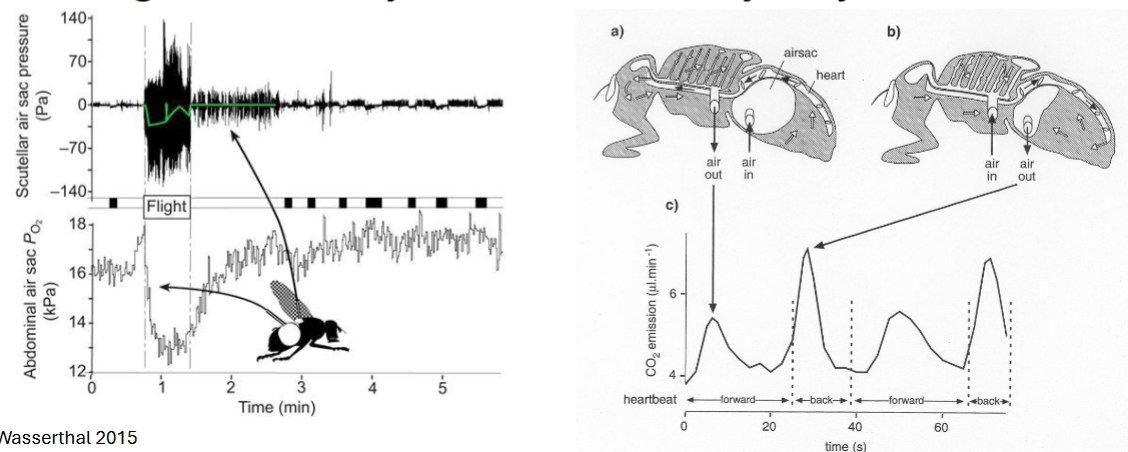
abdominal venting
used for “active” respiration
various sclerite movements compress trachea to force air exchange
physical “gills”
air bubble exchanges O2 with water
plastron layer
part of physical “gills,” cuticular hairs that capture a thin, constant layer of air that connects to spiracles

how do diving beetles use their physical “gills”?
(keep in mind they don’t actually have gills)
diving beetles trap bubbles under elytra, but must resurface due to differences in SA:V ratio
biological “gills”
air exchange across cuticle of gills
gills are appendages → flapped to increase water flow

siphon
a “snorkel” with hairs to prevent water entry

hemoglobin in respiratory systems
AN EXCEPTION — most insects lack hemoglobin
blood worms (larvae of some midges) live in lake sediment (extreme hypoxia)
possess free hemoglobin in blood to fix oxygen (thus its red color)