Chapter 8 - T Cell-Mediated Immunity
 Call Kai
 Learn
 Practice Test
 Spaced Repetition
 Match
 Flashcards
 Knowt Play
1/92

There's no tags or description
Looks like no tags are added yet.
93 Terms
Define what is meant by “T-cell activation.” This process is identified to be the first step in what type of immune response?
T-cell activation, also known as T-cell priming, refers to the process in which a naïve T cell encounters its specific antigen for the first time and becomes stimulated to proliferate and differentiate into an effector T cell.
Define what is meant by “T-cell activation.” This process is identified to be the first step in what type of immune response?
This process is the first step in: Primary Adaptive immune Response
It marks the beginning of a targeted immune reaction, enabling T cells to help eliminate pathogens through specialized roles (e.g., cytotoxicity, helper functions)
At the site of infection, which cell type is responsible for capturing antigens and transporting them? Where will these antigens be brought for their presentation to naïve T cells?
At the site of infection, dendritic cells are the primary cells responsible for capturing antigens. These are specialized, migratory antigen-presenting cells (APCs) that serve as sentinels in peripheral tissues. Once they take up the antigen, dendritic cells:
Mature and undergo changes that enhance their ability to present antigen.
Migrate from the site of infection via the lymphatic system.
Transport the antigens to secondary lymphoid tissues, such as:
Lymph nodes (for skin and peripheral tissue infections),
Spleen (for blood-borne infections),
Tonsils or BALT (for respiratory infections),
Peyer’s patches, appendix, or GALT (for gastrointestinal infections).
At the site of infection, which cell type is responsible for capturing antigens and transporting them? Where will these antigens be brought for their presentation to naïve T cells?
In these secondary lymphoid organs, dendritic cells present processed antigens on MHC molecules to naïve T cells, initiating T-cell activation and the adaptive immune response.
Immature vs. Mature Dendritic Cells: In the skin, what is the name of the cells we discussed in class to be our model for an immature dendritic cell?
Langerhans’ cells
Immature vs. Mature Dendritic Cells: What are the cellular characteristics of immature dendritic cells? (Hint: we mentioned 3)
Actively perform:
Receptor-mediated endocytosis (e.g., DEC-205)
Macropinocytosis
Antigen processing and MHC loading
Immature vs. Mature Dendritic Cells: What is the other term that mature dendritic cells can be known by? (Hint: morphology)
Interdigitating reticular cells
Immature vs. Mature Dendritic Cells: Once a dendritic cell becomes mature, it loses its ability to perform a mechanism it used to be able to when it was immature. What is this mechanism?
Phagocytosis and antigen uptake
Immature vs. Mature Dendritic Cells: Once mature, we describe dendritic cells to be purely ______ cells at this point. (Hint: loaded with three components that are required to activate naïve T cells; one is B7.)
Antigen-presenting
Immature vs. Mature Dendritic Cells: Which expresses DEC 205? Which expresses DC-SIGN?
DEC 205 – Immature dendritic cells
DC-SIGN – Mature dendritic cells
Immature vs. Mature Dendritic Cells: Upon reaching the secondary lymphoid tissue, where are dendritic cells observed to settle? During this arrival, are the dendritic cells considered to be mature or immature?
Settle in the T-cell areas (cortex) of the lymph node
Considered to be mature
Naïve T cells will try to find mature dendritic cells and establish interactions within the T-cell area of the lymph node. Explain what will happen to the T cell (its fate) if it successfully encounters and binds a peptide: MHC complex on the dendritic cell’s surface. What happens if the naïve T cell does NOT encounter and bind a peptide: MHC complex?
Successfully encounters and binds a peptide:MHC complex on a dendritic cell:
The T cell stays in the lymph node, becomes activated, and receives co-stimulatory signals (e.g., via CD28–B7 interaction).
The T cell undergoes proliferation and differentiation into effector T cells.
This process is known as T-cell activation or priming, and it begins the primary adaptive immune response.
Effector T cells will later exit the lymph node to perform immune functions, such as killing infected cells (CD8⁺) or helping other immune cells (CD4⁺ subsets).
Naïve T cells will try to find mature dendritic cells and establish interactions within the T-cell area of the lymph node. Explain what will happen to the T cell (its fate) if it successfully encounters and binds a peptide: MHC complex on the dendritic cell’s surface. What happens if the naïve T cell does NOT encounter and bind a peptide: MHC complex?
Does NOT encounter and bind a peptide: MHC complex:
The naïve T cell does not get activated and will leave the lymph node via the efferent lymph.
It will continue to recirculate through the blood and other secondary lymphoid tissues (e.g., other lymph nodes, spleen) in search of its specific antigen.
Make sure you are familiar with the types of effector T cells that migrate to sites of infection after their activation, as well as the types of T cells that remain in the secondary lymphoid tissues. (Hint: review slide 66)
SLIDE

Make sure you are familiar with the types of effector T cells that migrate to sites of infection after their activation, as well as the types of T cells that remain in the secondary lymphoid tissues.
Fate of a Naïve T Cell
If the naïve T cell does bind a peptide: MHC complex on the surface of a dendritic cell:
It remains in the lymph node.
It becomes activated.
The T cell proliferates and differentiates into a clone of effector T cells.
This marks the beginning of the adaptive immune response.
If the naïve T cell does not bind a peptide: MHC complex:
It does not become activated.
It exits the lymph node via the efferent lymphatic vessel.
It continues recirculating through the body, repeating this process until it eventually encounters its specific antigen.
Make sure you are familiar with the types of effector T cells that migrate to sites of infection after their activation, as well as the types of T cells that remain in the secondary lymphoid tissues.
Effector T Cells that Migrate to Sites of Infection:
CD8⁺ cytotoxic T cells – kill infected cells.
CD4⁺ TH1 cells – activate macrophages and enhance microbicidal activity.
CD4⁺ TH17 cells – recruit neutrophils via IL-17 secretion.
CD4⁺ Treg cells – modulate immune responses in peripheral tissues.
Effector T Cells that Remain in Secondary Lymphoid Tissues:
CD4⁺ TFH cells – stay in lymphoid follicles to help B cells differentiate into plasma or memory cells.
Some TH2 cells – assist B cells and do not necessarily migrate.
From the onset of an infection to the manifestation of a primary adaptive immune response, a delay of several days is experienced. Understand the reasons for why this is. (Hint: think about why pathogens and their associated antigens are “trapped” in a draining lymph node; there is a small number of initial lymphocytes that are specific for their antigen)
From the onset of an infection to the manifestation of a primary adaptive immune response, a delay of several days occurs due to several key factors:
Antigen Transport to Lymphoid Tissue:
The immune system does not initiate the adaptive immune response at the site of infection.
Instead, dendritic cells capture the pathogen and migrate to the draining secondary lymphoid tissues (e.g., lymph nodes, spleen, MALT).
This transport and maturation process takes time as dendritic cells move from immature antigen-capturing cells to mature antigen-presenting cells (APCs).
Low Frequency of Antigen-Specific T Cells:
Only 1 in 10⁴ to 1 in 10⁶ naïve T cells are specific for any given antigen.
These T cells must recirculate through lymphoid tissues to encounter the appropriate antigen presented by APCs.
T-Cell Activation and Proliferation:
Once a specific naïve T cell encounters its antigen presented on a peptide:MHC complex by a professional APC (mainly dendritic cells), it becomes activated.
The T cell then undergoes clonal expansion (driven by IL-2) and differentiates into effector T cells — a process that takes several days.
Trapping of Antigens in Draining Lymph Nodes:
Pathogens and their antigens are retained in draining lymph nodes, which helps ensure interaction with the appropriate lymphocytes but also contributes to the delay in the initial immune response.
This delay, although disadvantageous initially, is necessary for the precision and specificity of the adaptive immune response. It allows time for the system to match the invading pathogen with a small pool of specific lymphocytes and to expand that population effectively.
When a naïve T cell migrates from the blood into the lymphoid tissue, how does it enter? What is the name for this process by which naïve T cells leave the bloodstream to enter into the T-cell area? Identify the two chemokines used to guide this process.
How does a naïve T cell enter the lymphoid tissue?
Naïve T cells enter secondary lymphoid tissues (like lymph nodes) via the high endothelial venules (HEVs). These are specialized blood vessels that allow lymphocytes to leave the bloodstream and enter the lymph node cortex.
When a naïve T cell migrates from the blood into the lymphoid tissue, how does it enter? What is the name for this process by which naïve T cells leave the bloodstream to enter into the T-cell area? Identify the two chemokines used to guide this process.
What is the name of the process by which naïve T cells leave the bloodstream to enter into the T-cell area?
This process is known as homing.
When a naïve T cell migrates from the blood into the lymphoid tissue, how does it enter? What is the name for this process by which naïve T cells leave the bloodstream to enter into the T-cell area? Identify the two chemokines used to guide this process.
Which chemokines are used to guide this process?
Two critical chemokines guide this migration:
CCL21
CCL19
These chemokines are produced in the lymphoid tissue and bind to the chemokine receptor CCR7 on the surface of naïve T cells.
Review the 4 different types of leukocyte adhesion molecules (and what they bind to) that function in the initial interaction between the T cell and the vascular endothelium.
What 4?
Selectins
Vasular Addressins
Integrins
Immunoglobulin Superfamily
Review the 4 different types of leukocyte adhesion molecules (and what they bind to) that function in the initial interaction between the T cell and the vascular endothelium.
Selectins
Example: L-selectin on naïve T cells.
Binds to vascular addressins (CD34, GlyCAM-1)
Review the 4 different types of leukocyte adhesion molecules (and what they bind to) that function in the initial interaction between the T cell and the vascular endothelium.
Vascular Addressins:
Example: CD34, GlyCAM-1 on HEVs.
Bind to selectins.
Review the 4 different types of leukocyte adhesion molecules (and what they bind to) that function in the initial interaction between the T cell and the vascular endothelium.
Integrins:
Example: LFA-1, VLA-4 on T cells.
Bind to ICAM-1, ICAM-2 on APCs or endothelium.
VLA-4 guides effector T cells to inflamed tissues via VCAM-1.
Review the 4 different types of leukocyte adhesion molecules (and what they bind to) that function in the initial interaction between the T cell and the vascular endothelium.
Immunoglobulin Superfamily:
Examples: ICAM-1, ICAM-2, ICAM-3, LFA-3.
Interact with LFA-1, CD2 on T cells.
Review the 4 different types of leukocyte adhesion molecules (and what they bind to) that function in the initial interaction between the T cell and the vascular endothelium. Understand “homing” and “cell-to-cell adhesion,” which includes understanding the importance and the locations of the following:
selectins (L-selectin) & vascular addressins (CD34 & GlyCAM-1)
L-selectin: Expressed on naïve T cells (and B cells), facilitates homing to lymph nodes.
Vascular Addressins:
CD34 and GlyCAM-1 are expressed on high endothelial venules (HEVs) in lymph nodes.
They contain sulfated carbohydrates (e.g., sialyl-Lewis^x^) that bind to L-selectin.
Function: Initiate rolling interaction to slow down T cells for entry into lymph nodes.
Review the 4 different types of leukocyte adhesion molecules (and what they bind to) that function in the initial interaction between the T cell and the vascular endothelium. Understand “homing” and “cell-to-cell adhesion,” which includes understanding the importance and the locations of the following:
integrins (LFA-1) & immunoglobulin superfamily proteins (ICAM-1)
LFA-1: An integrin on the T cell surface; expression increases upon T cell activation.
ICAM-1: An immunoglobulin superfamily protein found on endothelial and antigen-presenting cells.
Function: After initial selectin-mediated rolling, chemokines activate LFA-1, which tightly binds to ICAM-1, allowing T cells to firmly adhere and migrate through endothelium or engage APCs.
Review the 4 different types of leukocyte adhesion molecules (and what they bind to) that function in the initial interaction between the T cell and the vascular endothelium. Understand “homing” and “cell-to-cell adhesion,” which includes understanding the importance and the locations of the following:
CCL21 & CCL19. What is the receptor for these chemokines?
CCL21 and CCL19: Chemokines produced in lymphoid tissue.
CCR7: The chemokine receptor on naïve T cells that binds CCL21 and CCL19.
Function: Directs naïve T cells to lymph node T-cell zones and enhances adhesion molecule activation.
Review the 4 different types of leukocyte adhesion molecules (and what they bind to) that function in the initial interaction between the T cell and the vascular endothelium. Understand “homing” and “cell-to-cell adhesion,” which includes understanding the importance and the locations of the following:
DC-SIGN & ICAM-3
DC-SIGN: An adhesion molecule on mature dendritic cells.
ICAM-3: Expressed on naïve T cells.
Function: Mediates the initial interaction between dendritic cells and T cells, enhancing antigen scanning.
Review the 4 different types of leukocyte adhesion molecules (and what they bind to) that function in the initial interaction between the T cell and the vascular endothelium. Understand “homing” and “cell-to-cell adhesion,” which includes understanding the importance and the locations of the following:
CCL18
Produced by: Mature dendritic cells.
Function: Attracts naïve T cells to areas in the lymph node where antigen presentation occurs.
Acts as part of the chemokine gradient guiding cell migration and interaction.
Explain the role of S1P and when its receptor is expressed. What would cause its expression to become suppressed?
Sphingosine-1-phosphate (S1P) plays a key role in the exit of T cells from lymphoid tissues, while its receptor—S1P receptor (S1PR1)—is crucial for sensing the S1P gradient between lymphoid tissues (low S1P) and the blood/lymph (high S1P).
Explain the role of S1P and when its receptor is expressed. What would cause its expression to become suppressed?
Role of S1P and Its Receptor
S1P is a signaling lipid found at high concentrations in the blood and lymph and at low concentrations in lymphoid tissues.
S1PR1 (S1P receptor 1) is expressed on naïve T cells, enabling them to exit the lymph nodes after they fail to encounter antigen.
When naïve T cells do not find their antigen, S1PR1 expression allows them to follow the S1P gradient out of the lymph node and resume circulation.
Explain the role of S1P and when its receptor is expressed. What would cause its expression to become suppressed?
When is the S1P Receptor Expressed?
Naïve T cells express S1PR1, enabling egress from lymphoid organs.
Upon antigen recognition and activation, S1PR1 expression is suppressed to retain the T cell in the lymph node for proliferation and differentiation.
Explain the role of S1P and when its receptor is expressed. What would cause its expression to become suppressed?
What Causes S1P Receptor Suppression?
TCR engagement with antigen:MHC complexes on dendritic cells causes a signaling cascade that:
Suppresses S1PR1 expression.
Retains the T cell in the lymph node to allow it time to proliferate and differentiate into effector cells.
This suppression ensures T cells remain localized at the site of antigen presentation instead of prematurely exiting the lymph node via the S1P gradient.
Professional Antigen-Presenting Cells (P-APCs): List the 3 cell types that fall into this category.
Dendritic cells
Macrophages
B cells
Professional Antigen-Presenting Cells (P-APCs): Know the distribution of these 3 cells in the lymph node.
Dendritic cells: Found primarily in the T-cell areas (cortex) where they present antigens to naïve T cells.
Macrophages: Scattered throughout the lymph node, including the marginal sinus and medullary regions, performing phagocytic and antigen-presenting roles.
B cells: Reside in follicles/germinal centers of the lymph node cortex, and present antigen primarily to helper T cells.
Professional Antigen-Presenting Cells (P-APCs): What molecules do these cells have that other cells don’t have?
All three express B7 co-stimulatory molecules (CD80/CD86) which bind CD28 on T cells, a requirement for full T-cell activation.
Only professional APCs express B7 constitutively or in response to microbial signals; non-P-APCs do not.
Professional Antigen-Presenting Cells (P-APCs): Do PAPCs utilize MHC class I, MHC class II, or both?
P-APCs express both:
MHC Class I: For presenting intracellular antigens to CD8 cytotoxic T cells.
MHC Class II: For presenting extracellular antigens to CD4 helper T cells.
Identify the components that make up the T-cell receptor complex. (You may need to revisit chapter 5 if you forgot)
TCR α and β Chains
These are the antigen-specific components of the TCR.
They recognize peptide:MHC complexes on antigen-presenting cells.
Each T cell expresses a unique α:β heterodimer that determines its antigen specificity.
CD3 Complex
The TCR α:β chains cannot signal on their own.
The CD3 complex provides the necessary intracellular signaling capability and consists of:
CD3γ
CD3δ
Two CD3ε chains
A ζ (zeta) chain homodimer
These chains have ITAMs (Immunoreceptor Tyrosine-based Activation Motifs) in their cytoplasmic tails that initiate signaling cascades upon TCR engagement.
Co-receptors
These stabilize interactions with MHC and are not part of the TCR itself but are required for full activation:
CD4: binds to MHC class II (helper T cells)
CD8: binds to MHC class I (cytotoxic T cells)
Additional Signaling Partners
The TCR complex works in conjunction with:
CD28 (on the T cell) binding to B7 (CD80/CD86) on professional APCs for co-stimulatory signaling.
CD28 and B7: What cell(s) is CD28 found on?
CD28 is found on naïve T cells (both CD4⁺ and CD8⁺)
CD28 and B7: What cell(s) is B7 found on?
B7 is found on professional antigen-presenting cells (P-APCs):
Dendritic cells (most potent)
Macrophages
B cells
CD28 and B7: What is the role of B7 during the activation of a naïve T cell?
B7 delivers the critical co-stimulatory signal (signal 2) by binding to CD28 on the T cell.
This signal is required in addition to TCR binding to peptide:MHC (signal 1) for full T-cell activation.
Without this co-stimulation, a T cell cannot properly activate, proliferate, or differentiate.
CD28 and B7: Is B7 always produced by dendritic cells, or only at certain times? Why?
B7 is not always expressed.
It is produced only during infections or when dendritic cells detect pathogen-associated molecular patterns (PAMPs) via innate immune receptors like TLRs.
Expression of B7 is induced by these microbial signals to ensure that T-cell activation occurs only in the context of infection, not in response to harmless or self-antigens.
CD28 and B7: Explain what happens when these two molecules come together and interact.
When CD28 binds to B7:
It delivers the second signal necessary for T-cell activation.
This interaction promotes:
IL-2 transcription and mRNA stabilization
Clonal expansion of T cells
Differentiation into effector cells
It amplifies IL-2 synthesis up to 100-fold, a key cytokine for T-cell proliferation.
CD28 and B7: In the case that B7 is NOT present, what would happen to the naïve T cell?
The T cell receives signal 1 (via TCR) but not signal 2 (via CD28:B7).
This leads to T-cell anergy:
Anergic T cells become nonresponsive
They do not produce IL-2
They do not proliferate or differentiate
They are functionally inert, contributing to peripheral tolerance.
Define clonal expansion and how it contributes to the activation of naïve T cells. (Hint: B7, CD28, and P-APCs are all involved at this point)
Clonal expansion is the rapid proliferation of antigen-specific T cells after their initial activation by an antigen-presenting cell (APC). It is a critical step in the development of an effective adaptive immune response.
Define clonal expansion and how it contributes to the activation of naïve T cells. (Hint: B7, CD28, and P-APCs are all involved at this point)
How Clonal Expansion Contributes to Naïve T-Cell Activation:
Antigen Encounter:
A naïve T cell encounters its specific antigen presented by a professional APC (P-APC)—usually a dendritic cell—within a secondary lymphoid organ (e.g., lymph node).
Co-stimulatory Signal Is Required:
TCR binding to peptide:MHC (signal 1) is not sufficient.
A second signal (co-stimulatory) is required:
CD28 (on T cell) binds to B7 (CD80/CD86) on the P-APC.
IL-2 Production and Response:
Upon receiving both signals, the T cell:
Produces the cytokine IL-2.
Upregulates high-affinity IL-2 receptors.
IL-2 binds its receptor and delivers a signal for the T cell to enter the cell cycle.
Proliferation:
The activated T cell divides many times, generating a clone of genetically identical effector T cells that all recognize the same antigen.
Effector Function:
These T cells then leave the lymphoid tissue to perform immune functions (e.g., killing infected cells, helping B cells, activating macrophages).
CTLA-4 and B7: What does CTLA-4 bind to? What is the result of this binding?
CTLA-4 binds to B7 molecules (CD80 and CD86) on professional antigen-presenting cells (P-APCs).
Result: Binding of CTLA-4 to B7 inhibits T-cell activation and limits cell proliferation.
CTLA-4 binds 20 times more strongly to B7 than CD28 does, outcompeting CD28 for B7 binding.
CTLA-4 and B7: What cell is responsible for putting-up CTLA-4 on its surface?
Activated T cells are responsible for expressing CTLA-4 on their surface.
CTLA-4 is not present on resting naïve T cells but is upregulated after T-cell activation.
CTLA-4 and B7: Differentiate between the effects of B7 and CTLA-4 in respect to one another. Which of the two will activate a T cell? Which of the two will limit activation? Understand this concept.
Molecule | Binds to | Effect | Role in T-cell Activation |
CD28 | B7 (CD80/CD86) | Activates T cell | Delivers co-stimulatory signal needed for activation and IL-2 production |
CTLA-4 | B7 (CD80/CD86) | Inhibits T cell | Delivers inhibitory signal that suppresses activation and limits proliferation |
CD28-B7 interaction: Required for initiating T-cell activation.
CTLA-4-B7 interaction: Acts as a brake on T-cell responses to prevent overactivation and maintain immune homeostasis.
Which molecule activates T cells and which limits activation?
CD28 activates T cells.
CTLA-4 limits or suppresses T-cell activation.
Understanding this balance is crucial:
CD28 ensures robust activation in response to pathogens.
CTLA-4 dampens the immune response to prevent tissue damage or autoimmunity once the pathogen threat is controlled.
What are ITAMs? What binds to them? Understand the importance of phosphorylation.
ITAMs (Immunoreceptor Tyrosine-based Activation Motifs) are specific signaling sequences found in the cytoplasmic tails of the CD3 chains (CD3γ, CD3δ, CD3ε, and CD3ζ) that are associated with the T-cell receptor (TCR).
What are ITAMs? What binds to them? Understand the importance of phosphorylation.
Once phosphorylated, ITAMs serve as docking sites for cytoplasmic protein tyrosine kinases, particularly:
ZAP-70 (ζ-chain-associated protein kinase 70)
ZAP-70 binds to the phosphorylated tyrosines on the ITAMs and initiates downstream signaling pathways.
What are ITAMs? What binds to them? Understand the importance of phosphorylation.
Phosphorylation of ITAMs is essential for T-cell activation:
When a TCR engages with a peptide:MHC complex, the TCR/CD3 complex undergoes conformational clustering.
This brings associated Lck kinase (bound to CD4 or CD8 co-receptors) into proximity with ITAMs.
Lck phosphorylates the tyrosine residues within ITAMs.
ZAP-70 binds to these phosphorylated ITAMs and becomes activated.
ZAP-70 activation leads to a cascade of intracellular signaling, ultimately resulting in:
Changes in gene transcription
IL-2 production
T-cell proliferation and differentiation
Review slide 44 (Figure 8.10). Synergy between signals produced from the T-cell receptor and co-receptor are efficient in activating a naïve T-cell. During this clustering event of TCRs, co-receptors, and co-stimulator receptors, understand how the following are involved:
During T-cell activation, TCRs, co-receptors (CD4 or CD8), and co-stimulatory receptors (e.g., CD28) cluster together at the immune synapse.
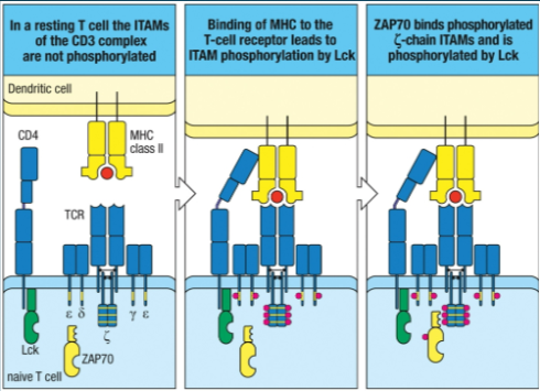
Review slide 44 (Figure 8.10). Synergy between signals produced from the T-cell receptor and co-receptor are efficient in activating a naïve T-cell. During this clustering event of TCRs, co-receptors, and co-stimulator receptors, understand how the following are involved:
CD4 and CD8(cytoplasmic tails)
These co-receptors bind to MHC class II (CD4) or MHC class I (CD8) molecules on the antigen-presenting cell.
Their cytoplasmic tails are associated with the tyrosine kinase Lck.
Lck phosphorylates ITAMs (see below), initiating intracellular signaling.
Review slide 44 (Figure 8.10). Synergy between signals produced from the T-cell receptor and co-receptor are efficient in activating a naïve T-cell. During this clustering event of TCRs, co-receptors, and co-stimulator receptors, understand how the following are involved:
ITAMs of the CD3 complex(ζ chain)
ITAMs (Immunoreceptor Tyrosine-based Activation Motifs) are motifs in the cytoplasmic tails of CD3γ, δ, ε, and especially the ζ chain.
Upon TCR binding to peptide: MHC, ITAMs are phosphorylated by Lck, creating docking sites for other signaling proteins.
Review slide 44 (Figure 8.10). Synergy between signals produced from the T-cell receptor and co-receptor are efficient in activating a naïve T-cell. During this clustering event of TCRs, co-receptors, and co-stimulator receptors, understand how the following are involved:
Lck and ZAP-70
Lck: Src-family tyrosine kinase bound to CD4/CD8. Phosphorylates ITAMs upon TCR activation.
ZAP-70: Binds to phosphorylated ITAMs on the CD3 ζ chain.
Once bound, ZAP-70 is phosphorylated and activated by Lck, initiating multiple downstream signaling cascades (e.g., activating transcription factors like NFAT, AP-1, NF-κB).
Review slide 44 (Figure 8.10). Synergy between signals produced from the T-cell receptor and co-receptor are efficient in activating a naïve T-cell. During this clustering event of TCRs, co-receptors, and co-stimulator receptors, understand how the following are involved:
How is “clonal selection” observed here?
Clonal selection refers to the specific activation of only those T cells whose TCRs recognize the presented antigen.
Once a T cell is activated by TCR + CD4/CD8 + CD28 signals:
It expresses IL-2 and IL-2 receptors (high-affinity).
IL-2 binding drives proliferation (clonal expansion) of this specific T-cell clone.
This results in a large population of antigen-specific effector T cells.
Review slide 45 (Figure 8.11)

Review slide 45 (Figure 8.11) that summarizes the intracellular signaling pathways upon the initial interaction between a naïve T cell and a dendritic cell. From this slide, you should be familiar with the following:
When is ZAP-70 activated and by which cell type?
ZAP-70 is activated in the naïve T cell after:
The TCR binds to peptide:MHC on a dendritic cell.
Lck, a kinase associated with CD4/CD8, phosphorylates ITAMs on CD3.
ZAP-70 binds to phosphorylated ITAMs and is itself phosphorylated by Lck, becoming active.
Review slide 45 (Figure 8.11) that summarizes the intracellular signaling pathways upon the initial interaction between a naïve T cell and a dendritic cell. From this slide, you should be familiar with the following:
PLC-γ
Activated ZAP-70 triggers signaling pathways that activate PLC-γ.
PLC-γ cleaves PIP2 (phosphatidylinositol bisphosphate) into two second messengers:
DAG (diacylglycerol)
IP3 (inositol trisphosphate)
Review slide 45 (Figure 8.11) that summarizes the intracellular signaling pathways upon the initial interaction between a naïve T cell and a dendritic cell. From this slide, you should be familiar with the following:
PIP2
PIP₂ is a membrane phospholipid that is cleaved by the enzyme PLC-γ1 (phospholipase C-gamma 1) into two second messengers:
DAG (diacylglycerol) – remains in the membrane.
IP₃ (inositol 1,4,5-trisphosphate) – diffuses into the cytosol.
These products initiate parallel signaling pathways that activate critical transcription factors.
Review slide 45 (Figure 8.11) that summarizes the intracellular signaling pathways upon the initial interaction between a naïve T cell and a dendritic cell. From this slide, you should be familiar with the following:
DAG & IP3
DAG:
Remains in the membrane and activates Protein Kinase C (PKC) and RasGRP, leading to activation of the transcription factor NFκB and AP-1.
IP3:
Increases intracellular Ca²⁺ levels, which activates calcineurin, leading to NFAT activation.
Review slide 45 (Figure 8.11) that summarizes the intracellular signaling pathways upon the initial interaction between a naïve T cell and a dendritic cell. From this slide, you should be familiar with the following:
NFκB, NFAT, and AP-1
NFκB
Activated via the DAG/PKCθ pathway.
DAG activates Protein Kinase C-θ, which leads to the degradation of IκB, an inhibitor of NFκB.
NFκB translocates to the nucleus, promoting transcription of genes involved in:
Cell survival
Cytokine production (like IL-2)
T cell proliferation
NFAT
Activated via the IP₃/Ca²⁺/calcineurin pathway.
IP₃ causes release of Ca²⁺ from the endoplasmic reticulum.
Increased cytosolic Ca²⁺ activates calcineurin, a phosphatase.
Calcineurin dephosphorylates NFAT, allowing it to enter the nucleus and initiate gene transcription, including IL-2 production.
AP-1
Formed by the combination of Fos and Jun proteins.
Activated downstream of Ras/MAPK signaling (triggered by DAG).
Works synergistically with NFAT to bind composite DNA elements and activate gene transcription.
Review slide 45 (Figure 8.11) that summarizes the intracellular signaling pathways upon the initial interaction between a naïve T cell and a dendritic cell. From this slide, you should be familiar with the following:
Understand what produces a change in gene expression and when this would occur during the intracellular signaling mechanism. Explain the importance of the cytokineIL-2 in this context and how “clonal expansion” is observed (proliferation & differentiation).
Defined as the proliferation of a single activated T cell into a large population of identical effector T cells.
Driven by:
TCR signal
CD28:B7 co-stimulation
IL-2 signaling
What are the components of the high-affinity IL-2 receptor? When IL-2 binds to its receptor, what is the result? If a B cell has been rendered anergic, will it make IL-2?
The high-affinity IL-2 receptor is composed of three chains:
IL-2Rα (CD25)
IL-2Rβ (CD122)
Common γ chain (γc, CD132)
Naïve T cells express only the β and γ chains, which form a low-affinity receptor.
What are the components of the high-affinity IL-2 receptor? When IL-2 binds to its receptor, what is the result? If a B cell has been rendered anergic, will it make IL-2?
Upon T cell activation, the α chain (CD25) is upregulated, allowing the assembly of the high-affinity IL-2 receptor.
What are the components of the high-affinity IL-2 receptor? When IL-2 binds to its receptor, what is the result? If a B cell has been rendered anergic, will it make IL-2?
If a T cell has been rendered anergic, will it make IL-2?
No, anergic T cells do not produce IL-2.
Anergy occurs when a TCR binds antigen without receiving co-stimulation (i.e., no B7-CD28 interaction).
As a result:
IL-2 transcription is not activated.
IL-2 mRNA is not stabilized.
No proliferation or differentiation occurs.
Anergic T cells are permanently non-responsive, even upon future encounters with the same antigen.
This is a mechanism of peripheral tolerance to prevent autoimmunity.
Review slide 55 (Figure 8.14) and slide 77 (Figure 8.21).

Review slide 55 (Figure 8.14) and slide 77 (Figure 8.21).

Review slide 55 (Figure 8.14) and slide 77 (Figure 8.21): Know the 5 types of effector CD4 T cells and their effector functions
CD4 T Cell Subtype | Cytokines Driving Differentiation | Transcription Factor | Secreted Cytokines | Effector Function |
TH1 | IL-12, IFN-γ | T-bet | IFN-γ | Activates macrophages to destroy intracellular pathogens |
TH2 | IL-4 | GATA3 | IL-4, IL-5, IL-13 | Activates eosinophils, basophils, mast cells; promotes IgE class switching |
TH17 | TGF-β + IL-6 or IL-21 | RORγT | IL-17, IL-22 | Recruits neutrophils, enhances inflammation |
TFH | IL-6 | Bcl6 | IL-21 | Helps B cells in germinal centers to produce antibodies and undergo isotype switching |
Treg | TGF-β | FoxP3 | IL-10, TGF-β | Suppresses immune responses, maintains self-tolerance |
Review slide 55 (Figure 8.14) and slide 77 (Figure 8.21): Know the cytokines & transcription factors) responsible for carrying out their effector functions.
CD4 T Cell Subtype | Cytokines Driving Differentiation | Transcription Factor | Secreted Cytokines | Effector Function |
TH1 | IL-12, IFN-γ | T-bet | IFN-γ | Activates macrophages to destroy intracellular pathogens |
TH2 | IL-4 | GATA3 | IL-4, IL-5, IL-13 | Activates eosinophils, basophils, mast cells; promotes IgE class switching |
TH17 | TGF-β + IL-6 or IL-21 | RORγT | IL-17, IL-22 | Recruits neutrophils, enhances inflammation |
TFH | IL-6 | Bcl6 | IL-21 | Helps B cells in germinal centers to produce antibodies and undergo isotype switching |
Treg | TGF-β | FoxP3 | IL-10, TGF-β | Suppresses immune responses, maintains self-tolerance |
Review slide 55 (Figure 8.14) and slide 77 (Figure 8.21): Know the cytotoxins for CD8 T cells.
CD8 T cells release cytotoxins stored in lytic granules:
Perforin: Forms pores in the target cell membrane.
Granzymes: Serine proteases that enter target cells and activate apoptosis via caspase pathways.
Granulysin: Antimicrobial; also contributes to apoptosis.
Serglycin: Acts as a scaffold for storing other cytotoxins in lytic granules.
Review slide 55 (Figure 8.14) and slide 77 (Figure 8.21): Do all effector T cells produce cytokines?
Yes, all effector T cells produce cytokines, though the type and combination varies by subtype:
CD4 T cells (TH1, TH2, TH17, TFH, Treg): Secrete distinct cytokine profiles that shape and regulate immune responses.
CD8 T cells: In addition to cytotoxins, they also produce IFN-γ, which:
Inhibits viral replication,
Enhances antigen presentation (via MHC I),
Activates macrophages.
Differentiate between a TH1 cell-mediated response and a TH2 cell-mediated response. Which is observed for Tuberculoid leprosy, and which is observed for Lepromatous leprosy?
TH1 vs TH2 Cell-Mediated Response: Comparison and Leprosy Forms
Feature | TH1 Response | TH2 Response |
Function | Activates macrophages to destroy ingested microbes | Activates B cells to produce antibodies, especially IgE |
Primary Role | Defense against intracellular pathogens (e.g., viruses, some bacteria) | Defense against extracellular parasites (e.g., helminths) |
Key Cytokines Produced | IFN-γ, IL-2 | IL-4, IL-5, IL-13 |
Transcription Factor | T-bet | GATA-3 |
Induced by | IL-12, IFN-γ (from DCs or NK cells) | IL-4 |
Macrophage Activation | Strong (via IFN-γ and CD40L) | Inhibited (IL-4 suppresses macrophage activation) |
Antibody Production | Helps class switch to opsonizing antibodies | Promotes class switching to IgE |
Inflammatory Response | Pro-inflammatory | Anti-inflammatory / allergic |
Differentiate between a TH1 cell-mediated response and a TH2 cell-mediated response. Which is observed for Tuberculoid leprosy, and which is observed for Lepromatous leprosy?
Leprosy Forms and T Helper Response
Form of Leprosy | Immune Response | T Helper Profile | Outcome |
Tuberculoid Leprosy | Strong CMI (cell-mediated immunity) | TH1-dominant | Bacteria are contained; disease is localized and less severe |
Lepromatous Leprosy | Poor CMI; high antibody levels | TH2-dominant | Bacteria proliferate; widespread lesions; more severe disease |
Explain how CD4 T cells can help activate naïve CD8 T cells and understand how IL-2 and its receptor are involved. Is this a paracrine or autocrine response?
Naïve CD8 T cells require stronger co-stimulation than CD4 T cells to become fully activated. There are two main pathways by which CD8 T cells can be activated:
Direct Activation (Optimal Conditions):
When the dendritic cell provides strong co-stimulation (via B7 binding to CD28), CD8 T cells can become activated directly. Upon activation:
CD8 T cells synthesize IL-2.
They also express the high-affinity IL-2 receptor (αβγ complex).
IL-2 binding triggers proliferation and differentiation into cytotoxic T lymphocytes (CTLs).
CD4 T Cell Help (Suboptimal Co-stimulation):
When co-stimulatory signals are insufficient, CD4 T cells assist CD8 T cell activation:
CD4 and CD8 T cells must interact with the same antigen-presenting cell (APC) (e.g., dendritic cell) displaying their respective peptide:MHC complexes.
Activated CD4 T cells secrete IL-2.
This IL-2 can be used by nearby CD8 T cells to enhance their activation and clonal expansion.
This is a paracrine response, because IL-2 is produced by CD4 T cells but acts on neighboring CD8 T cells.
Can effector T cells be activated in the absence of a co-stimulatory signal? Differentiate between when a T cell is naïve/resting versus when it’s an activated effector T cell. Know the cell-surface molecules associated in this context and their locations. (Hint: L-selectin; LFA-1; VLA-4; VCAM-1)
Yes, effector T cells can be activated in the absence of a co-stimulatory signal, unlike naïve T cells which require both TCR engagement and co-stimulation (usually via CD28–B7 interaction) to become activated.
Can effector T cells be activated in the absence of a co-stimulatory signal? Differentiate between when a T cell is naïve/resting versus when it’s an activated effector T cell. Know the cell-surface molecules associated in this context and their locations. (Hint: L-selectin; LFA-1; VLA-4; VCAM-1)
Naïve (Resting) T Cells:
Require co-stimulation (CD28 binding to B7 on professional APCs).
Surface molecules:
L-selectin (CD62L): allows homing to secondary lymphoid tissues by binding vascular addressins (e.g., GlyCAM-1, CD34) on HEVs.
Low levels of LFA-1 (integrin) and CD2.
No VLA-4 expression.
Location: Secondary lymphoid organs (e.g., lymph nodes), entering via HEVs.
Effector T Cells (Activated):
Do NOT require co-stimulation for activation.
Can recognize antigen on non-PAPCs (e.g., infected epithelial cells).
Surface molecules:
Lose L-selectin (no longer need to home to lymph nodes).
Upregulate LFA-1 and CD2 to strengthen adhesion with target cells.
Express VLA-4, which binds VCAM-1 on inflamed tissue endothelium.
Location: Can migrate into peripheral tissues, especially at sites of infection or inflammation.
Effector CD8 T cells: What is the only cell we discussed that can activate a CD8 T cell without any help?
Dendritic cells
These are the only professional antigen-presenting cells (P-APCs) capable of activating naïve CD8 T cells independently, thanks to their high expression of co-stimulatory molecules like B7 and efficient presentation of antigen on MHC class I.
Effector CD8 T cells: What is an additional way that CD8 T cells can become activated? (Hint: answer part a first to understand which cell type requires help in this situation)
With help from CD4 T cells
If co-stimulation is suboptimal, a CD4 T cell that recognizes the same antigen on the same APC can provide help. The CD4 T cell secretes IL-2, which then acts on the CD8 T cell, stimulating it to proliferate and differentiate into a cytotoxic T lymphocyte.
Effector CD8 T cells: Can CD8 T cells kill more than one cell? Or are they considered to be similar to neutrophils, in that they can only kill one cell and then die?
CD8 T cells can kill multiple target cells
Unlike neutrophils, which often die after one killing event, cytotoxic CD8 T cells are serial killers. They:Release lytic granules to kill an infected target.
Detach and move on to kill other infected cells.
Synthesize new lytic granules between killing events.
Effector CD8 T cells: Know the names of the cytotoxins associated with CD8 T cells and their roles.
Cytotoxin | Function |
Perforin | Forms pores in the target cell membrane, allowing entry of other cytotoxins |
Granulysin | Antimicrobial; also contributes to pore formation and apoptosis induction |
Serglycin | Scaffold that helps package and deliver cytotoxins within lytic granules |
Granzymes | Serine proteases that activate caspases in the target cell, initiating apoptosis |
JAKs and STATs: What type of receptors are being signaled?
Cytokine receptors are being signaled. These receptors usually consist of two or three chains, and they do not possess intrinsic kinase activity but rely on associated kinases for signaling.
JAKs and STATs: Do JAKs come first or STATs?
JAKs (Janus Kinases) come first. When a cytokine binds to its receptor, it induces receptor dimerization, which activates JAKs. These kinases then phosphorylate the receptor itself and also phosphorylate STATs (Signal Transducers and Activators of Transcription), allowing them to dimerize and enter the nucleus.
JAKs and STATs: What determines the genes that are activated as an end result of this pathway?
The specific cytokine that binds to the receptor determines which STATs are activated, and thus, which genes are transcribed. This means the outcome is cytokine-specific, allowing different cytokines to activate distinct transcriptional programs.
JAKs and STATs: Review slide 80 (Figure 8.22)

Differentiate between “Necrosis” and “Apoptosis.”
Necrosis
Definition: Uncontrolled cell death due to physical or chemical injury.
Process:
Cells swell, lyse, and disintegrate.
Cellular contents spill into the surrounding tissue.
Consequence: Often induces inflammation due to release of intracellular contents.
Appearance: Plasma membrane is poorly defined.
Apoptosis
Definition: Programmed cell death — a controlled, regulated process.
Process:
Cells shrink, condense, and form membrane-bound vesicles (apoptotic bodies).
The plasma membrane remains intact.
Consequence: Does not induce inflammation; apoptotic bodies are safely phagocytosed.
Induction by CD8 T Cells:
Triggered by cytotoxins: perforin, granulysin, granzymes, and serglycin.
Ensures safe removal of infected cells without releasing pathogens.
Identify the effector CD4 T cell that is responsible for activating macrophages, which provides an overall increase to its phagocytic capabilities. Explain the two signals that are needed for their activation.
The effector CD4 T cell responsible for activating macrophages is the TH1 (T helper 1) cell.
Activation of Macrophages by TH1 Cell
TH1 cells enhance the microbicidal activity of macrophages, especially against intracellular pathogens that survive inside vesicles (like Mycobacterium tuberculosis).
Identify the effector CD4 T cell that is responsible for activating macrophages, which provides an overall increase to its phagocytic capabilities. Explain the two signals that are needed for their activation.
Two Signals Required for Macrophage Activation:
IFN-γ (Interferon-gamma)
This is the primary signal.
Secreted by TH1 cells upon recognizing their antigen.
Increases phagocytic and killing capacity of macrophages.
CD40 Ligand (CD40L)
Expressed on the surface of the TH1 cell.
Binds to CD40 receptor on the macrophage.
Works in synergy with IFN-γ to fully activate macrophages.
Effects of Activation:
Enhanced fusion of phagosomes with lysosomes.
Increased production of reactive oxygen species (ROS), nitric oxide (NO), and proteolytic enzymes.
Improved ability to kill ingested microbes.
Which effector CD4 T cell prevents macrophages from becoming activated? What do these CD4 T cells activate instead?
The effector CD4 T cell that prevents macrophage activation is the TH2 cell.
Which effector CD4 T cell prevents macrophages from becoming activated? What do these CD4 T cells activate instead?
What Do TH2 Cells Activate Instead?
Rather than enhancing macrophage microbicidal activity (like TH1 cells), TH2 cells activate alternative immune responses suited for fighting extracellular parasites, such as:
B cells → promote antibody class switching, especially to IgE
Eosinophils, mast cells, and basophils
They also support tissue repair and inhibit inflammation.
Mechanism of Inhibition
TH2 cells secrete cytokines such as:
IL-4
IL-5
IL-10
IL-13
These cytokines can inhibit TH1 cell-mediated activation of macrophages, which helps prevent excessive inflammation and tissue damage.
How do TFH cells stimulate the proliferation and differentiation of naïve B cells? Explain the involvement of CD40 and CD40L.
TFH cells (T follicular helper cells) stimulate the proliferation and differentiation of naïve B cells primarily through cognate interaction involving:
CD40 and CD40L Interaction
CD40 is expressed on B cells.
CD40L (CD40 ligand) is expressed on activated TFH cells.
When a TFH cell recognizes antigen presented on MHC class II by a B cell:
CD40L on the TFH cell binds CD40 on the B cell, delivering a crucial co-stimulatory signal.
This interaction is necessary for:
B cell activation
Isotype switching
Affinity maturation
Formation of germinal centers
Differentiation into plasma cells and memory B cells
Linked Recognition (Cognate Interaction)
Even though a B cell may internalize an entire antigen via its BCR, it presents peptides on MHC II to the TFH cell.
The TFH cell doesn’t need to recognize the same epitope, just a peptide derived from the same antigen.
This ensures B cell help is only provided to those B cells presenting relevant antigen, which is a safeguard against inappropriate activation.