chapter 16: disorders of brain function
 Call Kai
 Learn
Learn Practice Test
 Spaced Repetition
 Match
Match Flashcards
 Knowt Play
1/100

There's no tags or description
Looks like no tags are added yet.
Name | Mastery | Learn | Test | Matching | Spaced | Call with Kai | Chat |
|---|
No analytics yet
Send a link to your students to track their progress
101 Terms
3 main parts of brain
cerebrum: divided into 2 hemispheres
cerebellum: little brain
brainstem: divided into midbrain, pons, & medulla oblongata
protective structures of brain
cranial meninges and associated spaces
carnial bones
brainstem
divided into midbrain, pons, and medulla oblong
cerebral circulation
it delivers oxygen and nutrients for its metabolic needs, and to remove CO2 + other waste products
picture of structural parts of brain

cranial meninges
dura mater: thick, tough membrane - outermost
arachnoid mater: lies directly below mater - middle
pia mater: very thin, delicate membrane covering brain surface - inner
circle of willis
network of brain arteries that provide alternative pathways for blood flow in case of blockage

brain injuries classified into two categories:
focal brain injury & global brain injury
focal brain injury
caused by localized neurologic deficits
symptoms correspond to the injured brain region
global brain injury
affects entire brain
usually results in altered consciousness
brain injuries manifested by:
changes in lvl of consciousness
alterations in cognitive function
motor dysfunction
sensory impairment
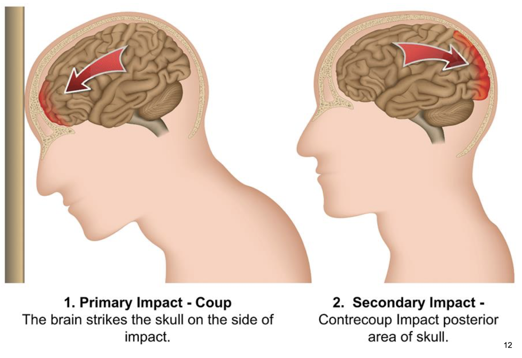
coup-contrecoup injury
type of injury in which brain damaged at both sites of impact due to rapid movement of brain within skull
coup injury
occurs directly beneath site where head is struck
person hit in forehead
frontal lobe impacts inside of skull at point of impact, contusion develops
contrecoup injury
occurs on side opposite of impact
after striking front of skull, brain rebounds backward
occipital region hits inner surface of skull = second contusion develops on opposite side (occipital lobe)
photo of primary and secondary impact
brain injury occurs when:
brain tissue is damaged by a physical force, loss of blood, lack of oxygen, infection, toxins
mechanism of injury depends on cause; there are two types:
primary brain injury and secondary brain injury
primary brain injury
immediate damage occurs at time of insult
ex: head trauma, causing bruising or tearing of brain tissue
mechanism of primary injury
mechanical disruption of neurons and glial cells
tearing of axons
direct injury to blood vessels
secondary brain injury
develops minutes to days after initial injury and often causes additional damage
ex: ischemia and hypoxia, excitotoxicity
ischemia and hypoxia (secondary brain injury)
reduced blood flow or oxygen delivery deprives neurons of oxygen and glucose
ATP production fails
ion pumps fail, causing cellular swelling + dysfunction
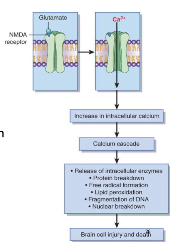
excitotoxicity (secondary brain injury)
injured neurons release excessive amounts of neurotransmitter glutamate
glutamate stimulates NMDA & AMPA receptors
excess calcium enters cells
result in activation of destructive enzymes, damage to cell membranes, proteins, and DNA
role of glutamate NMDA receptor in brain cell injury:
glutamate is principal excitatory NT in brain
prolonged ischemia results in glutamate transporters become immobilized and ineffective, extracellular glutamate accumulates
concussion
temporary disturbance of brain function caused by blow to head, resulting in disruption to delicate axonal fibers + white matter tracts that project cerebral cortex
no structural brain damage visible on imaging
temp neurological dysfunction
may involve brief loss of consciousness or none at all
signs & symptoms of concussion
headache, dizziness, confusion
amnesia
nausea and vomiting
blurred vision and sensitivity to light or noise
contusion
bruise of brain tissue caused by direct trauma, resulting in structural damage and localized bleeding
more severe than concussion
involves hemorrhage, edema, and tissue injury
often occurs beneath site of impact (coup injury) or opposite side of brain (contrecoup injury)
signs & symptoms of contusion
signs of increased intracranial pressure
altered lvl of consciousness, persistent headache, nausea, and vomiting
seizures, weakness, or paralysis
hematomas
result from injury to blood vessels and subsequent bleeding
blood can accumulate within diff compartments of skull
classified into four main types:
epidural, subdural, intracerebral
epidural hematoma
collection of blood between inner surface of skull and dura mater (outermost meningeal layer)
caused by rupture of middle meningeal artery
manifestations: an initial loss of consciousness, a temporary "lucid interval" of seeming recovery, and a subsequent rapid neurological decline
subdural hematoma
collection of blood between dura mater and arachnoid mater
results from tearing of bridging veins that connect cerebral cortex to dural venous sinuses
venous bleeding occurs more slowly than arterial bleeding seen in epidural hematomas
manifestations similar to epidural hematoma
intracerebral hematoma
collection of blood within brain tissue
results from severe movement of brain during head trauma or progression of cerebral contusion into hematoma
manifestations: vary depending on size + location of hematoma
picture of locations of hematomas

picture of mechanisms of bleeding in subdural hematoma

reticular activating system (RAS)
network of neurons located in brainsteam (midbrain, pons, and upper medulla)
plays role in regulating arousal, alertness, wakefulness, and consciousness
what sensory input does RAS receive and what does it receive
it receives sensory input from visual, auditory, and tactile sensory pathways!
what do these signals from RAS increase?
cortical activity = enabling awareness, attention, and wakefulness
level of consciousness (LOC)
degree of awareness of self + environment and ability to respond to stimuli
stimuli: full consciousness, confusion, lethargy, obtundation, stupor, coma (cant be awakened by any stimuli)
lethargy (LOC)
reduction in mental alertness where person appears drowsy
very slow in mental processes, motor activity, and speech
responds appropriately to painful stimuli
obtundation (LOC)
decreased alertness and interest in environment
requires repeated verbal stimulation to maintain attention
responses are slower and less accurate
stupor (LOC)
can only be aroused by vigorous or painful stimuli
minimal verbal responses
returns to unresponsive state when stimulation stops
glasgow coma scale (GCS)
neurological scoring system used to assess LOC after head injury or in critically ill patients
components of GCS include:
eye opening (E): scores 4-1
verbal response (V): scores 5-1
motor response (M): scores 6-1
total score:
GCS = E + V + M; ex: 3+4+6=13/15
max: 15 (fully alert)
min: 3 (deep coma)
abnormal flexion and extension are:
abnormal motor responses assessed in GCS!
decorticate and decerebrate posturing are:
indicators of severe neurological injury
postures help localize neurological lesions + assess severity
as coma progresses, motor tracts are interrupted at specific lvls initiate rigidity + abnormal postures
decorticate posture
appearance:
flexion of arms, wrists, and fingers
arms adducted toward body
lower extremities internally rotated, feet plantar flexed
cause:
lesions of cerebral hemispheres
damage to internal capsule

decerebrate posture
appearance:
arms rigid and extended
palms turned away from body
legs stiffly extended
feet plantar-flexed
cause:
lesions involve diencephalon, midbrain, and upper brainstem
associated w/ poorer prognosis than decorticate posturing

brain death
irreversible loss of function + entire brain, including brain stem
body cannot maintain internal homeostasis
no conscioussness, no brain reflexes (pupils don’t react to light), and no ability to breathe independently
cerebral death
irreversible loss of function of cerebral hemispheres; brainstem function is present
no awareness of higher mental function
brainstem may still control breathing, heart rate, and reflexes
intracranial pressure (ICP)
pressure that opposes blood entry into brain
total intracranial volume remains constant so normal adult ICP is 5-15 mmHg
ICP reflects balance between
brain balance (~80%)
cerebral blood volume (~10%)
cerebrospinal fluid (~10%)
increase in one component must be compensated by a decrease in one or both of the others; otherwise ICP rises!
monro-kellie hypothesis
brain tissue, blood, and CSF volumes reciprocally adjust to maintain a normal ICP
change in one component is balanced by equal + opposite effect in one or both of remaining components

blood reaches brain only when:
cerebral perfusion pressure (CPP) is adequate!
CPP = mean arterial pressure (MAP) - intracranial pressure (ICP)
mean arterial pressure (MAP):
pressure pushing blood into brain
when ICP rises:
compression of cerebral blood vessels occurs
CPP decreases
cerebral blood flow falls
brain tissues receives less oxygen and glucose
cerebral ischemia + neuronal injury develop
brain herniation
displacement of brain tissue from normal position due to increased ICP, causing it to move across rigid intracranial structures such as: falx cerebri, tentorium cerebelli, or foramen magnum
life-threatening neurological emergency
compression of vital brain structures and blood vessels
common causes of brain herniation
traumatic brain injury
intracerebral hemorrhage
brain tumors
types of brain herniation:
cingulate, uncal, central
cingulate (brain herniation)
cingulate gyrus is pushed beneath falx cerebri
may compress anterior cerebral artery
signs: unilateral or bilateral leg weakness
uncal (brain herniation)
medical temporal lobe (uncus) herniates through tentorial notch
compresses ipsilateral third cranial nerve
signs: dilated, fixed ipsilateral pupil, decreased consciousness
central (brain herniation)
downward displacement of diencephalon and brainstem
signs: progressive loss of consciousness, decorticate posturing
picture of brain herniation

hydrocephalus
condition in which excess cerebrospinal fluid accumulates within ventricles of brain
three types: noncommunicating, communicating, overproduction
noncommunicating (hydrocephalus)
CSF flow blocked within ventricular system
ex: aqueductal stenosis, tumors, congenital malformations
communicating (hydrocephalus)
CSF reaches subarachnoid space but not adequately absorbed
ex: meningitis, arachnoid villi dysfunction
overproduction (hydrocephalus)
overproduction of CSF
ex: choroid plexus tumors
characteristics of hydrocephalus:
increased amount of CSF in brain’s ventricles
enlargment of ventricles due to CSF accum
increased ICP which causes headaches, vomiting, or changes in vision
stroke
acute focal neurological deficit caused by vascular disorder resulting injury to brain tissue
occurs when blood flow to region of brain is interrupted or when cerebral bv ruptures
types: ischemic, hemorrhagic
ischemic stroke
acute neurological deficit causing brain tissue injury
cause: interruption or blockage of blood flow within cerebral artery
results in reduced oxygen + nutrient delivery to brain tissue

ischemic stroke results from:
thrombosis: the formation of a blood clot (called a thrombus) inside a blood vessel or heart chamber, which slows or blocks normal blood flow
embolism: when a traveling substance—such as a blood clot, fat, air bubble, or amniotic fluid—gets stuck in a blood vessel and blocks normal blood flow
transient ischemic attack (TIA)
temp interruption of cerebral blood flow
similar causes to ischemic stroke: atherosclerosis
symptoms (similar to stroke) resolve within minutes to hours
no perm brain damage occurs
picture of TIA and ischemic stroke

thrombotic stroke (ischemic stroke)
caused by blood clot formation within cerebral arteries
slow onset of neurological symptoms
associated w/ atherosclerosis
lacunar stroke (ischemic stroke)
small vessel ischemic stroke affecting deep brain structures (basal ganglia, brain stem)
often due to chronic hypertension causing small vessel disease
embolic stroke (ischemic stroke)
caused by traveling blood clot (embolus)
occurs when clot or debris travels through bloodstream to brain
sudden onset of neurological symptoms
emboli originate from: thrombus in left atrium or ventricle
hemorrhagic stroke
cerebral blood vessel rupture causes bleeding in and around the brain tissue and spaces
caused by rupture of cerebral bv, resulting in bleeding within or around brain
causes brain injury due to: comrpession, swelling (edema), increased ICP
two types: intracerebral, subarachnoid

intracerebral hemorrhage (hemorrhagic stroke)
bleeding directly into brain tissue
subarachnoid hemorrhage (hemorrhagic stroke)
bleeding into space arachnoid and pia mater, often due to ruptured aneurysm
picture of risk factors


picture of stroke manifestations !!

motor deficits with strokes:
most common consequence of a stroke
damage of corticospinal tract causes weakness on contralateral side of body
initial phase of stroke-related motor deficits
flaccidity (decreased muscle tone)
severe weakness
recovery phase of stroke-related motor deficits
6-8 weeks
hyperreflexia develops
spasticity replaces flaccidity
stroke-related dysathria
stroke-related, communication disorders after stroke
impaired articulation of speech sounds
changes in voice quality or pitch
results from weakness of muscles controlling pharynx, palate, tongue, lips
doesn’t affect language comprehension
stroke-related aphasia
stroke-related, communication disorders after stroke
decline in speech and language comprehension
seizure
sudden, transient disruption in brain electrical function caused by abnormal excessive discharges of cortical neurons
symptoms of disease, but no specific disease
associated w/ metabolic derangements, infections brain tumors, drug abuse, vascular lesions
single ep of abnormal neuronal discharge
divided into focal and generalized
epilepsy
recurrence of seizures and disorder for which no cause can be found
chronic neurological disorder
characterized by recurrent seizures
focal seizure
begin in specific (focal) area of one cerebral hemisphere
symptoms depend on location of affected brain region
may remain localized or spread to other areas
can classify into: focal aware, focal impaired awareness
focal aware seizures
consciousness remains intact
symptoms: depend on area, but can include: jerking of limb, tingling sensations, visual or auditory disturbances, emotional changes
focal impaired awareness seizures
altered or impaired consciousness
may involve automatisms like: lip smacking, chewing, repetitive hand movements, often followed by confusion
generalized seizures
begin simultaneously in both cerebral hemispheres
involve widespread brain activity from onset
usually affect consciousness + may produce bilateral motor symptoms
classifies into: absence, tonic, clonic, tonic-clonic, status epilepticus
absence generalized seizure
brief loss of awareness
common in children, usually last a few seconds
tonic generalized seizure
sudden muscle stiffening that may cause falls
clonic generalized seizure
repetitive rhythmic jerking movements
tonic-clonic generalized seizure
most common type
tonic phase: muscle stiffening
clonic phase: rhythmic jerking
often followed by postictal period of confusion + fatigue
status epilepticus generalized seizure
continuing/recurring seizures w/ incomplete recovery, unrelenting seizure activity that lasts 30 mins or more
medical emergency
seizure sequence
prodromal phase: occurs before some seizures
aura phase: earliest symptoms of seizure
common symptoms: unsual smells, taste, or sound
ictal phase (seizure event): period of active seizure
postictal phase: recovery phase following seizure
exhaustion, headache, weakness, confusion
dementia
chronic, progressive decline in cognitive function
decreased cortical function leads to range of cognitive and behavioral impairments, including:
impaired cognitive skills, impaired thinking, judgment, learning, memory loss, confusion, personality changes
common types of dementia: alzheimer’s disease, vascular dementia, lewy body dementia (dementia associated with advanced PD)
alzheimer’s disease
progressive cortical atrophy
accumulation of beta-amyloid plaques between neurons
formation of neurofibrillary tangles (tau proteins) inside neurons
neuronal damage + brain atrophy, especially in hippocampus, and reduced lvls of acetylcholine (neurotransmitter for memory)
cause unknown, but risk factors include advanced age, genetics/history
picture of alzheimer disease

early signs for alzheimer’s disease (AD)
impaired learning, poor judgment
mild memory loss of recent events
difficulty finding words
middle and advances stages for alzheimer’s disease (AD)
behavioral changes
increased confusion and disorientation
difficulty recognizing familiar people
gradual loss of memory and lack of concentration
decline of cognitive function, and language