BI 233: Digestive system histology
 Call Kai
 Learn
Learn Practice Test
 Spaced Repetition
 Match
Match Flashcards
 Knowt Play
1/34

There's no tags or description
Looks like no tags are added yet.
Name | Mastery | Learn | Test | Matching | Spaced | Call with Kai | Chat |
|---|
No analytics yet
Send a link to your students to track their progress
35 Terms
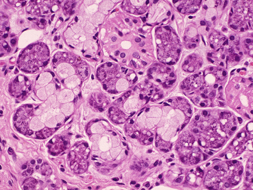

Salivary Glands

Salivary gland: red→Mucus Acini. Yello—>Salivary Duct. Blue—>Serous Demilunes

Salivary Gland: Green>Salivary Duct. Blue>Serous Demilunes

Mucus Acini cells of salivary glands


Serous Demilunes (Dark boarder around white cells)

Salivary Ducts (simple cuboidal)


Stomach

Stomach


Gastic Pit

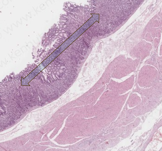
Mucosa layer of stomach (Simple Columnar E.)


What layer of the stomach? Which cells are found in this layer?
Green: Gastric Pit, Blue:Gastric Glands. Cells located within the Gastric Glands of the mucosa.
Mucous Neck Cells: Secrets Alkaline mucus coasts stomach and protects it from acids
Chief Cells:Secrets pepsinogen (converts into pepsin enzyme contacted with HCl to break down proteins). Gastric Lipase enzyme breaks down lipids into fatty acids in stomach Parietal cells: Produce HCl (Stomach Acid) and intrinsic factor (Vit. B12 absobtion) G Cells (Entroendocrine): Secret the hormone Gastrin into blood. Stimulates digestion


Submucosa of Stomach


#1a:Gastric Pit. #1b: Gastric Glands-cells (a&b part of Mucosa Layer). #2:Muscularis Mucosa #3:Submucosa

#1a:Simple Columnar E. of stomach Mucosa, Gastric pit (White crypt) #1b:Gastric glands #2:Submuosa of stomach #2:Muscularis Externa


Stomach 1)Mucosa 2)Submucosa 3)Muscularis Externa 4)Serosa

Serosa of stomach

Muscularis externa of Stomach


Duodenum-Small Intestine
Mucosa-Simple (Columnar E.): Vili and Intestinal Crypts
Submucosa: Duodenal Glands secret alkaline mucus
Muscularis Externa then Serosa


Villi of Duodenum (S. Intestine) in Mucosa.
Plica Circularis: Large+deep folds of Mucosa and Submucosa. Vili sit on too of plica circularis


Duodenal/Brunner’s Glands in Submucosa (beneath the Vili).
Secrets Alkaline mucous


Intestinal Crypts of Duadenum


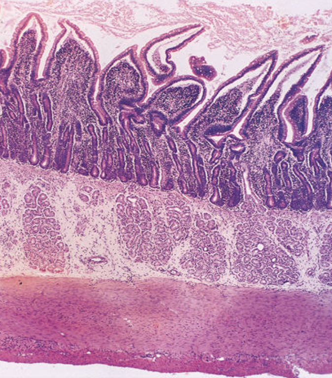

Duodenum


Ileum of S. Intestine
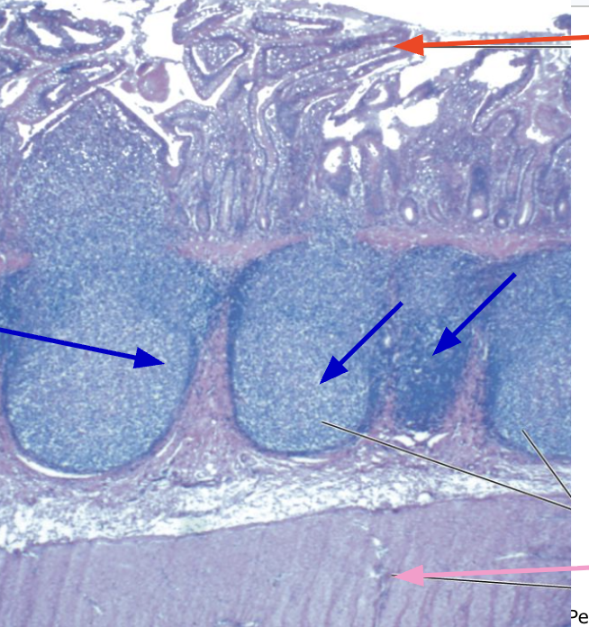
Ileum: red>Vili in mucosa. Blue>Peyer’s Patches in Submucosa. Pink.Muscularis Externa
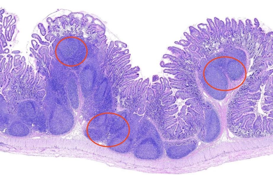
Peyer’s Patches of the Ileum (s. intestine) located in Submucosa


Small Intestine: Duodenum-Jejunum-Ilium

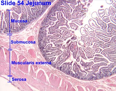

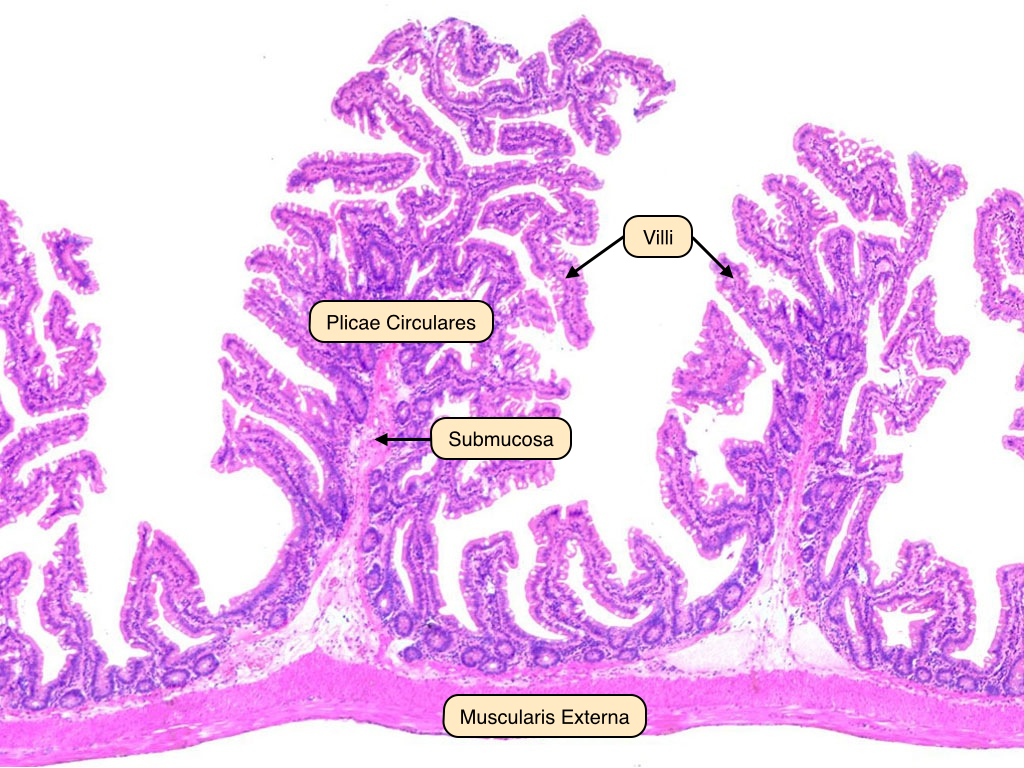
Jejunum (notice absence of Peyer’s patches and duodenal glands)

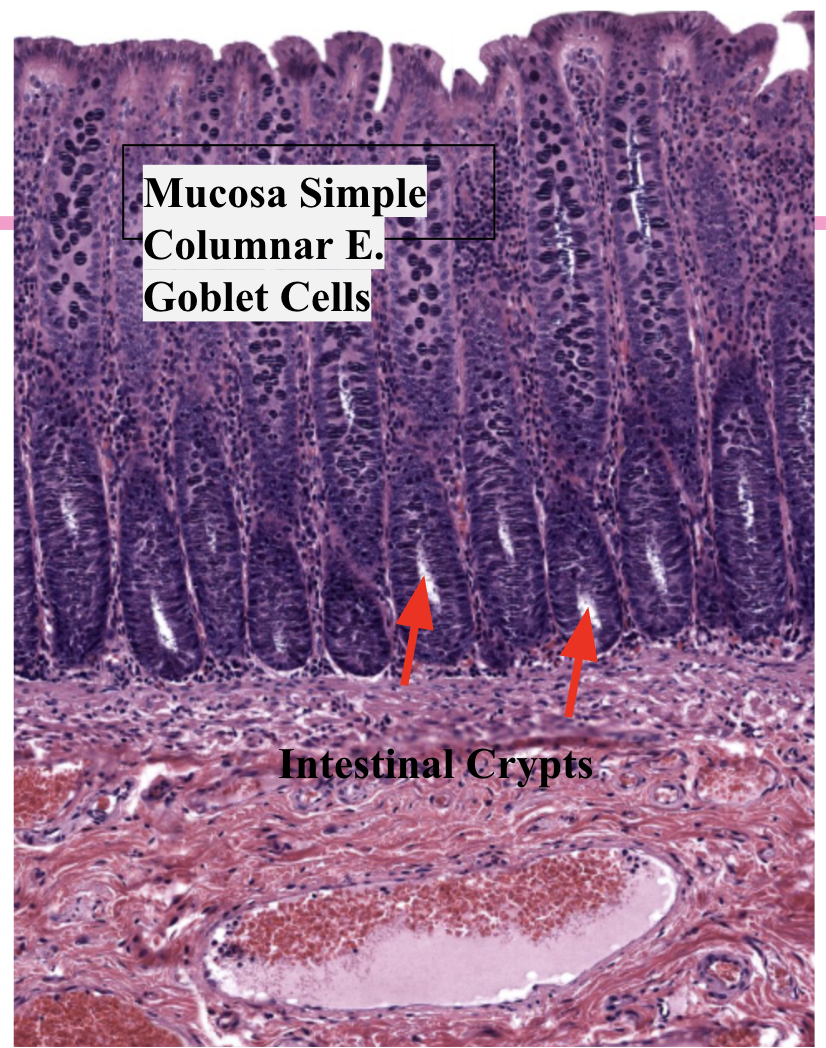
Large Intestine- Mucosa includes Simple Columnar E. with many goblet cells secreting mucus


L. Intestine: Intestinal Crypts (Deep white pockets)

Portal Triads of the liver- Found at corners of Lobules

Lobules of Liver


What cell?
Goblet cells secret mucus

Liver

Sinusoids of Liver

Hepatocyte cells of liver
